

Abstract
Rotaviruses are an important cause of severe diarrheal illness in children globally. We characterized rotaviruses sampled in humans, insectivores (shrews) and rodents from urban and rural regions of Zhejiang province, China. Phylogenetic analyses revealed seven genotypic constellations of human rotaviruses with six different combinations of G and P genotypes – G3P[8] (50.06%), G9P[8] (36.16%), G1P[8] (8.92%), G2P[4] (4.63%), G3P[3] (0.12%), and G3P[9] (0.12%). In rodents and shrews sampled from the same locality we identified a novel genotype constellation (G32-P[46]-I24-R18-C17-M17-A28-N17-T19-E24-H19), a novel P genotype (P[45]), and two different AU-1-like rotaviruses associated with a G3P[3] genotype combination. Of particular note was a novel rotavirus from a human patient that was closely related to viruses sampled from rodents in the same region, indicative of a local species jump. In sum, these data are suggestive of the cross-species transmission of rodent rotaviruses into humans and for reassortment among human and animal rotaviruses.
Keywords: Rotavirus, Evolution, Reassortment, Genotype, Human infection, Rodents, Shrews
Highlights
-
•
Rotaviruses are an important cause of severe diarrheal illness.
-
•
Although rotaviruses are associated with a diverse range of animals, relatively little attention has been directed toward rotaviruses in rodents.
-
•
However, as rodents often live in close proximity to humans and domestic animals, rodents may play an important role in the cross-species transmission of rotaviruses among animals and perhaps directly or indirectly to humans.
-
•
Our data suggest the direct spill-over of rodent rotaviruses in human populations, as well as the reassortment between human and zoonotic rotaviruses.
1. Introduction
Rotaviruses are an important cause of severe diarrheal illness in infants and young children globally, causing 453,000 deaths per year in those <5 years of age (Estes and Greenberg, 2013, Tate et al., 2008). Rotaviruses are members of the genus Rotavirus of the family Reoviridae and possess a genome comprising 11 double-stranded RNA segments that encodes 6 structural (VP1-VP4, VP6, VP7) and 6 non-structural (NSP1-NSP6) proteins. Of the documented species (rotavirus A-H) (Matthijnssens et al., 2012) and the tentative species (rotavirus I) (Mihalov-Kovács et al., 2015), rotavirus A (RVA) is responsible for the majority of seasonal endemic diarrheal disease in young children. Based on sequences of the VP7 and VP4 genes RVAs have been further classified into G and P genotypes, respectively. To date, at least 31 G and 44 P genotypes have been identified worldwide (Matthijnssens et al., 2011, Trojnar et al., 2013; Rotavirus Classification Working Group (RCWG), 2015). Recently, a uniform system for rotavirus nomenclature was established for RVA based on nucleotide identities of the 11 rotavirus genome segments and phylogenetic analysis (Matthijnssens et al., 2008). Globally, the most common combinations in humans are G1, G2, G3, G4 or G9 and the P[4] or P[8] genotypes, especially G1P[8] which accounts for approximately 31% of human strains globally (Bányai et al., 2012, Dóró et al., 2014). Notably, genotype distributions vary among geographic regions, and several unusual strain combinations have recently been identified (Dóró et al., 2014, Nordgren et al., 2012).
As well as humans, rotaviruses infect a wide range of vertebrates including domestic and wild mammals and birds (Estes and Greenberg, 2013). Rodentia (rodents) is the largest order of mammals, with approximately 2277 species distributed globally (Wilson and Reeder, 2005). It is therefore no surprise that rodents are also the largest zoonotic source of human infectious diseases (Luis et al., 2013, Meerburg et al., 2009). In addition to known pathogens, rodents are also an obvious source for the discovery of novel viruses, and new rodent arenaviruses, coronaviruses, and hantaviruses have been described recently (Gonzalez et al., 2007, Guo et al., 2013, Holmes and Zhang, 2015, Li et al., 2015, Wang et al., 2015). To date, however, relatively little attention has been directed toward the rotaviruses that might circulate in rodent populations (Everest et al., 2011, Firth et al., 2014, Greenwood and Sanchez, 2002, Linhares et al., 1986, Sachsenröder et al., 2014). However, as rodents (especially rats) often live in close proximity to humans and domestic animals, and at high densities, they may play an important role in the cross-species transmission of rotaviruses, including to human populations.
Zoonotic rotavirus infections in humans are not uncommon, and there are a growing number of reports describing the interspecies transmission of rotavirus among animals and from animals to humans (Ben Hadj Fredj et al., 2013, Bonica et al., 2015, Gautam et al., 2015, Martella et al., 2010, Medici et al., 2015, Zhou et al., 2015). Like influenza viruses, zoonotic rotaviruses can become increasingly “humanized” by reassortment with co-infecting human viruses (Cowley et al., 2013, Jeong et al., 2014, Matthijnssens et al., 2009, Matthijnssens et al., 2010, Theuns et al., 2015). Clearly, to better understand the evolution and emergence of rotaviruses it is important to determine the diversity, evolution and origins of rotaviruses in those animals that live in close proximity to humans, including rodents.
Wenzhou is located on the southwestern coast of Zhejiang province, China, and is a prefecture-level city (Figure S1). Wenzhou incorporates both urban and rural areas with a total population of 9.12 million, of which 1.69 million can be classed as pediatric. Wenzhou experiences a high level of rotavirus infections, totaling >65,000 cases each year. Longquan, also in Zhejiang province, is located 250 km west of Wenzhou, and is a county-level city with a population of approximately 280,000. More than 90% of the Longquan's total area is mountainous. Compared with Wenzhou, Longquan experiences a relatively low level of rotavirus infection. In this study we investigated the diversity of rotaviruses in humans and small mammals (rodents and shrews) in Wenzhou and Longquan, as well as the evolutionary relationships between these viruses and those circulating in the local human population.
2. Method and materials
2.1. Patient sampling
Between October 2013-December 2014 a total of 1099 stool specimens were collected from diarrheal patients visiting the Diarrheal Department of the Second Affiliated Hospital and Yuying Children's Hospital of Wenzhou Medical University, Wenzhou city, Zhejiang province, China. Patient demographic data and clinical symptoms, including any complications, were collected and evaluated. Signed individual written informed consent for fecal sample collection was obtained from the guardian of each patient.
2.2. Trapping of small animals and sample collection
Between March 2013–October 2014, 1865 small mammals (rodents and insectivores) were captured in Longwan, Lucheng, Ruian, and Wencheng counties/districts of Wenzhou city, as well as from Longquan city, as described previously (Guo et al., 2013) (Fig. S1). All animals were treated according to the “Rules for Implementation of Laboratory Animal Medicine” from the Ministry of Health, China. Trapped animals were identified by morphological examination and further verified by sequence analysis of the cytochrome b (Cyt-b) gene (Guo et al., 2013). All animals were anesthetized before they were sacrificed, and every effort was made to minimize suffering. Stool samples were collected from each animal and used for the detection of rotaviruses.
2.3. RT-PCR and sequencing
Total RNA was extracted from stool samples using the Bioteke fecal RNA isolation kit (Bioteke, Beijing, China) according to the manufacturer's instructions. All samples were screened for the presence of rotaviruses using nested RT-PCR. Two pairs of primers based on conserved regions of sequences of the VP7 segment of known group A rotaviruses were used (Table S1). RT-PCR was performed under the following conditions: incubation at 50 °C for 30 min and 94 °C for 3 min, 40 cycles of denaturation at 94 °C for 40 s, annealing at 46 °C for 40 s, and extending at 72 °C for 60 s. The second amplification was performed as follows: incubation at 94 °C for 3 min, 40 cycles of denaturation at 94 °C for 40 s, annealing at 46 °C for 40 s, and extending at 72 °C for 50 s. Both the VP7 and VP4 genes were recovered from each of the rotavirus positive samples. Additionally, at least one complete genome sequence of each genotype was recovered by RT-PCR using primers based on conserved regions of known viruses. All primer sequences are described in Table S2.
PCR products purified using the QIAquick Gel Extraction kit (Qiagen) were sent to Shanghai Sangon Biological Engineering Technology and Services Co., Ltd. (Shanghai, China) for sequencing. They were sequenced with the PCR primers in both directions on an ABI Prism 3130 automated capillary sequencer (Applied Biosystems, Foster City, CA). Additionally, primer walking was performed to obtain the complete sequence of large genomic segments. Sequencing results were compared with the non-redundant nucleotide database at GenBank using Blastn to confirm they were of rotavirus origin. All rotavirus sequences obtained here from humans and animals have been deposited in GenBank and assigned accession numbers KU243375-KU243694 (Table S3).
2.4. Phylogenetic analysis
In addition to the sequences recovered here, reference sequences that cover the phylogenetic diversity of rotaviruses were compiled for evolutionary analyses. The following rotavirus data set sizes were used: VP7=84 sequences; VP4=70 sequences; VP1=72 sequences; VP2=73 sequences; VP3=72 sequences; VP6=76 sequences; NSP1=77 sequences; NSP2=76 sequences; NSP3=75 sequences; NSP4=73 sequences; NSP5=74 sequences. The 5′ and 3′ terminal sequences of each segment complementary to the primers were excluded.
All viral genome sequences obtained in this study were manually edited using the Seqman program implemented in the DNAStar v7.1 software package (DNASTAR, Inc., USA). Complete coding regions were aligned using the ClustalW method implemented in MEGA v5.05 (Tamura et al., 2011). DNAStar was also used to calculate nucleotide and amino acid identities.
Phylogenetic trees of these data were inferred using the maximum likelihood (ML) method implemented in the PhyML v3.0 package (Guindon et al., 2009). The best-fit nucleotide substitution model was determined using jModeltest (Posada 2008). Accordingly, the VP1, VP2, VP3, NSP1 and NSP4 segments were analyzed using the HKY+I+Γ model; the VP4, VP6, NSP2 and NSP3 segments were analyzed using the GTR+Γ model; VP7 was analyzed using the HKY+Γ model; while NSP5 was analyzed using the GTR+I+ Γ model. In addition, 1000 bootstrap replicates were used to assess the support for individual nodes on each tree.
3. Results
3.1. Human RVA cases and clinical epidemiology
During 2013-2014 we studied a total of 1099 diarrheal patients who visited the Diarrheal Department of the Second Affiliated Hospital and Yuying Children's Hospital, Wenzhou city. Of these, 863 (78.53%) were confirmed as experiencing rotavirus infection based on RT-PCR targeting the viral VP7 gene. None of these patients were hospitalized. Of the patients with confirmed rotavirus infection, 99.07% were natives of Wenzhou, 0.58% were from surrounding areas, while 0.35% were from the “floating” (i.e. transient) population from other Chinese provinces. Their ages ranged from 2 months to 12 years and 66.5% were male.
3.2. Diversity of rotaviruses circulating in children with diarrhea in Zhejiang province
To better understand the genetic diversity of rotaviruses in human patients, nucleotide sequences of both the VP7 and VP4 gene segments were recovered from each of rotavirus positive samples ( Table 1). Based on phylogenetic analysis of the VP7 segment sequences ( Fig. 1), we determined that these infections were caused by RVAs at the following frequencies: G1 (8.92%), G2 (4.63%), G3 (50.06%), and G9 (36.16%). This analysis also revealed that G3 viruses (55.45%) were the most common in 2013, but decreased substantially in frequency (9.8%) in 2014, at which point G9 viruses became the predominant genotype (67.64%). Among the P genotypes, P[8] was at the highest prevalence (95.14%), followed by P[4] (4.63%), while P[3] and P[9] were only rarely detected (0.12% and 0.12%, respectively). Overall, the G3P[8] combination (50.06%) was the predominant genotype, followed by G9P[8] (36.16%), G1P[8] (8.92%), G2P[4] (4.63%), G3P[3] (0.12%), and G3P[9] (0.12%) (Table 1).
Table 1.
The G-P combinations of rotaviruses identified in patients from Zhejiang province, China, during 2013–2014.
| Year | PCR positive/ Total (%) | Genotype (%) |
|||||
|---|---|---|---|---|---|---|---|
| G1P[8] | G2P[4] | G3P[8] | G9P[8] | G3P[3] | G3P[9] | ||
| 2013 | 761/987 (77.10) | 60 (7.88) | 34 (4.47) | 422 (55.45) | 243 (31.93) | 1 (0.13) | 1(0.13) |
| 2014 | 102/112 (91.07) | 17 (16.67) | 6 (5.88) | 10 (9.8) | 69 (67.64) | 0 (0) | 0(0) |
| Total | 863/1099 (78.53) | 77 (8.92) | 40 (4.63) | 432 (50.06) | 312 (36.16) | 1 (0.12) | 1 (0.12) |
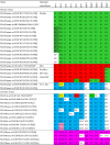
Fig. 1.

Phylogenetic analyses of the VP7 and VP4 segments of rotaviruses identified in humans (red) and rodents and shrews (blue) sampled in Zhejiang province, China. Genotypes are indicated on the right-hand side of the figure, with those newly described here shown in green. The sequences covered nt 49-1034 in VP7 and nt 10-2337 in VP4 with reference to strain RVA/Human-tc/USA/Wa/1974/G1P[8]. The trees are mid-pointed rooted for clarity only and all horizontal branches are drawn to a scale of nucleotide substitutions per site. Bootstrap support values (>70%) are also shown. (For interpretation of the references to color in this figure legend, the reader is referred to the web version of this article.)
3.3. Rotaviruses in local rodents and insectivores
To understand the role played by local small mammals in the evolution and cross-species transmission of rotaviruses, we performed a molecular epidemiological surveillance of rotaviruses in rodents and shrews in the same geographic region as the human cases described above. Accordingly, a total of 1865 small animals including 1416 rodents from 10 species and 449 insectivores of a single species (Suncus murinus, the Asian house shrew) were captured from Longquan and Wenzhou and screened by RT-PCR (Table S4). This resulted in the detection of rotaviruses in 7 species of rodents, comprising Rattus norvegicus (7/385, 1.82%), R. tanezumi (1/171, 0.59%), R. losea (1/200, 0.5%), Niviventer confucianus (1/109, 0.92%), Mus musculus (1/35, 2.86%), M. fortis (3/168, 1.79%), and Apodemus agrarius (1/332, 0.3%), with a total prevalence of 1.06% across all species. Similarly, 12 Asian house shrews tested positive for rotavirus RNA, with an overall prevalence of 2.67%.
The nucleotide sequences of the VP7 and VP4 genes were also recovered from each of the rotavirus positive samples (Table S5). Phylogenetic analysis of the VP7 genes revealed the presence of two rotavirus genotypes in rodents and insectivores; G3 (51.72%) and a novel genotype (48.28%) which the RCWG designated as genotype G32 (Fig. 1). Similarly, there were three P genotypes identified in these small mammals; P[3] (24.14%) and two novel genotypes (48.28% and 20.69%) that the RCWG designated as P[45] and P[46], respectively. The G32P[46] combination (48.28%) was the predominant genotype, followed by G3P[3] (24.14%), G3P[45] (20.69%).
3.4. Evolutionary relationship among rotaviruses sampled from humans and small mammals
Phylogenetic analysis revealed that all VP7 gene sequences from human rotaviruses fell into the G1, G2, G3, and G9 genotypes (Fig. 1). Within the G1 genotype, those rotaviruses sampled from Wenzhou formed two separated lineages, showing a close evolutionary relationship to viruses sampled outside of China. In the G2 genotype, the viruses identified in Wenzhou were closely related to each other and formed a single distinct lineage, most closely related to human viruses from the USA. As noted previously (Matthijnssens et al., 2009), the genotype G3 viruses did not form a monophyletic group. In the VP7 gene tree, the G3 viruses identified here and previously were classified into two groups, separated by other genotypes, including the newly identified genotype G32 and the G14. The first group included most human G3 viruses identified here. Notably, within this group, viruses could be further subdivided into two lineages. One lineage comprised the virus RVA/Human/CHN/WZ606/2013/G3P[9] that clustered with the human virus RVA/Human-tc/CHN/L621/2006/G3P[9] from China, as well as a number of animal (feline and porcine) rotaviruses, and therefore indicative of cross-species transmission. The second lineage consisted of only human viruses identified here and previously in and outside of China. The second group, which was more closely related to genotype G14, mainly comprised animal viruses from diverse species and small number of human viruses, again indicative of inter-host transmission. Of particular importance was that the virus RVA/Human-wt/CHN/WZ101/2013/G3P[3] from Wenzhou was closely related to those viruses identified in mice and rats from neighboring Longquan, compatible with the idea that the human virus originated in the local animal population.
All VP7 gene sequences from rodents and shrews were classified into four groups in the VP7 gene tree (Fig. 1). The first group included the viruses identified in shrews and mice in Wenzhou. Notably, these viruses comprised a novel genotype – G32 – that was clearly distinct from known G genotypes (<82% nucleotide similarity). Within the second group, the virus RVA/Rat-wt/CHN/RA108/2013/G3P[3] identified in a rat from Ruian was the most distinct, falling in a basal position. The remaining rotaviruses sampled from mice and rats in Wenzhou and Longquan formed two lineages. The viruses RVA/Rat-wt/CHN/WC179/2013/G3P[45] and RVA/Rat-wt/CHN/RA116/2013/G3P[45] identified in rats from Ruian and Wencheng were closely related to each other and clustered with viruses previously identified in horses, rats, cats, and humans. As described above, the rotaviruses sampled from mice and rats in Longquan were closely related to the human virus RVA/Human-wt/CHN/WZ101/2013/G3P[3]. Further analysis of the antigenic regions of VP7 revealed that there were no amino acid differences between the human and rodent viruses (Table S6).
In the VP4 gene tree (Fig. 1), all human viral sequences from Wenzhou fell into the P[3], P[4], P[8], and P[9] genotypes, while all animal viruses sampled from Wenzhou fell into the P[3] genotype and the two novel P types identified here – P[45] and P[46] – which were clearly distinct from known P genotypes (<74% and <75% nucleotide similarities). Within the P[8] genotype, the human rotaviruses from Wenzhou formed three lineages separated by human viruses identified from geographic regions both inside and outside of China. In the P[4] genotype the human viruses from Wenzhou were closely related to each other and formed a clade with human rotaviruses from the USA. Within the P[9] genotype the Chinese viruses identified here and previously were closely related to cat rotaviruses, indicative of their zoonotic origin. Notably, within the P[3] type, the human virus RVA/Human-wt/CHN/WZ101/2013/G3P[3] was again most closely related to those viruses sampled in mice and rats from Longquan.
In the case of the animal rotaviruses, although the sequences of the RVA/Rat-wt/CHN/WC179/2013/G3P[45] and RVA/Rat-wt/CHN/RA116/2013/G3P[45] viruses were most closely related to those of the P[10] and P[12] genotypes, the phylogenetic distance (>26% nucleotide difference) was such that the RCWG assigned them to a novel P genotype (P[45]). Similarly, and consistent with the pattern seen in the VP7 gene, the cluster of viruses from shrews and mice forms another novel genotype that we denote P[46]. In addition, viruses from mice and rats in Longquan still clustered together with the human virus RVA/Human-wt/CHN/WZ101/2013/G3P[3] in the P[3] genotype. Finally, the rat virus RVA/Rat-wt/CHN/RA108/2013/G3P[3] formed a distinct lineage within the P[3] genotype, and showed a close relationship with a rat virus previously sampled in Germany (Sachsenröder et al., 2014).
3.5. Genotype constellations of rotaviruses in Zhejiang province
To better characterize those rotaviruses identified in humans and small mammals in Zhejiang province, the remaining segments of representative of each virus were sequenced, from which we were able to define their full genotype constellations ( Table 2). Three genotype constellations were identified within the human viruses: (i) Wa-like, (ii) Wa-like x DS-1-like reassortant, (iii) AU-1-like (Table 2). All the Wa-like viruses identified here possessed the same genomic backbone (I1-R1-C1-M1-A1-N1-T1-E1-H1) (genotypes identified in the trees shown in Fig. 2, Fig. 3, Fig. 4). To date, only a small number of human Wa-like x DS-1-like reassortants have been described (Matthijnssens and Van Ranst, 2012). Interestingly, although we did not identify any DS-1-like viruses, two genetic constellations of Wa-like x DS-1-like reassortants were observed, with the H1 genotype identified in three viruses and the C1, N1, T1, and H1 genotypes identified in another. Notably, one AU-1-like strain possessed the A9 and H6 genotypes.
Table 2.
Genotypic constellations of human and zoonotic rotaviruses form Zhejiang province, China. The colors highlight constellations generated by reassortment.
 |
Note: NA, not available; *, reference strains..
Fig. 2.

Phylogenetic analyses of the VP1, VP2, VP3 and VP6 segments of rodent and shrew rotaviruses (blue) and human rotaviruses (red) sampled in Zhejiang province, China. Genotypes are indicated on the right-hand side of the figure, with those newly described here shown in green. The sequences covered nt 19-3285 in VP1, nt 17-2689 in VP2, nt 50-2557 in VP3, and nt 24-1339 in VP6 with reference to strain RVA/Human-tc/USA/Wa/1974/G1P[8]. The trees are mid-pointed rooted for clarity only and all horizontal branches are drawn to a scale of nucleotide substitutions per site. Bootstrap support values (>70%) are also shown. (For interpretation of the references to color in this figure legend, the reader is referred to the web version of this article.)
Fig. 3.

Phylogenetic analyses of the NSP1, NSP2, NSP3 and NSP4 segments of rodent and shrew rotaviruses (blue) and human rotaviruses (red) sampled in Zhejiang province, China. Genotypes are indicated on the right-hand side of the figure, with those newly described here shown in green. The sequences covered nt 32-1492 in NSP1, nt 47-1000 in NSP2, nt 35-967 in NSP3, and nt 42-732 in NSP4 with reference to strain RVA/Human-tc/USA/Wa/1974/G1P[8]. The trees are mid-pointed rooted for clarity only and all horizontal branches are drawn to a scale of nucleotide substitutions per site. Bootstrap support values (>70%) are also shown. (For interpretation of the references to color in this figure legend, the reader is referred to the web version of this article.)
Fig. 4.

Phylogenetic analysis of the NSP5 segment of rodent and shrew rotaviruses (blue) and human rotaviruses (red) sampled in Zhejiang province, China. Genotypes are indicated on the right-hand side of the figure, with those newly described here shown in green. The sequences covered was nt 22-594 in NSP5 with reference to strain RVA/Human-tc/USA/Wa/1974/G1P[8]. The tree is mid-pointed rooted for clarity only and all horizontal branches are drawn to a scale of nucleotide substitutions per site. Bootstrap support values (>70%) are also shown. (For interpretation of the references to color in this figure legend, the reader is referred to the web version of this article.)
In the phylogenetic trees estimated for the VP1-VP3 and VP6 genes ( Fig. 2) and the NSP1-NSP5 genes ( Fig. 3, Fig. 4), the human Wa-like viruses (I1-R1-C1-M1-A1-N1-T1-E1-H1) from Wenzhou were closely related to those previously identified in humans from a variety of geographic locations. Notably, they only clustered together in the NSP5 gene tree (Fig. 4) and were separated by viruses sampled from other locations in the remaining 8 gene trees (Fig. 2, Fig. 3). Hence, these data reveal that there have been multiple independent introductions of these viruses into Wenzhou. Both types of Wa-like x DS-1-like reassortant viruses (Table 1) clustered together in the NSP4 gene tree and with viruses with the genotype constellation (G9-P[4]-I2-R2-C2-M2-A2-N2-T2-E6-H2) in the NSP2 gene tree. However, they exhibited distinct topologies in the VP1-VP3, NSP1, and NSP3 gene trees, and showed great phylogenetic variation in the VP6 gene tree. Additionally, in the VP2 and NSP3 gene trees, the human virus RVA/Human-wt/CHN/WZ193/2013/G2P[4] was closely related to those sampled from Wenzhou and other parts of China. More importantly, in the NSP5 gene tree all Wa-like x DS-1-like viruses exhibited a close evolutionary relationship with Wa-like viruses also sampled from Wenzhou, suggesting that reassortment events may have occurred locally. Remarkably, all nine segments of the AU-like virus strain RVA/Human-wt/CHN/WZ101/2013/G3P[3] were most closely related to those identified in local rodents that showed the same reassortment pattern (Table 2). Finally, five segments of the AU-like virus RVA/Human-wt/CHN/WZ606/2013/G3P[9] were closely related to those viruses sampled from animals, while the remaining four segments were closely related to human viruses from China (Wang et al., 2013a, Wang et al., 2013b). Hence, these very close phylogenetic relationships clearly indicate the interspecies transmission of RVA from animals into humans.
A variety of genotype constellations were characterized in the animal rotaviruses sampled here. AU-like viruses were found in both the mouse and rat viruses sampled from Longquan and Wenzhou; they possessed at least two different genotype constellations (I3-R3-C3-M3-A9-N3-T3-E3-H6) and (I3-R3-C3-M10-A22-N3-T3-E3-H13) that were associated with G3P[3] and G3P[45] combinations, respectively. Importantly, rodent viruses with the genotype constellation I3-R3-C3-M3-A9-N3-T3-E3-H6 were closely related to the human virus RVA/Human-wt/CHN/WZ101/2013/G3P[3] in all 11 segments (Fig. 2, Fig. 3, Fig. 4), with 94.7% to 99.4% sequence similarity. Also of note was that a novel genotype constellation (G32-P[46]-I24-R18-C17-M17-A28-N17-T19-E24-H19) was identified in shrews and mice in Wenzhou, and was distinct from the known rotaviruses in all 11 gene trees (Fig. 2, Fig. 3, Fig. 4), with genetic differences ranging from 74% to 89%.
4. Discussion
Rotavirus-associated diarrheal disease is a major public health problem in China, with several million cases reported in children each year (Liu et al., 2014, Nan et al., 2014, Zhang et al., 2015). The co-circulation of different G and P genotypes of human RVA has been described in multiple regions globally (Bányai et al., 2012, Dóró et al., 2014, Liu et al., 2014, Nan et al., 2014, Santos and Hoshino, 2005). We observed high levels of genetic diversity in rotaviruses sampled in Zhejiang province. Indeed, analysis of the 11 genome segments revealed the co-circulation of Wa-like, Wa-like x DS-1 reassortant, and AU-1-like viruses in patients from a single hospital in Wenzhou.
Globally, genotypes G1, G2, G3, G4, G9, and G12 combined with P[4], P[6], and P[8] are commonly detected in humans, with G1P[8] the most prevalent (31%) in the pre-rotavirus vaccine era (Bányai et al., 2012). However, the dominant G and P genotypes, as well as their combinations, exhibit both temporal and geographic variation, even on a restricted scale as in this study (Zeller et al., 2010). In China, the most commonly detected G and P types are G1 (>30%) and G3 (>30%), and P[8] (>50%), with the combination G3P[8] (>32%) the most prevalent (Liu et al., 2014, Nan et al., 2014, Li et al., 2014). Similar to previous studies, G3P[8] was most commonly sampled in humans during 2013–2014 in Wenzhou. However, we observed an increasing incidence of G9 (to 67.64% in 2014).
One of the most striking observations of our study was the diversity of rotaviruses found in rodents and insectivores in Zhejiang, including a novel genotype constellation identified in rodent and shrews that was distinct in all 11 gene trees (Fig. 1, Fig. 2, Fig. 3, Fig. 4). According to the criteria for genotype demarcation in the genus Rotavirus proposed by the RCWG (Matthijnssens et al., 2011), this virus is sufficiently genetically distinct that it should be recognized as a distinct genotype constellation. Furthermore, one novel G (G32) and two novel P genotypes (P[45] and P[46]) were identified in rodents captured in Wenzhou, with >20% nucleotide differences from any recognized VP7 and VP4 genes. Finally, at least two different AU-1-like viruses were circulating in rodents and insectivores in this geographic region (Table 2).
The AU-1-like genotype constellation (I3-R3-C3-M3-A3/A12-N3-T3-E3-H3/H6) is believed to be of feline/canine RVA origin (Matthijnssens and Van Ranst, 2012), and AU-1-like reassortants have also been reported in cats (Matthijnssens et al., 2011). Overall, compared with Wa-like and DS-1-like viruses, AU-1-like and AU-1-like reassortant viruses are at low prevalence in humans (Matthijnssens and Van Ranst, 2012). Recently, the genotype constellation G3-P[3]-I8-R3-C3-M3-A9-N3-T3-E3-H6 was identified in bats sampled from China (He et al., 2013). In this study, three types of AU-1-like genotype constellations were identified in rodents (Table 2), indicative of a diverse host range of AU-1-lke genotype constellations with multiple reassortment events involving bat, feline species, and rodents.
The potential for novel animal rotaviruses to cross species boundaries and infect humans is clearly of public health importance, and comparable to the process that commonly occurs with hantaviruses and influenza viruses (Holmes and Zhang, 2015, Wang et al., 2013a, Wang et al., 2013b, Yoon et al., 2014). Strikingly, our phylogenetic analyses identified one human virus (RVA/Human-wt/CHN/WZ101/2013/G3P[3]) that was very closely related to those rotaviruses sampled from rodents in the same geographic region, strongly suggestive of animal to human cross-species transmission. Despite the relatively low prevalence of RVAs in rodents and insectivores, such that the likelihood of zoonotic transmission from these animals may be small, additional work to determine whether this rodent virus has infected other humans in this part of China will be important. Because of the close relationship between RVA/Human-wt/CHN/WZ101/2013/G3P[3] and those viruses found in bats, dogs, cats and rodents, this may represent a true animal rotavirus genotype constellation. In addition, although it clustered with human AU-1-like viruses in nine of the segments (G3, P[3], I3, R3, C3, M3, N3, T3, E3) (Table 2), its NSP1 and NSP5 clearly fell into the A9 and A6 groups, respectively, suggesting that it may be a reassortant.
Previous studies have revealed the presence of multiple RVAs in rodents (Everest et al., 2011, Firth et al., 2014, Greenwood and Sanchez, 2002, Linhares et al., 1986, Sachsenröder et al., 2014). Our study is of note because rat, mouse and shrew populations were found to harbor (i) known genotypes of RVAs, (ii) novel G and P genotypes, and (iii) a novel genotype constellation. In sum, these data suggest that rodents and insectivores may constitute an important reservoir for rotaviruses, including their potential cross-species transmission to humans. As rodents and shrews often live in close contact with humans, as well as with farm and companion animals, they clearly provide an important nexus between wildlife communities and human populations.
Acknowledgements
This work was supported by the National Natural Science Foundation of China (Grants 81290343, 81273014), Mega Project of Research on the Prevention and Control of HIV/AIDS, Viral Hepatitis Infectious Diseases (Grant 2014ZX10004001-005), State Key Laboratory for Infectious Disease Prevention and Control (2015SKLID302), Edward C. Holmes is supported by a National Health and Medical Research Council Australia Fellowship (AF30).
Footnotes
Supplementary data associated with this article can be found in the online version at doi:10.1016/j.virol.2016.04.017.
Appendix A. Supplementary material
Fig. S1.

Map of Zhejiang province, China, showing the location of the trapping sites in Wenzhou and Longquan.
Supplementary material
Supplementary material
Supplementary material
Supplementary material
Supplementary material
Supplementary material
References
- Bányai K., László B., Duque J., Steele A.D., Nelson E.A., Gentsch J.R., Parashar U.D. Systematic review of regional and temporal trends in global rotavirus strain diversity in the prerotavirus vaccine era: insights for understanding the impact of rotavirus vaccination programs. Vaccine. 2012;30S:A122–A130. doi: 10.1016/j.vaccine.2011.09.111. [DOI] [PubMed] [Google Scholar]
- Ben Hadj Fredj M., Heylen E., Zeller M., Fodha I., Benhamida-Rebai M., Van Ranst M., Matthijnssens J., Trabelsi A. Feline origin of rotavirus strain, Tunisia, 2008. Emerg. Infect. Dis. 2013;19:630–634. doi: 10.3201/eid1904.121383. [DOI] [PMC free article] [PubMed] [Google Scholar]
- Bonica M.B., Zeller M., Van Ranst M., Matthijnssens J., Heylen E. Complete genome analysis of a rabbit rotavirus causing gastroenteritis in a human infant. Viruses. 2015;7:844–856. doi: 10.3390/v7020844. [DOI] [PMC free article] [PubMed] [Google Scholar]
- Cowley D., Donato C.M., Roczo-Farkas S., Kirkwood C.D. Novel G10P[14] rotavirus strain, northern territory, Australia. Emerg. Infect. Dis. 2013;19:1324–1327. doi: 10.3201/eid.1908.121653. [DOI] [PMC free article] [PubMed] [Google Scholar]
- Dóró R., László B., Martella V., Leshem E., Gentsch J., Parashar U., Bányai K. Review of global rotavirus strain prevalence data from six years post vaccine licensure surveillance: is there evidence of strain selection from vaccine pressure? Infect. Genet. Evol. 2014;28:446–461. doi: 10.1016/j.meegid.2014.08.017. [DOI] [PMC free article] [PubMed] [Google Scholar]
- Estes M., Greenberg H.B. Rotaviruses. In: Knipe D.M., Howley P.M., Cohen J.I., Griffin D.E., Lamb R.A., Martin M.A., Racaniello V.P., Roizman B., editors. 6th ed. vol. 2. Lippincott, Williams and Wilkins; Philadelphia, PA: 2013. pp. 1347–1401. (Fields Virology). [Google Scholar]
- Everest D.J., Duff J.P., Higgins R.J. Rotavirus in a wild English red squirrel (Sciurus vulgaris) identified by electron microscopy. Vet. Rec. 2011;169:160. doi: 10.1136/vr.d4963. [DOI] [PubMed] [Google Scholar]
- Firth C., Bhat M., Firth M.A., Williams S.H., Frye M.J., Simmonds P., Conte J.M., Ng J., Garcia J., Bhuva N.P., Lee B., Che X., Quan P.L., Lipkin W.I. Detection of zoonotic pathogens and characterization of novel viruses carried by commensal Rattus norvegicus in New York City. MBio. 2014;5 doi: 10.1128/mBio.01933-14. e01933-14. [DOI] [PMC free article] [PubMed] [Google Scholar]
- Gautam R., Mijatovic-Rustempasic S., Roy S., Esona M.D., Lopez B., Mencos Y., Rey-Benito G., Bowen M.D. Full genomic characterization and phylogenetic analysis of a zoonotic human G8P[14] rotavirus strain detected in a sample from Guatemala. Infect. Genet. Evol. 2015;33:206–211. doi: 10.1016/j.meegid.2015.05.004. [DOI] [PMC free article] [PubMed] [Google Scholar]
- Gonzalez J.P., Emonet S., de Lamballerie X., Charrel R. Arenaviruses. Curr. Top. Microbiol. Immunol. 2007;315:253–288. doi: 10.1007/978-3-540-70962-6_11. [DOI] [PMC free article] [PubMed] [Google Scholar]
- Greenwood A.G., Sanchez S. Serological evidence of murine pathogens in wild grey squirrels (Sciurus carolinensis) in North Wales. Vet. Rec. 2002;150:543–546. doi: 10.1136/vr.150.17.543. [DOI] [PubMed] [Google Scholar]
- Guindon S., Delsuc F., Dufayard J.F., Gascuel O. Estimating maximum likelihood phylogenies with PhyML. Methods. Mol. Biol. 2009;537:113–137. doi: 10.1007/978-1-59745-251-9_6. [DOI] [PubMed] [Google Scholar]
- Guo W.P., Lin X.D., Wang W., Tian J.H., Cong M.L., Zhang H.L., Wang M.R., Zhou R.H., Wang J.B., Li M.H., Xu J., Holmes E.C., Zhang Y.Z. Phylogeny and origins of hantaviruses harbored by bats, insectivores, and rodents. Plos. Pathog. 2013;9:e1003159. doi: 10.1371/journal.ppat.1003159. [DOI] [PMC free article] [PubMed] [Google Scholar]
- He B., Yang F., Yang W., Zhang Y., Feng Y., Zhou J., Xie J., Feng Y., Bao X., Guo H., Li Y., Xia L., Li N., Matthijnssens J., Zhang H., Tu C. Characterization of a novel G3P[3] rotavirus isolated from a lesser horseshoe bat: a distant relative of feline/canine rotaviruses. J. Virol. 2013;87:12357–12366. doi: 10.1128/JVI.02013-13. [DOI] [PMC free article] [PubMed] [Google Scholar]
- Holmes E.C., Zhang Y.Z. The evolution and emergence of hantaviruses. Curr. Opin. Virol. 2015;10:27–33. doi: 10.1016/j.coviro.2014.12.007. [DOI] [PubMed] [Google Scholar]
- Jeong S., Than V.T., Lim I., Kim W. Whole-genome analysis of a rare human Korean G3P[9] rotavirus strain suggests a complex evolutionary origin potentially involving reassortment events between feline and bovine rotaviruses. Plos. One. 2014;9:e97127. doi: 10.1371/journal.pone.0097127. [DOI] [PMC free article] [PubMed] [Google Scholar]
- Li K., Lin X.D., Wang W., Shi M., Guo W.P., Zhang X.H., Xing J.G., He J.R., Wang K., Li M.H., Cao J.H., Jiang M.L., Holmes E.C., Zhang Y.Z. Isolation and characterization of a novel arenavirus harbored by Rodents and Shrews in Zhejiang province, China. Virology. 2015;476:37–42. doi: 10.1016/j.virol.2014.11.026. [DOI] [PubMed] [Google Scholar]
- Li Y., Wang S.M., Zhen S.S., Chen Y., Deng W., Kilgore P.E., Wang X.Y. Diversity of rotavirus strains causing diarrhea in <5 years old Chinese children: a systematic review. Plos. One. 2014;9 doi: 10.1371/journal.pone.0084699. e84699. [DOI] [PMC free article] [PubMed] [Google Scholar]
- Linhares A.C., Pereira J.D., Nakauth C.M., Gabbay Y.B. Rotavirus infection in wild marsupials (Didelphis marsupialis) of the Amazon region. Trans. R. Soc. Trop. Med. Hyg. 1986;80:20–24. doi: 10.1016/0035-9203(86)90186-0. [DOI] [PubMed] [Google Scholar]
- Liu N., Xu Z., Li D., Zhang Q., Wang H., Duan Z.J. Update on the disease burden and circulating strains of rotavirus in China: a systematic review and meta-analysis. Vaccine. 2014;32:4369–4375. doi: 10.1016/j.vaccine.2014.06.018. [DOI] [PubMed] [Google Scholar]
- Luis A.D., Hayman D.T., O’Shea T.J., Cryan P.M., Gilbert A.T., Pulliam J.R., Mills J.N., Timonin M.E., Willis C.K., Cunningham A.A., Fooks A.R., Rupprecht C.E., Wood J.L., Webb C.T. A comparison of bats and rodents as reservoirs of zoonotic viruses: are bats special? Proc. Biol. Sci. 2013;280:20122753. doi: 10.1098/rspb.2012.2753. [DOI] [PMC free article] [PubMed] [Google Scholar]
- Martella V., Bányai K., Matthijnssens J., Buonavoglia C., Ciarlet M. Zoonotic aspects of rotaviruses. Vet. Microbiol. 2010;140:246–255. doi: 10.1016/j.vetmic.2009.08.028. [DOI] [PubMed] [Google Scholar]
- Matthijnssens J., Ciarlet M., Heiman E., Arijs I., Delbeke T., McDonald S.M., Palombo E.A., Iturriza-Gómara M., Maes P., Patton J.T., Rahman M., Van Ranst M. Full genome-based classification of rotaviruses reveals a common origin between human Wa-like and porcine rotavirus strains and human DS-1-like and bovine rotavirus strains. J. Virol. 2008;82:3204–3219. doi: 10.1128/JVI.02257-07. [DOI] [PMC free article] [PubMed] [Google Scholar]
- Matthijnssens J., Ciarlet M., McDonald S.M., Attoui H., Bányai K., Brister J.R., Buesa J., Esona M.D., Estes M.K., Gentsch J.R., Iturriza-Gómara M., Johne R., Kirkwood C.D., Martella V., Mertens P.P., Nakagomi O., Parreño V., Rahman M., Ruggeri F.M., Saif L.J., Santos N., Steyer A., Taniguchi K., Patton J.T., Desselberger U., Van Ranst M. Uniformity of rotavirus strain nomenclature proposed by the Rotavirus Classification Working Group (RCWG) Arch. Virol. 2011;156:1397–1413. doi: 10.1007/s00705-011-1006-z. [DOI] [PMC free article] [PubMed] [Google Scholar]
- Matthijnssens J., Otto P.H., Ciarlet M., Desselberger U., Van Ranst M., Johne R. VP6-sequence-based cutoff values as a criterion for rotavirus species demarcation. Arch. Virol. 2012;157:1177–1182. doi: 10.1007/s00705-012-1273-3. [DOI] [PubMed] [Google Scholar]
- Matthijnssens J., Rahman M., Ciarlet M., Zeller M., Heylen E., Nakagomi T., Uchida R., Hassan Z., Azim T., Nakagomi O., Van Ranst M. Reassortment of human rotavirus gene segments into G11 rotavirus strains. Emerg. Infect. Dis. 2010;16:625–630. doi: 10.3201/eid1604.091591. [DOI] [PMC free article] [PubMed] [Google Scholar]
- Matthijnssens J., Potgieter C.A., Ciarlet M., Parreño V., Martella V., Bányai K., Garaicoechea L., Palombo E.A., Novo L., Zeller M., Arista S., Gerna G., Rahman M., Van Ranst M. Are human P[14] rotavirus strains the result of interspecies transmissions from sheep or other ungulates that belong to the mammalian order Artiodactyla? J. Virol. 2009;83:2917–2929. doi: 10.1128/JVI.02246-08. [DOI] [PMC free article] [PubMed] [Google Scholar]
- Matthijnssens J., Van Ranst M. Genotype constellation and evolution of group A rotaviruses infecting humans. Curr. Opin. Virol. 2012;2:426–433. doi: 10.1016/j.coviro.2012.04.007. [DOI] [PubMed] [Google Scholar]
- Medici M.C., Tummolo F., Bonica M.B., Heylen E., Zeller M., Calderaro A., Matthijnssens J. Genetic diversity in three bovine-like human G8P[14] and G10P[14] rotaviruses suggests independent interspecies transmission events. J. Gen. Virol. 2015;96:1161–1168. doi: 10.1099/vir.0.000055. [DOI] [PubMed] [Google Scholar]
- Meerburg B.G., Singleton G.R., Kijlstra A. Rodent-borne diseases and their risks for public health. Crit. Rev. Microbiol. 2009;35:221–270. doi: 10.1080/10408410902989837. [DOI] [PubMed] [Google Scholar]
- Mihalov-Kovács E., Gellért Á., Marton S., Farkas S.L., Fehér E., Oldal M., Jakab F., Martella V., Bányai K. Candidate new rotavirus species in sheltered dogs. Hungary. Emerg. Infect. Dis. 2015;21:660–663. doi: 10.3201/eid2104.141370. [DOI] [PMC free article] [PubMed] [Google Scholar]
- Nan X., Jinyuan W., Yan Z., Maosheng S., Hongjun L. Epidemiological and clinical studies of rotavirus-induced diarrhea in China from 1994-2013. Hum. Vaccin. Immunother. 2014;10:3672–3680. doi: 10.4161/21645515.2014.979691. [DOI] [PMC free article] [PubMed] [Google Scholar]
- Nordgren J., Nitiema L.W., Sharma S., Ouermi D., Traore A.S., Simpore J., Svensson L. Emergence of unusual G6P[6] rotaviruses in children, Burkina Faso, 2009-2010. Emerg. Infect. Dis. 2012;18:589–597. doi: 10.3201/eid1804.110973. [DOI] [PMC free article] [PubMed] [Google Scholar]
- Posada D. jModelTest: phylogenetic model averaging. Mol. Biol. Evol. 2008;25:1253–1256. doi: 10.1093/molbev/msn083. [DOI] [PubMed] [Google Scholar]
- Rotavirus Classification Working Group (RCWG). 2015. Newly assigned genotypes. 〈https://rega.kuleuven.be/cev/viralmetagenomics/virus-classification/rcwg〉.
- Sachsenröder J., Braun A., Machnowska P., Ng T.F., Deng X., Guenther S., Bernstein S., Ulrich R.G., Delwart E., Johne R. Metagenomic identification of novel enteric viruses in urban wild rats and genome characterization of a group A rotavirus. J. Gen. Virol. 2014;95:2734–2747. doi: 10.1099/vir.0.070029-0. [DOI] [PMC free article] [PubMed] [Google Scholar]
- Santos N., Hoshino Y. Global distribution of rotavirus serotypes/genotypes and its implication for the development and implementation of an effective rotavirus vaccine. Rev. Med. Virol. 2005;15:29–56. doi: 10.1002/rmv.448. [DOI] [PubMed] [Google Scholar]
- Tamura K., Peterson D., Peterson N., Stecher G., Nei M., Kumar S. MEGA5: Molecular evolutionary genetics analysis using maximum likelihood, evolutionary distance and maximum parsimony methods. Mol. Biol. Evol. 2011;28:2731–2739. doi: 10.1093/molbev/msr121. [DOI] [PMC free article] [PubMed] [Google Scholar]
- Tate J.E., Burton A.H., Boschi-Pinto C., Steele A.D., Duque J., Parashar U.D. WHO-coordinated Global Rotavirus Surveillance Network., 2012. Lancet Infect. Dis. 2008;12:136–141. doi: 10.1016/S1473-3099(11)70253-5. estimate of worldwide rotavirus-associated mortality in children younger than 5 years before the introduction of universal rotavirus vaccination programmes: a systematic review and meta-analysis. [DOI] [PubMed] [Google Scholar]
- Theuns S., Heylen E., Zeller M., Roukaerts I.D., Desmarets L.M., Van Ranst M., Nauwynck H.J., Matthijnssens J. Complete genome characterization of recent and ancient Belgian pig group A rotaviruses and assessment of their evolutionary relationship with human rotaviruses. J. Virol. 2015;89:1043–1057. doi: 10.1128/JVI.02513-14. [DOI] [PMC free article] [PubMed] [Google Scholar]
- Trojnar E., Sachsenroder J., Twardziok S., Reetz J., Otto P.H., Johne R. Identification of an avian group A rotavirus containing a novel VP4 gene with a close relationship to those of mammalian rotaviruses. J. Gen. Virol. 2013;94:136–142. doi: 10.1099/vir.0.047381-0. [DOI] [PubMed] [Google Scholar]
- Wang W., Lin X.D., Zhou R.H., Wang M.R., Wang C.Q., Ge S., Mei S.H., Li M.H., Shi M., Holmes E.C., Zhang Y.Z. Discovery, diversity and evolution of novel coronaviruses sampled from rodents in China. Virology. 2015;474:19–27. doi: 10.1016/j.virol.2014.10.017. [DOI] [PMC free article] [PubMed] [Google Scholar]
- Wang W., Wang M.R., Lin X.D., Guo W.P., Li M.H., Mei S.H., Li Z.M., Cong M.L., Jiang R.L., Zhou R.H., Holmes E.C., Plyusnin A., Zhang Y.Z. Ongoing spillover of Hantaan and Gou hantaviruses from rodents is associated with hemorrhagic fever with renal syndrome (HFRS) in China. Plos. Negl. Trop. Dis. 2013;7:e2484. doi: 10.1371/journal.pntd.0002484. [DOI] [PMC free article] [PubMed] [Google Scholar]
- Wang Y.H., Pang B.B., Zhou X., Ghosh S., Tang W.F., Peng J.S., Hu Q., Zhou D.J., Kobayashi N. Complex evolutionary patterns of two rare human G3P[9] rotavirus strains possessing a feline/canine-like H6 genotype on an AU-1-like genotype constellation. Infect. Genet. Evol. 2013;16:103–112. doi: 10.1016/j.meegid.2013.01.016. [DOI] [PubMed] [Google Scholar]
- Wilson D.E., Reeder D.M. 3rd ed. The Johns Hopkins University Press; Baltimore: 2005. Mammal Species of the World. A Taxonomic and Geographic Reference; pp. 745–751. [Google Scholar]
- Yoon S.W., Webby R.J., Webster R.G. Evolution and ecology of influenza A viruses. Curr. Top. Microbiol. Immunol. 2014;385:359–375. doi: 10.1007/82_2014_396. [DOI] [PubMed] [Google Scholar]
- Zeller M., Rahman M., Heylen E., De Coster S., De Vos S., Arijs I., Novo L., Verstappen N., Van Ranst M., Matthijnssens J. Rotavirus incidence and genotype distribution before and after national rotavirus vaccine introduction in Belgium. Vaccine. 2010;28:7507–7513. doi: 10.1016/j.vaccine.2010.09.004. [DOI] [PubMed] [Google Scholar]
- Zhang J., Duan Z., Payne D.C., Yen C., Pan X., Chang Z., Liu N., Ye J., Ren X., Tate J.E., Jiang B., Parashar U.D. Rotavirus-Specific and Overall Diarrhea Mortality in Chinese Children Younger than 5 Years; 2003 to 2012. Pediatr. Infect. Dis. J. 2015;34:e233–e237. doi: 10.1097/INF.0000000000000799. [DOI] [PMC free article] [PubMed] [Google Scholar]
- Zhou X., Wang Y.H., Ghosh S., Tang W.F., Pang B.B., Liu M.Q., Peng J.S., Zhou D.J., Kobayashi N. Genomic characterization of G3P[6], G4P[6] and G4P[8] human rotaviruses from Wuhan, China: Evidence for interspecies transmission and reassortment events. Infect. Genet. Evol. 2015;33:55–71. doi: 10.1016/j.meegid.2015.04.010. [DOI] [PubMed] [Google Scholar]
Associated Data
This section collects any data citations, data availability statements, or supplementary materials included in this article.
Supplementary Materials
Supplementary material
Supplementary material
Supplementary material
Supplementary material
Supplementary material
Supplementary material