

Members
| CEA | CEACAM5, CD66e |
| CEACAM1 | CD66a, Cell-CAM 105, BGP, biliary glycoprotein, NCA-160 |
| CEACAM3 | CD66d, CGM1 |
| CEACAM4 | CGM7 |
| CEACAM6 | CD66c, NCA, NCA-90, CGM6 |
| CEACAM7 | CGM2 |
| CEACAM8 | CD66b, CGM6, CGM8, NCA-95 |
Family
Immunoglobulin superfamily
Structure

| Molecular weights | ||
| Amino acids | CEA | 702 |
| CEACAM1 | 526 | |
| CEACAM3 | 252 | |
| CEACAM6 | 344 | |
| CEACAM8 | 349 | |
| Polypeptide | CEA | 76795 |
| CEACAM1 | 57560 | |
| CEACAM3 | 27077 | |
| CEACAM6 | 37161 | |
| CEACAM8 | 38154 | |
| SDS-PAGE reduced | CEA | 180–200 kDa |
| CEACAM1 | 140–180 kDa | |
| CEACAM3 | 35 kDa | |
| CEACAM6 | 90–95 kDa | |
| CEACAM8 | 95–100 kDa | |
| Carbohydrate | ||
| N-linked sites | CEA | 28 |
| CEACAM1 | 20 | |
| CEACAM3 | 2 | |
| CEACAM6 | 12 | |
| CEACAM8 | 11 | |
| O-linked sites | ||
| Gene location | 19q13.1–19q13.2 | |
| Gene structure | The CEA family comprises at least 28 separate genes. | |
Alternative forms
CEACAM1, CEACAM3 and CEACAM7 are alternatively spliced. The largest isoforms are shown here.
Structure
The human CEA family is part of a cluster of at least 28 genes divided into two functional groups. By genomic mapping, the CEACAM subgroup contains seven members and the PSG (pregnancy-specific glycoprotein) subgroup of secreted molecules contains 11 members. The remaining genes are thought to be pseudogenes1. Within the best characterized CEACAM members, CEACAM1 and CEACAM3 encode type 1 transmembrane proteins while CEACAM8, CEACAM6 and CEA are GPI anchored in the membrane. All members possess an N-terminal V-type Ig domain followed by between 0 and 6 C2-type Ig domains. Apart from CEACAM3, the extracellular domains are extensively N-glycosylated. CEACAM1 and CEACAM3 have putative tyrosine phosphorylation sites in their cytoplasmic domains, which could bind signalling components such as the tyrosine phosphatases SHP-1 and SHP-2; CEACAM1 can associate with the cytoplasmic tyrosine kinases Src, Lyn and Hck3, 4. Alternative splicing results in CEACAM1, 3 and 7 isoforms with varying numbers of Ig domains and/or shorter cytoplasmic domains1. Further structural and sequence information on other CEACAM family members and members of the PSG family can be found in refs 1 and 2.
Ligands
The CEACAM family can mediate homophilic cell–cell adhesion and in certain combinations, heterophilic interactions with other family members5. Binding is via the V-type domain6, 7. In addition, CEACAM1 and CEACAM6 have been reported to bind E-selectin, CEACAM1 and CEACAM3 can act as a receptors for Neisseria gonorrhoeae and Neisseria meningitidis 8, murine CEACAM1 and CEACAM2 are receptors for murine coronaviruses9, and CEACAM6 can bind galectins.
Function
Binding assays indicate a role for CEACAM family members in mediating adhesion between granulocytes and/or between granulocytes and epithelial cells, and as microbial receptors. In addition, signalling via CEACAM1 and CEACAM3 cytoplasmic domains4 may regulate the adhesive activity of the β2 integrins10 and the cytolytic function of intraepithelial lymphocytes11. Different splice variants of CEACAM1 and 3 display different bacterial tropism and invasion4. Importantly, members of the CEACAM family are strongly down-regulated in malignancies, implicating these receptors as putative tumour suppressors4. It should be noted that Cell-CAM 105 originally identified in rats and described as a homophilic adhesion molecule involved in the formation and maintenance of hepatocyte polarization and exhibiting ecto-ATPase activity12,13, is CEACAM1.
Distribution
CEACAM1 and CEACAM6 are abundant on granulocytes and epithelial cells, CEACAM8 and CEACAM3 are restricted to granulocytes, and CEA is mostly found on epithelial cells.
Disease association
OMIM CEA, 114890; CEACAMl, 109770; CEACAM6, 163980.
CEACAM1 and CEA are strongly down-regulated in colon and other carcinomas. Evidence that CEACAM proteins can act as tumour suppressors comes from studies in which transfection of CEACAM1 in carcinoma cells resulted in an inhibition of tumour development in nude mice and conversely down-regulation in benign cells resulted in increased tumourigenicity4. CEA levels in serum are used routinely as clinical markers in the diagnosis and serial monitoring of cancer patients for recurrent disease or response to therapy.
Knockout
MGI:1347245 (CEACAM1)
Amino acid sequence of human CEA

In CEA the C-terminus is proteolytically cleaved and a GPI anchor attached. However, the site of cleavage has not been unambigously determined.
Amino acid sequence of human CEACAM1

Amino acid sequence of human CEACAM3

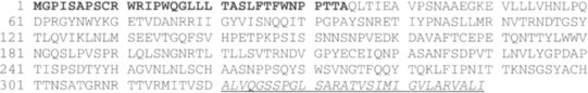
Amino acid sequence of human CEACAM6

The sequences sequences underlined and in italics are cleaved off to form mature CEACAM6 and a GPI anchor is added.
Amino acid sequence of human CEACAM8
The sequences sequences underlined and in italics are cleaved off to form mature CEACAM8 and a GPI anchor is added.
Database accession
References
- 1.Beauchemin N. Exp. Cell Res. 1999;252:243–249. doi: 10.1006/excr.1999.4610. [DOI] [PubMed] [Google Scholar]
- 2.http://www.uni-freiburg.de/cea
- 3.Skubitz K.M. J. Immunol. 1995;155:5382–5390. [PubMed] [Google Scholar]
- 4.Abrink B. Curr. Opin. Cell Biol. 1997;9:616–626. doi: 10.1016/S0955-0674(97)80114-7. [DOI] [PMC free article] [PubMed] [Google Scholar]
- 5.Benchimol S. Cell. 1989;57:327–334. doi: 10.1016/0092-8674(89)90970-7. [DOI] [PubMed] [Google Scholar]
- 6.Teixeira A.M. Blood. 1994;84:211–219. [PubMed] [Google Scholar]
- 7.Yamanaka T. Biochem. Biophys. Res. Commun. 1996;219:842–847. doi: 10.1006/bbrc.1996.0320. [DOI] [PubMed] [Google Scholar]
- 8.Virji M. Mol. Microbiol. 1996;22:929–939. doi: 10.1046/j.1365-2958.1996.01548.x. [DOI] [PubMed] [Google Scholar]
- 9.Dveksler G.S. J. Virol. 1991;65:6881–6891. doi: 10.1128/jvi.65.12.6881-6891.1991. [DOI] [PMC free article] [PubMed] [Google Scholar]
- 10.Skubitz K.M. J. Leukocyte Biol. 1996;60:106–117. doi: 10.1002/jlb.60.1.106. [DOI] [PubMed] [Google Scholar]
- 11.Morales V.N. J. Immunol. 1999;163:1363–1370. [PubMed] [Google Scholar]
- 12.Lin S.H. J. Biol. Chem. 1989;264:14408–14414. [PubMed] [Google Scholar]
- 13.Sippel C.J. J. Biol. Chem. 1996;271:33095–33104. doi: 10.1074/jbc.271.51.33095. [DOI] [PubMed] [Google Scholar]