Abstract
Campylobacter jejuni has caused several campylobacteriosis outbreaks via raw milk consumption. This study reports follow-up of a milk-borne campylobacteriosis outbreak that revealed persistent C. jejuni contamination of bulk tank milk for seven months or longer. Only the outbreak-causing strain, representing sequence type (ST) 883, was isolated from milk, although other C. jejuni STs were also isolated from the farm. We hypothesized that the outbreak strain harbors features that aid its environmental transmission or survival in milk. To identify such phenotypic features, the outbreak strain was characterized for survival in refrigerated raw milk and in aerobic broth culture by plate counting and for biofilm formation on microplates by crystal violet staining and quantification. Furthermore, whole-genome sequences were studied for such genotypic features. For comparison, we characterized isolates representing other STs from the same farm and an ST-883 isolate that persisted on another dairy farm, but was not isolated from bulk tank milk. With high inocula (105 CFU/ml), ST-883 strains survived in refrigerated raw milk longer (4–6 days) than the other strains (≤3 days), but the outbreak strain showed no outperformance among ST-883 strains. This suggests that ST-883 strains may share features that aid their survival in milk, but other mechanisms are required for persistence in milk. No correlation was observed between survival in refrigerated milk and aerotolerance. The outbreak strain formed a biofilm, offering a potential explanation for persistence in milk. Whether biofilm formation was affected by pTet-like genomic element and phase-variable genes encoding capsular methyltransferase and cytochrome C551 peroxidase warrants further study. This study suggests a phenotypic target candidate for interventions and genetic markers for the phenotype, which should be investigated further with the final aim of developing control strategies against C. jejuni infections.
Introduction
Campylobacter jejuni, which is the leading cause of bacterial gastroenteritis worldwide, is asymptomatically carried in the digestive tract of numerous wild and domesticated bird and mammal species. Human infection is usually acquired by the consumption of contaminated poultry meat, water, raw milk, or contact with animal feces. C. jejuni is prevalent in cattle and the consumption of raw cow’s milk has mediated several campylobacteriosis outbreaks [1].
C. jejuni grows optimally at 37‒42°C under microaerobic conditions and cannot tolerate drying and atmospheric levels of oxygen. Despite fastidious growth requirements, C. jejuni possesses mechanisms to survive in stress conditions, which play a role in host colonization, transmission in the environment, and survival in the food chain to cause human infection [2–4]. Such mechanisms include defense against atmospheric levels of oxygen and reactive oxygen species, heat shock, low pH, osmotic stress, and nutrient-poor environments. C. jejuni can survive, but not proliferate, in nutrient-poor cold waters for months [5].
One strategy for survival in harsh conditions is biofilm formation. C. jejuni can form biofilm on a variety of abiotic surfaces and coexist with other species in polymicrobial biofilms [6]. Indeed, secondary colonization of existing biofilms by C. jejuni has been suggested to occur on poultry farms [7]. C. jejuni biofilms have not been reported on dairy farms to our knowledge, but milking equipment could potentially allow biofilm formation like other food production and processing environments.
Biofilm formation by C. jejuni is a complex process involving several gene functions, not yet fully elucidated. As suggested, genetic mechanisms behind the biofilm-forming phenotype may even vary between different C. jejuni lineages: ST-21 CC and ST-45 CC [8]. Biofilm formation has been associated with surface proteins, flagella, and quorum sensing in mutational studies. Furthermore, shifted expression levels have been observed in biofilm-grown C. jejuni towards iron uptake, oxidative stress defense, and membrane transport [3].
Inter-strain variability has been observed both in the ability to cope with environmental stresses and in niche adaptation. As revealed by multilocus sequence typing (MLST), certain C. jejuni lineages (such as ST-21 CC) are found more often in human infection and in the food chain, whereas others (such as ST-45 CC) are often present in environmental sources with less clinical impact [3,4]. Furthermore, host specificity is common among lineages in wildlife, whereas lineages in livestock (such as ST-21 CC and ST-45 CC) show more generalist nature and often coexist in farm environments. A few livestock-associated host-specialist lineages are also known, such as ST-61 CC in cattle [9,10]. Better understanding of the survival strategies and transmission patterns of C. jejuni strains in farm environments is required to develop control strategies that would lessen the disease burden of this pathogen.
In 2012, a follow-up study of a campylobacteriosis outbreak revealed persistent C. jejuni contamination in bulk tank milk on a Finnish dairy farm. Interestingly, only the outbreak strain was isolated from milk, although other strains were detected on the farm simultaneously. We hypothesized that the outbreak strain possesses survival mechanisms to aid its on-farm transmission or survival in milk. We studied the outbreak strain for survival in refrigerated raw milk, aerotolerance, biofilm formation, antimicrobial susceptibility, and genomic content to explore mechanisms behind persistence. Ultimately, we aimed to determine whether and, if so, why certain C. jejuni strains pose a higher health risk in milk production settings to aid the development of enhanced control strategies.
Results
C. jejuni in bulk tank milk and milk filters
Samples were collected at the dairy farm from bulk tank milk and milk filters 11 times during six months after the outbreak (December 2012 to June 2013). Simultaneously, rigorous hygienic measures were applied to eliminate C. jejuni contamination from the farm. Despite hygienic measures, C. jejuni was isolated from 10/11 milk samples (91%) and 10/21 milk filter samples (48%), being detected from milk or milk filters in all 11 samplings. The concentration of thermotolerant Campylobacter in the milk samples ranged from 0.007 to 35 MPN/ml. All C. jejuni isolates matched the outbreak type pattern in pulsed-field gel electrophoresis (PFGE) studies, suggesting that the strain persistently contaminated bulk tank milk for seven months or longer.
C. jejuni in cattle feces and the farm environment
Cattle feces on the farm were sampled for C. jejuni twice within two months of the outbreak (in December 2012 and January 2013), and samples were collected from the farm environment throughout the six-month monitoring period. C. jejuni was isolated from 25/39 fecal samples (64%). Isolates from 12 fecal samples represented the outbreak type, while six other pulsotypes were detected from 13 fecal samples. Two cows carried the outbreak type in both samplings, whereas other pulsotypes were detected sporadically among cow specimens. C. jejuni of the outbreak type was isolated from only 3/54 environmental samples (5%) taken from the milk room and a feeding table, while a sporadic type was isolated from an udder cloth. Altogether, eight C. jejuni pulsotypes were detected on the farm, but interestingly only the outbreak type was isolated from bulk tank milk and milk filters and occurred repeatedly.
Multilocus sequence typing of C. jejuni farm isolates
In seven-loci MLST, the outbreak type represented sequence type (ST) ST-883 and clonal complex (CC) ST-21 CC. Other farm pulsotypes represented were ST-45 (ST-45 CC), ST-50 (ST-21 CC), ST-58 (unassigned CC), and ST-61 (ST-61 CC). ST-50 was isolated from the udder cloth and the other three STs (ST-45, ST-58, and ST-61) from cattle.
Genomic epidemiology of ST-58 and ST-883 isolates was further studied using whole-genome multilocus sequence typing (wgMLST). Other STs were not studied because they were represented by only a single isolate. ST-58 farm isolates of this study (n = 3) were compared with ST-58 isolates from the UK (n = 34), representing publicly available ST-58 genomes. All ST-58 isolates were obtained from ruminant sources from 2011 to 2018, suggesting host-specificity. In the allelic profile size of 1064, two ST-58 farm isolates (ST58_2012–12_C1 and ST58_2013–01_C3) appeared within close pairwise distance (PWD) of 1 (0.1%). One farm isolate (ST58_2012–12_C2), however, appeared unrelated (PWD 4.7%), considering PWD of the closest UK isolate (5.5%) with the farm isolates of this study. Thus, two clones were recognized among the ST-58 isolates of this study.
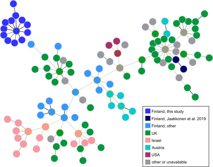
ST-883 isolates of this study (n = 40) were compared with globally collected ST-883 isolates (n = 137). The dataset included ST-883 isolates (n = 5) that were found to persist on another Finnish dairy farm for 11 months or longer without being detected from bulk tank milk in weekly samplings [11]. In the allelic profile size of 718, the ST-883 isolates of this study appeared within the maximum PWD of 4 (0.6%). Closest outgroup isolate (IN_Cj_FI_109) appeared within PWD of 4.9% and was isolated from cattle in Finland in 2003. Isolates from the other Finnish dairy farm appeared within PWD of 21% from the isolates of this study. Finnish isolates were generally dispersed within the minimum spanning tree, showing no evidence of geographic circulation of the outbreak clone (Fig 1). When comparing only ST-883 isolates of this study, the maximum PWD was 5 (0.5%) in the allelic profile size of 1032. Therefore, ST-883 isolates of this study appeared similar in wgMLST, suggesting that the farm was contaminated by a single clone of ST-883, and the outbreak originated from the dairy farm. As revealed by MLST and further wgMLST analysis, altogether six clones were recognized among the isolates from the outbreak farm.
Fig 1. Minimum spanning tree from wgMLST comparison of ST-883 C. jejuni isolates.
Dairy farm isolates from this study (n = 40) are compared with globally collected ST-883 isolates (n = 137), including dairy farm isolates from Jaakkonen et al. [11]. Nodes are colored by country. Black and dark gray links indicate short allelic distances of 1 and 2 loci, respectively, in the profile size of 718 loci.
Survival in refrigerated raw milk
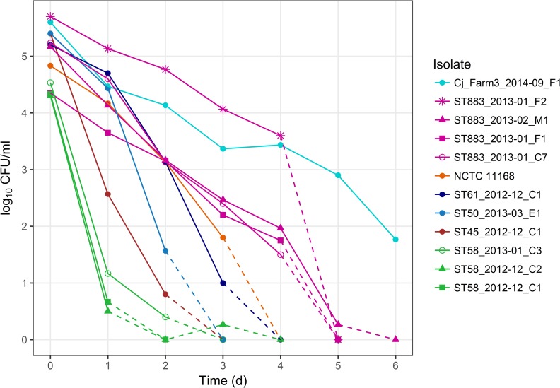
Persistent contamination of bulk tank milk was hypothesized to be due to prolonged survival of the outbreak strain in refrigerated raw milk. Indeed, outbreak type (ST-883) isolates from bulk tank milk (ST883_2013–02_M1), cattle (ST883_2013–01_C7), and milk filters (ST883_2013–01_F1 and ST883_2013–01_F2) survived in milk for four to five days, whereas the other farm isolates survived for only three days or less (Fig 2). Survival for three days was observed for ST-61 isolate and the control strain NCTC 11168. ST-45, ST-50, and one ST-58 isolate survived for two days. Two ST-58 isolates reached the quantification limit already within one day. However, a milk filter isolate (Cj_Farm3_2014–09_F1) of ST-883 from the other Finnish dairy farm survived in milk longest, at least for six days, despite not being detected in bulk tank milk in the longitudinal study [11]. Survival in refrigerated raw milk varied within the lineage ST-21 CC and even within the same ST, indicating unconserved traits behind survivability.
Fig 2. Survival of C. jejuni farm isolates in refrigerated raw milk.
Mean colony counts of three experiments are shown at time points 0 d (maximum standard error of the mean ±0.6), 1 d (±0.7), 2 d (±1.0), 3 d (±0.7), 4 d (±1.0), 5 d (±0.3), and 6 d (±0.1). Dashed lines indicate the decrease of C. jejuni counts below the quantification limit (0 log10 CFU/ml): between 0 and 3 d (ST-58 isolates), between 2 and 3 d (ST-50 and ST-45 isolates), between 3 and 4 d (ST-61 isolate and control strain NCTC 11168), and between 4 and 6 d (ST-883 isolates). ST-883 isolate (Cj_Farm3_2014–09_F1) from another dairy farm [11] could be quantified at every time point for 6 d. Summary statistics of the data are presented in Table A in S1 Datasets.
Aerotolerance
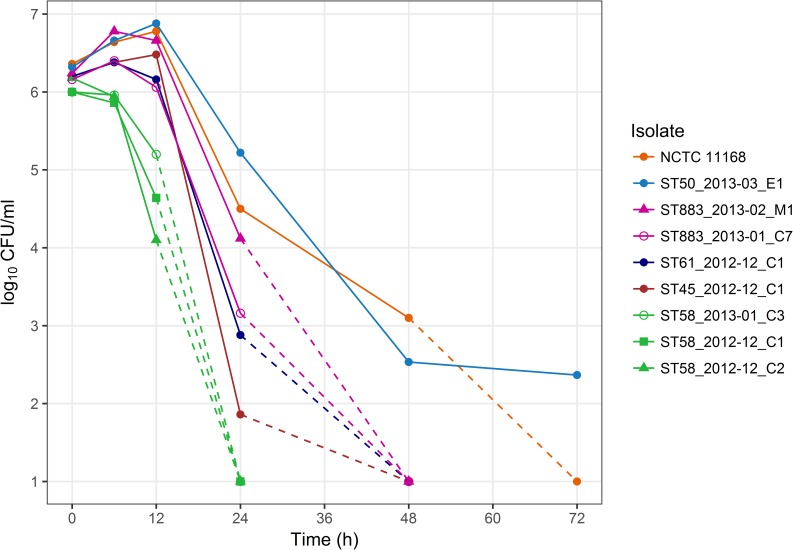
Aerotolerance of C. jejuni could enhance environmental fitness, thus aiding transmission in the farm environment and survival in bulk tank milk and milk filters. As defined by Oh et al. [12], aerotolerant strains survive after 12 h and hyper-aerotolerant strains after 24 h of aerobic shaking. In our study, all representative farm isolates survived after 12 h of aerobic shaking in five experiments (Fig 3). Survival after 24 h was observed in five experiments for ST-21 CC isolates: ST-883 isolate from bulk tank milk, ST-50 isolate from an udder cloth, and the control strain NCTC 11168. In addition, survival in two or three of five experiments was detected for ST-883, ST-61, and ST-45 cattle isolates after 24 h and survival in one of three experiments for ST-50 and NCTC 11168 after 48 h. Interestingly, isolates from bulk tank milk (outbreak type ST-883) and an udder cloth (ST-50) showed hyper-aerotolerance consistently, as opposed to cattle isolates. Survival under aerobic shaking conditions did not, however, correlate with survival in refrigerated raw milk (Pearson coefficient 0.23, P = 0.56), and other mechanisms were thus suspected to contribute to survival in milk.
Fig 3. Survival of C. jejuni farm isolates in broth cultures under aerobic shaking at 41.5°C.
Mean colony counts of five experiments are shown at time points 0 h (maximum standard error of the mean ±0.2), 6 h (±0.3), 12 h (±0.5), and 24 h (±1.2) and mean colony counts of three experiments at 48 h (±2.1), and 72 h (±1.4). Dashed lines indicate the decrease of C. jejuni counts below the quantification limit (1 log10 CFU/ml): between 12 and 24 h (ST-58 isolates), between 24 and 48 h (ST-883, ST-61, and ST-45 isolates), and later (ST-50 isolate and control strain NCTC 11168). Summary statistics of the data are presented in Table B in S1 Datasets.
Biofilm formation
Persistence of the outbreak strain in bulk tank milk could possibly also be explained by biofilm formation in the milking machine or milk tank, enhancing the survival and transmission of Campylobacter. As an indicator for biofilm, rinsing water of the milking machine was analyzed twice for Campylobacter (in May and June 2013). No Campylobacter were detected from the water samples, despite simultaneous isolation of C. jejuni from bulk tank milk.
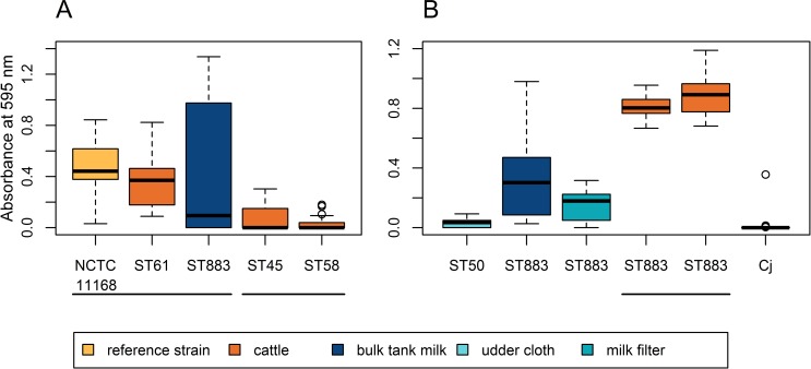
Biofilm formation of representative farm isolates was examined in monocultures on polystyrene microplates. The outbreak strain formed biofilm during 48-h incubation in higher quantities (P≤0.039) than four cattle isolates (Fig 4A). No difference was observed in biofilm quantities (P≥0.14) between the outbreak strain, control strain NCTC 11168, and one cattle isolate (ST-61). Although biofilm formation could not be detected from the milking machine, the outbreak strain was able to form biofilm in laboratory settings.
Fig 4. Biofilm formation of C. jejuni farm isolates on polystyrene microplates during 48-h incubation.
Boxplots show median (bold horizontal line), 25% quartile, and 75% quartile biofilm quantities of 18 replicates, indicated by the absorbance of crystal violet stain. Biofilm quantities that fall outside the box by the maximum of 1.5 times the box height (or interquartile range) are shown as whiskers, and the quantities that fall outside the whiskers are shown as circles, indicating possible outliers. Horizontal lines group pairs of means that are not significantly different from each other (t test with no assumption of equal variances on transformed data, P>0.05). Experiment setups A and B are shown respectively in panes A and B. (A) Comparison of the milk isolate (ST883_2013–02_M1) with cattle isolates of other STs. Two ST-58 isolates showed no difference to ST58_2012–12_C1 and are thus omitted from the plot. (B) Comparison of the milk isolate (ST883_2013–02_M1) with ST-50 isolate from an udder cloth and with ST-883 isolates from milk filter (ST883_2013–01_F1) and cattle (ST883_2013–01_C7 and ST883_2012–12_C6), and with ST-883 milk filter isolate (Cj_Farm3_2014–09_F1) from another dairy farm [11], all representing ST-21 CC.
To further explore whether biofilm formation could explain survival in milk, we studied biofilm formation of the outbreak type (ST-883) isolates from different sample materials (Fig 4B). Indeed, cattle isolates formed biofilm during 48-h incubation in higher quantities (P<2.7×10⁻⁶) than isolates from milk and milk filters. In addition, more variation in biofilm formation was observed between the replicates of the milk isolate (95% CI: ±0.12) than the replicates of the cattle isolates (95% CI: ±0.07). The milk isolate formed biofilm in an on/off manner between replicate cultures and in higher quantities than the milk filter isolate (P = 0.03). Interestingly, ST-883 milk filter isolate (Cj_Farm3_2014–09_F1) from the other Finnish dairy farm formed no biofilm, suggesting that biofilm formation could contribute to the persistence of the outbreak strain in bulk tank milk.
Comparative genomics
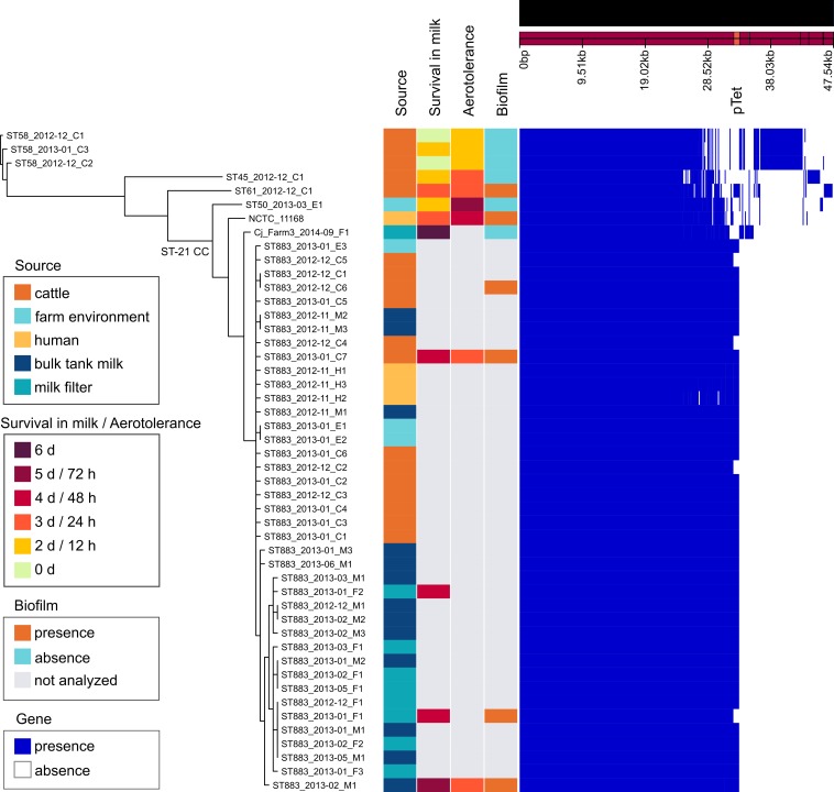
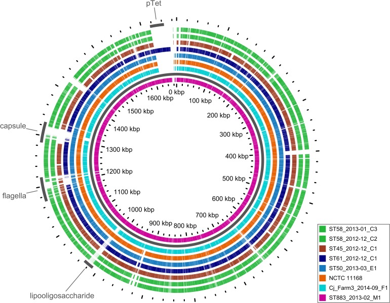
To recognize potential genotypic features behind the surviving phenotype of the outbreak strain, draft genomes of 46 dairy farm isolates, which represented both the outbreak type (40 isolates) and other STs (6 isolates), were studied for genomic content. Comparison included the reference strain NCTC 11168, representing ST-43, and the milk filter isolate (Cj_Farm3_2014–09_F1) from another Finnish dairy farm, representing ST-883 [11]. Gene content of the outbreak strain closely resembled that of the reference strain NCTC 11168, both representing ST-21 CC (Figs 5 and 6). Of 1620 Prokka-annotated genes of NCTC 11168, the outbreak strain shared 1592 genes (98.2%). Other farm strains shared 97.6% (ST-50) to 88.6% (ST-58) of the reference strain genes. ST-883 isolate from the other dairy farm shared 96.8% of the reference strain genes and 95.0% of the outbreak strain genes (Fig 5).
Fig 5. Genomic comparison of C. jejuni isolates from the outbreak farm.
The reference strain NCTC 11168 (RefSeq accession no. NC_002163.1) and ST-883 isolate (Cj_Farm3_2014–09_F1) from another dairy farm [11] are included in the comparison. (A) Left pane: Approximation of maximum-likelihood phylogeny based on the nucleotide alignment of 1356 core genes from Roary using FastTree (version 2.1.9) with the GTR+CAT model [13]. Branches with support <1 are not shown and recombinations are not masked. Names of the farm isolates indicate: ST, sampling time (year-month), sample source (C, E, H, M, or F; see legend), and isolate number. Middle pane: sample source and phenotype. Right pane: presence and absence of genes according to Roary.
Fig 6. BLAST atlas of the representative farm strains and NCTC 11168 against the outbreak strain (ST883_2013–02_M1).
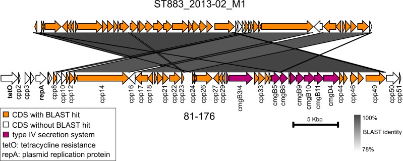
Most strikingly, the outbreak strain harbored a pTet-like element that showed 99.7% nucleotide sequence identity (coverage 93%) with the pTet plasmid of strain 81–176 (Fig 7). The pTet-like element was present in 36 (90%) of the outbreak type isolates and also in the ST-61 cattle isolate with an identical nucleotide sequence, suggesting horizontal transfer between these farm strains (Figs 5 and 6). Compared with the pTet plasmid of strain 81–176, the pTet-like element of the farm strains lacked 12 genes, including genes that encode tetracycline resistance and plasmid replication protein (Fig 7). Concordantly, the farm isolates were susceptible to tetracycline, along with all other tested antimicrobial agents. The pTet-like element harbored 43 predicted genes, including genes that encode the complete type IV secretion system. Six predicted genes were missing from the pTet plasmid of strain 81–176. These genes were annotated to encode hypothetical proteins and resembled (BLAST identity >99.4%, coverage 100%) those in previously sequenced C. jejuni plasmids.
Fig 7. Nucleotide sequence comparison of pTet.
The pTet-like element from the outbreak type isolate (ST883_2013–02_M1) is compared against the pTet plasmid from the C. jejuni strain 81–176 (RefSeq accession no. NC_006135.1). Arrows indicate predicted coding sequences (CDS).
Excluding the pTet-like element, no unique gene content was detected in the outbreak strain (Fig 6). Opposed to the reference strain NCTC 11168, functional di-/tripeptide transporter (DtpT; locus tag Cj0654c in NCTC 11168, RefSeq accession no. NC_002163.1) was annotated in the outbreak strain, which harbored two adjacent dtpT genes, one intact (dtpT_1) and one fragmented (dtpT_2). Organization of these genes varied among the farm strains: ST-45 strain harbored two intact genes, whereas ST-50 and ST-61 harbored intact dtpT_1 and fragmented dtpT_2 gene. ST-883 isolate from the other dairy farm harbored dtpT genes identical to the outbreak strain. The presence of dtpT genes was further studied within the global genealogy of C. jejuni, comprising 1159 isolates of non-human origin and 46 isolates from this study (Fig in S1 Appendix). The presence of intact dtpT_2 was associated with a few clonal complexes: ST-45 CC, ST-283 CC, and ST-42 CC. However, clonal complexes were not associated with dtpT_1. Intact dtpT_1 was most abundant among isolates from ruminants (87.7%), but was frequently observed also among isolates from other host taxa (≥40.0%) (Table in S1 Appendix).
Outbreak type isolates were further studied for genomic adaptation to their isolation source by analyzing single-nucleotide polymorphisms (SNPs), insertions, and deletions. Phase variation was observed in genes related to the capsule, flagella, and oxidative stress response (Table 1). Interestingly, a higher proportion of cattle isolates harbored a phase-variable, fragmented variant of cytochrome C551 peroxidase (Cj0020c) and an intact variant of capsular methyltransferase (Cj1420c) than isolates from milk, suggesting reversible adaptation by oxidative stress response and capsular variation inside or outside the cattle host. Gene organization in the capsular locus of the outbreak strain resembled that of the reference strain NCTC 11168.
Table 1. SNPs, insertions, and deletions among the outbreak type isolates (n = 39) against the in-group reference ST883_2013–01_C5, isolated from cattle1.
| Location in reference ST883_2013–01_C5 | Sequence variation | Proportion (%) of isolates representing an alternate variant by sample source | |||||||||||
|---|---|---|---|---|---|---|---|---|---|---|---|---|---|
| Function | Prokka annotation | Locus tag in NCTC 11168 (NC_002163.1) | Locus tag | Contig | Position | Type | Reference | Alternate | Gene fragmentation by variation in | Human (n = 3) | Cattle (n = 12) | Milk (n = 13) | Milk filter or environment (n = 11) |
| oxidative stress response | cytochrome C551 peroxidase | Cj0020c | KBNHAAAG_01697 | 14 | 66849 | del | CAAAATTC | CAAATTC | reference | 100 | 25 | 100 | 100 |
| - | iron-binding protein | Cj0045c | KBNHAAAG_00019 | 1 | 22262 | del | TCCCCCCCCCCCATAT | TCCCCCCCCCCATAT | reference | 0 | 17 | 8 | 9 |
| - | rod shape-determining protein MreB | Cj0276 | KBNHAAAG_00237 | 3 | 13418, 13512, 13527, or 13892 | snp2 | G, A, G, or A | A, T, A, or C | - | 100 | 0 | 8 | 0 |
| flagella | N-acetyltransferase | Cj1296 | KBNHAAAG_01245 | 8 | 122950 | del | CGGGGGGGGGGAGGTTA | CGGGGGGGGGAGGTTA | alternate | 100 | 25 | 8 | 9 |
| hypothetical protein | Cj1305c | KBNHAAAG_01254 | 8 | 129282 | del | ACCCCCCCCCCATAA | ACCCCCCCCATAA | reference | 67 | 42 | 69 | 45 | |
| hypothetical protein | Cj1306c | KBNHAAAG_01256 | 8 | 130522 | ins | ACCCCCCCCATA | ACCCCCCCCCATA | reference | 0 | 33 | 85 | 36 | |
| hypothetical protein | Cj1318 | KBNHAAAG_01269 | 8 | 141128 | ins | TGGGGGGGGTAT | TGGGGGGGGGGTAT | reference | 33 | 33 | 31 | 36 | |
| pseudaminic acid biosynthesis protein PseA | Cj1324 | KBNHAAAG_01277 | 8 | 146844 | ins | TTTAG | TATTAG | alternate | 0 | 33 | 0 | 0 | |
| isomerase | Cj1330 | KBNHAAAG_01283 | 8 | 151640 | snp3 | C | T | - | 0 | 0 | 69 | 64 | |
| protein PseD | Cj1335 | KBNHAAAG_01288 | 8 | 157973 | ins | TGGGGGGGGGTAT | TGGGGGGGGGGTAT | reference | 0 | 8 | 15 | 9 | |
| hypothetical protein | Cj1342c | KBNHAAAG_01293 | 9 | 5364 | del | ACCCCCCCCCATA | ACCCCCCCCATA | alternate | 0 | 8 | 23 | 45 | |
| capsule | methyltransferase | Cj1420c | KBNHAAAG_01367 | 9 | 82963 | del | ACCCCCCCCCTGT | ACCCCCCCCTGT | alternate | 67 | 8 | 62 | 55 |
| hypothetical protein | Cj1429c | KBNHAAAG_01377 | 11 | 6050 | del | TCCCCCCCCCCCATTA | TCCCCCCCCCCATTA | reference | 33 | 33 | 38 | 55 | |
| aminotransferase | Cj1437c | KBNHAAAG_01385 | 12 | 3713 | del | TCCCCCCCCCCGCCAGT | TCCCCCCCCCGCCAGT | reference | 0 | 25 | 8 | 9 | |
| D-arabinose 5-phosphate isomerase KpsF | Cj1443c | KBNHAAAG_01391 | 12 | 12376 | snp | G | T | - | 0 | 25 | 0 | 9 | |
1Only coding loci with variation in four (10%) or more isolates are shown.
2Each isolate (n = 4) harbored one SNP in a unique position.
3SNP was observed in isolates sampled in January 2013 and thereafter.
Discussion
This outbreak-related dairy farm study revealed persistent C. jejuni contamination of bulk tank milk for seven months or longer. The outbreak and the persisting milk contamination were caused by a single clone of sequence type ST-883 (ST-21 CC), which was also prevalent in the dairy herd and shed in feces repeatedly by two cows. In addition to the outbreak clone, five other clones were sporadically detected in the herd (ST-45, ST-58, and ST-61; CCs other than ST-21 CC) and in an udder cloth (ST-50; ST-21 CC), but not in bulk tank milk. They represented both generalist (ST-21 CC and ST-45) and cattle-associated (ST-61 and ST-58) lineages of C. jejuni. As previously suggested, simultaneous isolation of several C. jejuni genotypes in livestock-associated environments is common due to their inability to competitively exclude each other [10].
Persistent contamination by C. jejuni has been reported in bulk tank milk previously. Bianchini et al. [14] noted persistent milk contamination due to subclinical mastitis caused by C. jejuni (ST-38; ST-48 CC). Mammary excretion of C. jejuni could not be excluded in our study, as no milk samples were collected directly from cow specimens. However, our study revealed strain-specific characteristics that were potentially linked to survival in milk despite the contamination route. In addition, long-lasting milk contamination by C. jejuni (ST-50) was previously associated with poorly fitting milking liners, which allowed suction of fecal material-containing air inside the milking machine during milking [15,16]. In our study, rigorous hygienic measures were applied to eliminate milk contamination, and these measures included changing of milking liners. None of these measures showed an effect, and we could not identify the contamination route. Continuous application of hygienic measures yielded low counts of total bacteria (2000–15 000 cells/ml) and somatic cells (115 000–237 000 cells/ml) in milk despite the continued presence of C. jejuni. Inefficiency of hygienic measures against C. jejuni persistence in dairy cattle herds was also reported in a longitudinal study [11]. In that study, C. jejuni ST-883 persisted on a dairy farm for 11 months or longer, but was not isolated from bulk tank milk in weekly samplings. Lack of C. jejuni isolation from milk in the longitudinal study further raised the question of whether survival in milk, and thus detection in milk, is strain-dependent.
Indeed, survival in refrigerated raw milk varied between and among STs in this study, demonstrating variation between strains. The outbreak type isolates survived for four to five days in refrigerated milk, whereas other isolates from the outbreak farm survived only for three days or less in an experiment with high inocula (105 CFU/ml). With natural detection levels of the outbreak strain observed in this study (0.007–35 MPN/ml), contamination events by poor-surviving, sporadic strains likely remained undetected by the time that the sample shipment reached the laboratory and analyses were initiated, usually within 24 h. However, the ST-883 isolate from the longitudinal study [11] survived in refrigerated milk even longer, at least six days, than the outbreak strain, suggesting that survival in refrigerated raw milk cannot alone explain persistence of the outbreak strain in bulk tank milk.
In both laboratory settings and primary production of milk, milk-contaminating C. jejuni is exposed to oxygen species. Oxygen species in raw cow’s milk may be produced by the action or metabolism of other microbes (lactic acid bacteria), somatic cells (leukocytes), or antibacterial enzymes (mainly lactoperoxidase) present in milk [17–20]. Atmospheric oxygen is also introduced in milk by stirring. Oxygen tolerance has previously been shown to vary between C. jejuni isolates, and higher proportions of aerotolerant isolates have been reported among ST-21 CC, which is prevalent in food processing environments and among clinical isolates [12]. Therefore, we hypothesized that enhanced oxygen tolerance of C. jejuni could contribute to survival in milk or in the farm environment. No correlation was, however, observed between survival under aerobic shaking and in refrigerated raw milk among the farm isolates of this study, and other mechanisms were thus suggested to contribute to survival in milk.
Isolates from milk (ST-883) and an udder cloth (ST-50) showed hyper-aerotolerance consistently, unlike cattle isolates, which could indicate adaptation of C. jejuni after oxygen exposure in the environment. Concordantly, the majority of the outbreak-type cattle isolates and none of the isolates from other sources, including milk, showed impairment of cytochrome C551 peroxidase gene (Cj0020c) due to phase variation. Phase variation is a rapid adaption mechanism of C. jejuni, yielding reversible genotypes [21]. C. jejuni harbors two homologs of cytochrome C551 peroxidase gene, Cj0020c and Cj0358, the former of which has been associated with oxygen stress defense and chick colonization [22,23]. Our results further demonstrate that function of this gene may be reversibly adapted to survival inside and outside the cattle host, respectively, from low to high oxygen tensions.
Although oxygen tolerance did not explain the survival of C. jejuni in refrigerated raw milk, it may still be a consequence of other adaptation mechanisms such as biofilm formation. In biofilm formation, cells first adhere to a surface, aggregated, and cell metabolism is then adapted to biofilm lifestyle: towards iron uptake, oxidative stress defense, and membrane transport [24]. Indeed, adaption by sample source was observed in biofilm formation of the outbreak type isolates in this study. Cattle isolates formed biofilm in higher quantities than isolates from milk and milk filter in microaerobic conditions at 41.5°C. More variation, appearing in an on/off manner, was observed in biofilm quantities among replicates of the milk isolate than among replicates of the cattle isolates. These results suggest that the milk isolate was reversibly adapted to survival in milk at the cost of biofilm formation or surface adhesion.
The observed on/off variation in biofilm quantities between replicates could be due to phase variation. The outbreak type milk isolate harbored phase-variable, impaired capsular methyltransferase gene (Cj1420c) compared with the cattle isolates harboring an intact gene variant. The capsule has previously been reported to contribute to biofilm formation in ST-45 CC using a genome-wide association approach, although the role of this methyltransferase gene remains unclear [8]. Moreover, cytochrome C551 peroxidase gene (Cj0020c) was associated with biofilm formation in ST-21 CC [8]. Therefore, further experiments should be conducted to elucidate the role of these genes in biofilm formation.
Biofilm-forming strains from the outbreak farm (ST-883 and ST-61) also harbored an identical pTet-like element that lacked genes for tetracycline resistance and replication, suggesting horizontal transfer and replication within the chromosome. The element contained genes that encode a type IV secretion system, which has previously been reported in both plasmid and chromosomal locations in Campylobacter fetus species associated with cattle host and to enable conjugative transfer of macromolecules [25]. Type IV secretion system-mediated cell contact has also been suggested to control biofilm formation in Helicobacter pylori [26].
In addition to the secretion system, the pTet-like element carried other genes of the pTet plasmid, most of them with unknown function. As no tetracycline resistance gene was present, the element could provide C. jejuni with another fitness advantage that exceeds the metabolic cost of carrying this genomic element. Four outbreak type isolates (10%) from cattle and a milk filter (ST883_2013–01_F1) had lost the element either on the farm or in the laboratory. All milk isolates harbored the pTet-like element, raising the question of whether this element could contribute to survival in milk or biofilm formation. Outbreak type isolates with and without the pTet-like element survived in refrigerated raw milk equally long, indicating that this element does not affect survival in refrigerated milk. Interestingly, an outbreak type isolate lacking the pTet-like element formed less biofilm than the other outbreak isolates, suggesting that the element may contribute to biofilm formation. Growing evidence from various other pathogenic enterobacteria has also suggested that the presence of plasmids may enhance biofilm formation [27–29]. Further studies should, however, be conducted to understand the mechanisms behind these observations in more detail.
Interestingly, the outbreak strain harbored an intact di-/tripeptide transporter gene (dtpT), which shared an identical nucleotide sequence to the ST-883 strain from the other dairy farm [11]. Peptide transporters are essential for the growth of the lactic acid bacterium Lactococcus lactis in milk, and DtpT plays a role in peptide-dependent signaling of L. lactis [30,31]. Furthermore, Listeria monocytogenes strains that persisted in the dairy environment showed higher expression levels of dtpT gene than non-persisting strains [32]. As gene expression was not investigated in our study, it remains inconclusive whether phenotypic differences arose from differential expression patterns. Within the global genealogy of C. jejuni, prevalence of the gene dtpT_1 was highest among C. jejuni isolates from ruminants, although the gene was also common among isolates from other host taxa. Therefore, it remains to be elucidated whether dtpT_1 provides a fitness advantage to C. jejuni in bovine host and farm environments. C. jejuni relies on amino acids in its energy metabolisms and compensates growth restriction in the host by peptide transport [33]. This provides rationale for further studies on the role of peptides and peptide transporters also in the survival of C. jejuni in milk.
In conclusion, this study reports persistent contamination of bulk tank milk for seven months or longer, which was recognized during a campylobacteriosis outbreak and was caused by a single C. jejuni clone of ST-883. Together with previous findings [11], we conclude that ST-883 strains are able to persist on dairy farms and may thus pose a higher health risk in milk production settings than some other C. jejuni STs. ST-883 strains survived in refrigerated raw milk longer than the other C. jejuni STs in this study, suggesting that these strains may share some features that promote their survival in milk. Candidate phenotypic and genetic markers were identified here. This study further demonstrates that C. jejuni may pose a health risk to raw milk consumers despite good on-farm hygiene and emphasizes the importance of avoiding campylobacteriosis by heat treatment of raw milk before consumption.
Materials and methods
Campylobacteriosis outbreak
In November 2012, a campylobacteriosis outbreak was recognized among people who had visited or consumed raw drinking milk from a Finnish dairy farm. Two children had been hospitalized with bloody diarrhea and a culture-confirmed C. jejuni infection. A questionnaire to regular milk-purchasing customers and party-attendees (total response rate 97%) revealed 18/62 (29%) diarrheic respondents, of whom 11/14 (79%) had a culture-confirmed C. jejuni infection and 12/16 (75%) had drunk raw milk. C. jejuni was isolated from 7 dairy farm samples (88%), including bulk tank milk, replaceable in-line milk filters of the milking machine, milk room surfaces, and cattle feces. No C. jejuni was isolated from drinking water in the barn. The C. jejuni farm isolates were indistinguishable from the patient isolates by PFGE, thus supporting the dairy farm as the source of the outbreak.
On-farm samplings and hygiene measures
The outbreak-associated dairy farm was located in western Finland and housed 40 cows in pipeline milking. The farm was sampled to trace back the outbreak source in November 2012 and to monitor on-farm hygiene during the following six months. Samplings were conducted by a municipal veterinary officer, entitled by law on foodborne outbreak investigations [34], and thus required no ethics approval. The farm was sampled for bulk tank milk (n = 11) and milk filters (n = 21) weekly during a three-month follow-up (December 2012 to March 2013) and once at six and seven months after the outbreak (May and June 2013). During the follow-up period rigorous on-farm hygienic measures were applied to eliminate C. jejuni contamination. These measures included acid treatment of the milking machine and milk tank, replacement of milking machine components, and disinfection of the drinking troughs, feeding tables, and stalls.
Samples from cattle feces (n = 39) were taken twice within two months of the outbreak (in December 2012 and January 2013). Five of thirty-three cows were sampled in both samplings. Samples from the farm environment (n = 54) were collected throughout the six-month monitoring period. The samples comprised rinsing water of the milking machine (n = 2), udder cloths (n = 2), and swabs from the milking liners (n = 12), milk room (n = 10), drinking troughs (n = 19), and feeding surfaces (n = 9).
Sample handling and test portions
Samples were chilled immediately and laboratory analyses were initiated within 24 h of sampling. Bulk tank milk was analyzed as five subsamples of 25 ml. One to four milk filters were collected during the 48 h preceding the milk sampling, refrigerated and moistened with buffered peptone water separately, and analyzed simultaneously with the milk samples. Fecal samples were collected from rectum and analyzed as 10-g test portions. Swab samples were taken with a moistened sponge (Polywipe; Medical Wire and Equipment, Corsham, Wiltshire, UK). A water sample (8 liters) was filtered through 0.45-μm-pore-size membrane filters (GN-6 Metricel Membrane; Pall Corporation, Ann Arbor, MI, USA), and the pooled filters were examined for Campylobacter.
Analysis of farm samples
During outbreak investigation the farm samples were cultured for thermotolerant Campylobacter according to NMKL 119:1990 [35] at SeiLab (Seinäjoki, Finland), and Campylobacter isolates were further characterized at the national reference laboratory for thermotolerant Campylobacter in food, the Finnish Food Authority (Helsinki, Finland). Follow-up samples were cultured for thermotolerant Campylobacter at the Finnish Food Authority according to NMKL 119:2007 with the exception of enrichment for 24 h [36]. Campylobacter in milk samples were quantified by the most probable number method using three dilutions and five replicates (3×5 MPN) [37]. Species of suspect Campylobacter isolates were determined biochemically or by matrix-assisted time-of-flight spectroscopy (MALDI Biotyper, reference library version 4.0.0.1, 5627 main spectra libraries, Bruker Daltonik, Bremen, Germany). Bulk tank milk was monitored for total bacteria and somatic cell counts at the dairy laboratory in separate samplings from this study, using flow cytometry (BactoScan FC; Foss, Hillerød, Denmark) and fluoro-opto-electronic methods [38].
Subtyping of C. jejuni isolates
C. jejuni isolates from each positive sample were subtyped by PFGE with SmaI digestion [39]: two or three isolates per milk or milk filter sample and one isolate per fecal or environmental sample. PFGE fingerprints were analyzed using BioNumerics software (version 6.6; Applied Maths, Sint-Martens-Latem, Belgium). Pulsotypes were designated by a difference of one or more bands.
Whole-genome sequencing, assembly, and multilocus sequence typing
Based on pulsotypes, representative isolates of the outbreak type (n = 40) and other pulsotypes (n = 6) were subjected to whole-genome sequencing. The outbreak type isolates represented patient isolates (n = 3), milk isolates during the outbreak (n = 3), all fecal (n = 13) and environmental isolates (n = 3), and each bulk tank milk (n = 10) and milk filter sampling (n = 8) during the follow-up period of six months.
Paired-end sequencing (read length of 100-bp, 150-bp, or 250-bp) was performed on MiSeq or HiSeq platform (Illumina, San Diego, CA, USA), preceded by the extraction of genomic DNA (PureLink Genomic DNA Mini Kit, Life Technologies, Thermo Fisher Scientific, Carlsbad, CA, USA or DNeasy Blood and Tissue Kit, Qiagen, Hilden, Germany) and preparation of genomic libraries (Nextera XT or Nextera Flex Kit, Illumina). Sequencing reads were subjected to quality control, de novo assembly, and MLST using INNUca pipeline (version 4.0.2) (Table C in S1 Datasets) [40]. MLST types were derived from the pubMLST database [41,42]. Within the INNUca pipeline, contamination of reads and assembled contigs were checked with Kraken 2 software against the MiniKraken2_v1_8GB database (retrieved on 13 November 2018) [43,44].
Whole-genome multilocus sequence typing
Genome assemblies of the farm isolates were further compared by wgMLST with globally isolated genomes of the same MLST ST, using chewbbaca software (version 2.0.16) and INNUENDO schema for C. jejuni [45,46]. Outbreak type genomes of this study (n = 40) were compared with 137 unique ST-883 genomes available from the INNUENDO database (n = 66), the BIGS database (n = 66; data retrieved on 27 January 2019), and another Finnish dairy farm (n = 5) [11,41,46] (Table D in S1 Datasets). ST-58 genomes of this study (n = 3) were compared with 34 genomes available from the BIGS database (n = 34; data retrieved on 21 March 2019) (Table E in S1 Datasets). After allele calling and extraction of core loci with chewbbaca, minimum spanning trees and distance matrices were calculated using PHYLOViZ Online (http://online.phyloviz.net) and visualized with PHYLOViZ version 2.0, which uses the goeBURST algorithm (Fig 1) [47,48]. Genomes obtained from the BIGS database were regarded as reference material to explore genomic diversity within the farm isolates only.
Phenotypic characterization
A representative isolate of each pulsotype (n = 8) was selected for phenotypic characterization, including survival in refrigerated raw milk, aerotolerance, biofilm formation, and antimicrobial susceptibility testing. These representatives comprised one isolate of each ST (ST-45, ST-50, and ST-61), all three ST-58 isolates, and two ST-883 isolates (ST883_2013–02_M1 and ST883_2013–01_C7). ST-883 representatives were isolated from milk and cattle, ST-50 from an udder cloth, and other STs from cattle. NCTC 11168 (representing ST-43 and ST-21 CC in MLST) was used as a control strain in all analyses. Additional isolates of the outbreak type (ST-883) from milk filters and cattle were studied for survival in refrigerated raw milk and for biofilm formation, together with an ST-883 isolate (Cj_2014–09_F1) from milk filters of another dairy farm [11].
Isolates were freshly inoculated from glycerol stocks stored at −70°C and, unless stated otherwise, grown under microaerobic conditions (5% oxygen, 10% CO2) on ovine blood agar plates at 37°C for 48 h or in Mueller Hinton (MH) broth at 41.5°C without shaking. To obtain inocula of 106 colony-forming units (CFU) /ml for phenotypic analyses, a single colony was inoculated into 10 ml of MH broth, incubated overnight for 16‒18 h, and diluted with fresh broth 10-fold (ST-58 isolates) or 100-fold (other isolates). Colony counts were determined from 1 ml of broth by serial dilutions and plating. All experiments were repeated at least three times, and uninoculated broth was used as a negative control.
Survival in refrigerated raw milk
Packaged, organic raw milk of the indigenous cattle breed of Finland (Kaskikansa, Saloniemen Juustola, Laitila, Finland) was purchased from retail and stored in aliquots at −70°C on the day of delivery. Overnight cultures were diluted in 100 ml of thawed raw milk to a concentration of 105 CFU/ml in a 250-ml glass flask with a screw cap. Inoculated raw milk was incubated under aerobic conditions at 4°C without shaking. Colony counts were determined on CCDA selective medium (Oxoid, Thermo Fisher Scientific, Wesel, Germany) once a day for six days: after 0 h, 24 h, 48 h, 72 h, 96 h, 120 h, and 144 h of incubation.
Aerotolerance
Aerotolerance studies were adapted from a protocol presented elsewhere [12]. Overnight cultures were diluted in 10 ml of MH broth to a concentration of 106 CFU/ml. Cultures were incubated under aerobic conditions at 41.5°C with shaking (200 rpm). Colony counts were determined on blood agar plates for three days: after 0 h, 6 h, 12 h, 24 h, 48 h, and 72 h of incubation.
Biofilm formation
Biofilm formation assay was modified from an earlier protocol [49]. For biofilm formation, plates were incubated at 41.5°C. Overnight cultures were diluted in MH broth to a concentration of 106 CFU/ml, and 75 μl of the culture was transferred to a polystyrene microplate (96 wells, flat bottom, TC-treated, sterile; Corning 3598; Sigma-Aldrich, Germany) in six technical replicates. The microplate was incubated under microaerobic conditions at 41.5°C for 48 h without shaking. The culture broth was then discarded, and the microplate was rinsed with phosphate-buffered saline (PBS), pH 7.4. The microplate was dried at room temperature for 20 min, followed by staining with 100 μl of 1% crystal violet in aqueous solution. After discarding the staining solution and rinsing the microplate with PBS, stained biofilms were eluted with 100 μl of 10% acetic acid and 30% methanol solution (experiment setup A) or 20% acetone and 80% ethanol solution (experiment setup B). Eluted stains were then quantified by measuring absorbance at 595 nm and subtracting the background reading of uninoculated broth.
Subtracted readings were transformed by square root to better meet normality assumption and analyzed using Welch’s analysis of variance for heteroscedastic data. Pairwise two-tailed t tests were then applied with no assumption of equal variances, adjusting P values with the Benjamini-Hochberg method. Data were analyzed in R software (version 3.4.4) [50].
Antimicrobial susceptibility
Antimicrobial susceptibility was tested by microdilution for erythromycin, ciprofloxacin, tetracycline, streptomycin, gentamicin, and nalidixic acid (VetMIC Camp EU, National Veterinary Institute, Uppsala, Sweden).
Comparative genomics
Contigs of the genome assemblies were ordered with Mauve (version 2.3.1) against the reference genome NCTC 11168 (RefSeq accession no. NC_002163.1) [21,51,52]. The reference genome was included in further analyses with all 46 genomes from the farm, in addition to the ST-883 isolate from another dairy farm (Cj_Farm3_2014–09_F1) [11]. Genomes were annotated with Prokka (version 1.13) and subjected to pangenome analysis with Roary (version 3.8.0) [53,54]. Roary was run with the option not to split paralogs.
Functional annotations of representative isolates were additionally explored with RAST and SEED Viewer [55]. Geneious (version 10.2.2; Biomatters, Auckland, New Zealand) and Artemis Comparison Tool (version 18.0.2) were used for manual inspection and alignment of genomic regions of interest [56]. BLAST program MegaBLAST was used for nucleotide sequence comparisons [57]. Illustrations were rendered using Phandango (Fig 5), GView (Fig 6), and Easyfig (Fig 7) [58–60]. The following BLAST cut-offs were used for GView: e-value <10−10, identity >80%, and length >100 nt and Easyfig: e-value <1.
Nucleotide sequences of suspected plasmid origin were characterized by BLAST comparisons against the NCBI database (accessed 11 June 2019) and against the plasmids of the reference strain 81–176, pTet (RefSeq accession no. NC_006135.1), and pVir (NC_005012.1) [61–64]. The presence or absence of pTet genes was confirmed by mapping reads against the reference.
The presence of dtpT genes was studied in 1205 C. jejuni genomes, comprising 46 genomes from this study and 1159 genomes of non-human origin from the INNUENDO database [46]. The genomes were annotated with Prokka, and Prokka-annotated nucleotide coding sequences were screened against the reference genes from the strain ST45_2012–12_C1 (genes dtpT_1 and dtpT_2; locus tags BELOJCJC_00581 and BELOJCJC_00582) using ABRicate (version 0.9.3) [65]. The reference genes shared nucleotide sequence identity of 64.9% with each other and represented the longest gene variants (1554 and 1530 nt, respectively) among the isolates of this study. The genes were considered present within the queried genomes with gene coverage of 100% and fragmented with coverage below 100%.
Genomes of the outbreak type (n = 40) were further analyzed for SNPs, insertions, and deletions using Snippy (version 3.2-dev) [66]. In the analysis, INNUca-trimmed reads of 39 genomes were mapped against an in-group reference genome (ST883_2013–01_C5), annotated by Prokka. The in-group reference was selected based on the highest number of coding sequences.
Supporting information
(PDF)
(XLSX)
Acknowledgments
We thank the farm for cooperation and all officials who participated in the outbreak investigation or the follow-up investigations on the farm. Furthermore, we thank Animal Health ETT for their expertise in the follow-up investigations. At the Finnish Food Authority and University of Helsinki, we acknowledge especially Kirsi-Maria Eklund, Satu Olkkola, Alejandra Culebro, and Mirko Rossi for their contributions to obtaining sequencing data or during the early stages of the study. Antti Mikkelä (Finnish Food Authority) and Saara Salmenlinna (National Institute for Health and Welfare, Helsinki, Finland) are acknowledged, respectively, for providing help in biofilm analyses and for obtaining the human data and critically reviewing the manuscript. The Finnish Institute for Molecular Medicine (Helsinki, Finland) and CSC–IT Center for Science Ltd. (Espoo, Finland) are thanked for providing, respectively, sequencing service and computing resources. Furthermore, we are grateful to institutes that submit genomic data to public repositories. This publication made use of the PubMLST website (https://pubmlst.org/) developed by Keith Jolley, sited at the University of Oxford, and funded by Wellcome Trust.
Data Availability
Sequencing reads were submitted to the European Nucleotide Archive under project accession no. PRJEB35057. Assembled contigs and Prokka-annotated references (ST45_2012-12_C1 and ST883_2013-01_C5) are available from the Zenodo repository (https://doi.org/10.5281/zenodo.3490627).
Funding Statement
This study received financial support from the Walter Ehrström Foundation (AJ, MH; http://www.maitohygienialiitto.fi/walter-ehrstromin-saatio) and the Finnish Foundation of Veterinary Research (AJ; https://www.sels.fi/). The funders had no role in study design, data collection and analysis, decision to publish, or preparation of the manuscript.
References
- 1.Kaakoush NO, Castano-Rodriguez N, Mitchell HM, Man SM. Global epidemiology of Campylobacter infection. Clin Microbiol Rev 2015. July;28(3):687–720. 10.1128/CMR.00006-15 [DOI] [PMC free article] [PubMed] [Google Scholar]
- 2.Crofts AA, Poly FM, Ewing CP, Kuroiwa JM, Rimmer JE, Harro C, et al. Campylobacter jejuni transcriptional and genetic adaptation during human infection. Nat Microbiol 2018. 04;3(4):494–502. 10.1038/s41564-018-0133-7 [DOI] [PMC free article] [PubMed] [Google Scholar]
- 3.Bronowski C, James CE, Winstanley C. Role of environmental survival in transmission of Campylobacter jejuni. FEMS Microbiol Lett 2014. July/01;356(1):8–19. 10.1111/1574-6968.12488 [DOI] [PubMed] [Google Scholar]
- 4.Yahara K, Méric G, Taylor AJ, de Vries SPW, Murray S, Pascoe B, et al. Genome-wide association of functional traits linked with Campylobacter jejuni survival from farm to fork. Environ Microbiol 2017. January/01; 2018/08;19(1):361–380. 10.1111/1462-2920.13628 [DOI] [PubMed] [Google Scholar]
- 5.Gonzalez M, Hänninen ML. Effect of temperature and antimicrobial resistance on survival of Campylobacter jejuni in well water: application of the Weibull model. J Appl Microbiol 2012. August;113(2):284–293. 10.1111/j.1365-2672.2012.05342.x [DOI] [PubMed] [Google Scholar]
- 6.Teh AHT, Lee SM, Dykes GA. Does Campylobacter jejuni form biofilms in food-related environments? Appl Environ Microbiol 2014. 09;80(17):5154–5160. 10.1128/AEM.01493-14 [DOI] [PMC free article] [PubMed] [Google Scholar]
- 7.Hanning I, Jarquin R, Slavik M. Campylobacter jejuni as a secondary colonizer of poultry biofilms. J Appl Microbiol 2008. October;105(4):1199–1208. 10.1111/j.1365-2672.2008.03853.x [DOI] [PubMed] [Google Scholar]
- 8.Pascoe B, Méric G, Murray S, Yahara K, Mageiros L, Bowen R, et al. Enhanced biofilm formation and multi-host transmission evolve from divergent genetic backgrounds in Campylobacter jejuni. Environ Microbiol 2015. November/01; 2019/04;17(11):4779–4789. 10.1111/1462-2920.13051 [DOI] [PMC free article] [PubMed] [Google Scholar]
- 9.Sheppard SK, Colles FM, McCarthy ND, Strachan NJC, Ogden ID, Forbes KJ, et al. Niche segregation and genetic structure of Campylobacter jejuni populations from wild and agricultural host species. Mol Ecol 2011. 08;20(16):3484–3490. 10.1111/j.1365-294X.2011.05179.x [DOI] [PMC free article] [PubMed] [Google Scholar]
- 10.Sheppard SK, Maiden MCJ. The evolution of Campylobacter jejuni and Campylobacter coli. Cold Spring Harb Perspect Biol 2015. 08;7(8):a018119 10.1101/cshperspect.a018119 [DOI] [PMC free article] [PubMed] [Google Scholar]
- 11.Jaakkonen A, Castro H, Hallanvuo S, Ranta J, Rossi M, Isidro J, et al. Longitudinal study on Shiga toxin-producing Escherichia coli and Campylobacter jejuni on Finnish dairy farms and in raw milk. Appl Environ Microbiol 2019. February 1;85:e02910–18. 10.1128/AEM.02910-18 [DOI] [PMC free article] [PubMed] [Google Scholar]
- 12.Oh E, McMullen L, Jeon B. High prevalence of hyper-aerotolerant Campylobacter jejuni in retail poultry with potential implication in human infection. Front Microbiol 2015;6:1263 10.3389/fmicb.2015.01263 [DOI] [PMC free article] [PubMed] [Google Scholar]
- 13.Price MN, Dehal PS, Arkin AP. FastTree 2 –approximately maximum-likelihood trees for large alignments. PLoS One 2010. March/10;5(3):e9490 10.1371/journal.pone.0009490 [DOI] [PMC free article] [PubMed] [Google Scholar]
- 14.Bianchini V, Borella L, Benedetti V, Parisi A, Miccolupo A, Santoro E, et al. Prevalence in bulk tank milk and epidemiology of Campylobacter jejuni in dairy herds in northern Italy. Appl Environ Microbiol 2014. 03;80(6):1832–1837. 10.1128/AEM.03784-13 [DOI] [PMC free article] [PubMed] [Google Scholar]
- 15.Schildt M, Savolainen S, Hänninen M. Long-lasting Campylobacter jejuni contamination of milk associated with gastrointestinal illness in a farming family. Epidemiol Infect 2006. 04;134(2):401–405. 10.1017/S0950268805005029 [DOI] [PMC free article] [PubMed] [Google Scholar]
- 16.de Haan CPA, Kivistö RI, Hakkinen M, Corander J, Hänninen ML. Multilocus sequence types of Finnish bovine Campylobacter jejuni isolates and their attribution to human infections. BMC Microbiol 2010. July/26;10:200 10.1186/1471-2180-10-200 [DOI] [PMC free article] [PubMed] [Google Scholar]
- 17.Quigley L, O'Sullivan O, Stanton C, Beresford TP, Ross RP, Fitzgerald GF, et al. The complex microbiota of raw milk. FEMS Microbiol Rev 2013. September/01; 2018/09;37(5):664–698. 10.1111/1574-6976.12030 [DOI] [PubMed] [Google Scholar]
- 18.Piard J, C., Desmazeaud M. Inhibiting factors produced by lactic acid bacteria. 1. Oxygen metabolites and catabolism end-products. Lait 1991;71(5):525–541. [Google Scholar]
- 19.Alhussien MN, Dang AK. Milk somatic cells, factors influencing their release, future prospects, and practical utility in dairy animals: an overview. Vet World 2018. 05;11(5):562–577. 10.14202/vetworld.2018.562-577 [DOI] [PMC free article] [PubMed] [Google Scholar]
- 20.Ekstrand B. Antimicrobial factors in milk ‐ a review. Food Biotechnol 1989. January/01;3(2):105–126. [Google Scholar]
- 21.Parkhill J, Wren BW, Mungall K, Ketley JM, Churcher C, Basham D, et al. The genome sequence of the food-borne pathogen Campylobacter jejuni reveals hypervariable sequences. Nature 2000. February/01;403(6770):665–668. 10.1038/35001088 [DOI] [PubMed] [Google Scholar]
- 22.Atack JM, Kelly DJ. Oxidative stress in Campylobacter jejuni: responses, resistance and regulation. Future Microbiol 2009. August/01; 2019/04;4(6):677–690. 10.2217/fmb.09.44 [DOI] [PubMed] [Google Scholar]
- 23.Hendrixson DR, DiRita VJ. Identification of Campylobacter jejuni genes involved in commensal colonization of the chick gastrointestinal tract. Mol Microbiol 2004. April/01; 2019/06;52(2):471–484. 10.1111/j.1365-2958.2004.03988.x [DOI] [PubMed] [Google Scholar]
- 24.Sampathkumar B, Napper S, Carrillo CD, Willson P, Taboada E, Nash JHE, et al. Transcriptional and translational expression patterns associated with immobilized growth of Campylobacter jejuni. Microbiology 2006;152(2):567–577. [DOI] [PubMed] [Google Scholar]
- 25.Graaf-van Bloois L,van der, Miller WG, Yee E, Gorkiewicz G, Forbes KJ, Zomer AL, et al. Campylobacter fetus subspecies contain conserved type IV secretion systems on multiple genomic islands and plasmids. PLoS One 2016. April/06;11(4):e0152832 10.1371/journal.pone.0152832 [DOI] [PMC free article] [PubMed] [Google Scholar]
- 26.Wong EHJ, Ng CG, Chua EG, Tay ACY, Peters F, Marshall BJ, et al. Comparative genomics revealed multiple Helicobacter pylori genes associated with biofilm formation in vitro. PLoS One 2016. November/21;11(11):e0166835 10.1371/journal.pone.0166835 [DOI] [PMC free article] [PubMed] [Google Scholar]
- 27.Ma T, Fu J, Xie N, Ma S, Lei L, Zhai W, et al. Fitness cost of blaNDM-5-carrying p3R-IncX3 plasmids in wild-type NDM-free Enterobacteriaceae. Microorganisms 2020;8(3):377. [DOI] [PMC free article] [PubMed] [Google Scholar]
- 28.Wang H, Palmer J, Flint S. Function of pYV plasmid on biofilm formation of Yersinia enterocolitica ERL032123 in the presence of Ca2+. J Food Prot 2019. October;82(10):1683–1687. 10.4315/0362-028X.JFP-19-018 [DOI] [PubMed] [Google Scholar]
- 29.Shi H, Zhou X, Zou W, Wang Y, Lei C, Xiang R, et al. Co-occurrence of biofilm formation and quinolone resistance in Salmonella enterica serotype typhimurium carrying an IncHI2-type oqxAB-positive plasmid. Microb Pathog 2018. October 2018;123:68–73. 10.1016/j.micpath.2018.06.006 [DOI] [PubMed] [Google Scholar]
- 30.Lamarque M, Aubel D, Piard J, Gilbert C, Juillard V, Atlan D. The peptide transport system Opt is involved in both nutrition and environmental sensing during growth of Lactococcus lactis in milk. Microbiology 2011;157(6):1612–1619. [DOI] [PubMed] [Google Scholar]
- 31.Garai P, Chandra K, Chakravortty D. Bacterial peptide transporters: Messengers of nutrition to virulence. Virulence 2016. August/09;8(3):297–309. 10.1080/21505594.2016.1221025 [DOI] [PMC free article] [PubMed] [Google Scholar]
- 32.Cabrita P, Trigo MJ, Ferreira RB, Brito L. Differences in the expression of cold stress–related genes and in the swarming motility among persistent and sporadic strains of Listeria monocytogenes. Foodborne Pathog Dis 2015. July/01; 2019/06;12(7):576–584. 10.1089/fpd.2014.1918 [DOI] [PubMed] [Google Scholar]
- 33.Vorwerk H, Mohr J, Huber C, Wensel O, Schmidt-Hohagen K, Gripp E, et al. Utilization of host-derived cysteine-containing peptides overcomes the restricted sulphur metabolism of Campylobacter jejuni. Mol Microbiol 2014. September/01; 2019/06;93(6):1224–1245. 10.1111/mmi.12732 [DOI] [PubMed] [Google Scholar]
- 34.Ministry of Agriculture and Forestry of Finland. Food Act 23/2006 [English translation]. 2006; In: Finlex [Internet]. Available from: https://www.finlex.fi/en/laki/kaannokset/2006/en20060023_20110352.pdf.
- 35.Nordic Committee on Food Analysis. NMKL 119:1990. Thermotolerant Campylobacter Detection, semi-quantitative and quantitative determination in foods and drinking water. 2nd ed Lyngby, Denmark: Nordic Committee on Food Analysis; 1990. [Google Scholar]
- 36.Nordic Committee on Food Analysis. NMKL 119:2007. Thermotolerant Campylobacter Detection, semi-quantitative and quantitative determination in foods and drinking water. 3rd ed Lyngby, Denmark: Nordic Committee on Food Analysis; 2007. [Google Scholar]
- 37.International Organization for Standardization. ISO 7218:2007/Amd.1:2013 Microbiology of food and animal feeding stuffs—General requirements and guidance for microbiological examinations. 3rd ed Geneva, Switzerland: International Organization for Standardization; 2013. [Google Scholar]
- 38.International Organization for Standardization. ISO 13366–2:2006 (IDF 148–2:2006). Milk–Enumeration of somatic cells–Part 2: Guidance on the operation of fluoro-opto-electronic counters. 2nd ed Geneva, Switzerland: International Organization for Standardization; 2006. [Google Scholar]
- 39.Centers for Disease Control and Prevention. Standard operating procedure for PulseNet PFGE of Campylobacter jejuni. Atlanta, Georgia: Centers for Disease Control and Prevention; 2013. [Google Scholar]
- 40.Machado MP, Halkilahti J, Jaakkonen A, Silva DN, Mendes I, Nalbantoglu Y, et al. INNUca. In: GitHub [Internet]. Available from: https://github.com/B-UMMI/INNUca.
- 41.Jolley KA, Bray JE, Maiden MCJ. Open-access bacterial population genomics: BIGSdb software, the PubMLST.org website and their applications. Wellcome Open Res 2018 15 October 2018;3:124. [DOI] [PMC free article] [PubMed] [Google Scholar]
- 42.Dingle KE, Colles FM, Wareing DRA, Ure R, Fox AJ, Bolton FE, et al. Multilocus sequence typing system for Campylobacter jejuni. J Clin Microbiol 2000. October/16;39(1):14–23. [DOI] [PMC free article] [PubMed] [Google Scholar]
- 43.Wood DE, Salzberg SL. Kraken: ultrafast metagenomic sequence classification using exact alignments. Genome Biol 2014. March/03;15(3):R46 10.1186/gb-2014-15-3-r46 [DOI] [PMC free article] [PubMed] [Google Scholar]
- 44.Wood D, Lu J, Langmead B. Kraken 2 software and MiniKraken2 databases. 2018; Available from: https://ccb.jhu.edu/software/kraken2/.
- 45.Silva M, Machado MP, Silva DN, Rossi M, Moran-Gilad J, Santos S, et al. chewBBACA: A complete suite for gene-by-gene schema creation and strain identification. Microb Genom 2018;4(3):000166. [DOI] [PMC free article] [PubMed] [Google Scholar]
- 46.Rossi M, Silva MS, Ribeiro-Gonçalves BF, Silva DN, Machado MP, Oleastro M, et al. INNUENDO whole genome and core genome MLST schemas and datasets for Campylobacter jejuni (Version 1.0). 2018; In: Zenodo [Internet]. Available from: 10.5281/zenodo.1322564. [DOI] [Google Scholar]
- 47.Nascimento M, Sousa A, Ramirez M, Francisco AP, Carrico JA, Vaz C. PHYLOViZ 2.0: providing scalable data integration and visualization for multiple phylogenetic inference methods. Bioinformatics 2017. January/01;33(1):128–129. 10.1093/bioinformatics/btw582 [DOI] [PubMed] [Google Scholar]
- 48.Francisco AP, Bugalho M, Ramirez M, Carriço JA. Global optimal eBURST analysis of multilocus typing data using a graphic matroid approach. BMC Bioinformatics 2009. May/18;10(1):152. [DOI] [PMC free article] [PubMed] [Google Scholar]
- 49.Oh E, Jeon B. Role of alkyl hydroperoxide reductase (AhpC) in the biofilm formation of Campylobacter jejuni. PLoS One 2014. January 31;9(1):e87312 10.1371/journal.pone.0087312 [DOI] [PMC free article] [PubMed] [Google Scholar]
- 50.R Core Team. R: a language and environment for statistical computing. Vienna, Austria: R Foundation for Statistical Computing; 2018. [Google Scholar]
- 51.Darling ACE, Mau B, Blattner FR, Perna NT. Mauve: multiple alignment of conserved genomic sequence with rearrangements. Genome Res 2004. July/01;14(7):1394–1403. 10.1101/gr.2289704 [DOI] [PMC free article] [PubMed] [Google Scholar]
- 52.NCBI Reference Sequence. Campylobacter jejuni subsp. jejuni NCTC 11168 = ATCC 700819 chromosome, complete genome. 2000; In: GenBank [Internet]. Available from: https://www.ncbi.nlm.nih.gov/nuccore/NC_002163.1.
- 53.Seemann T. Prokka: rapid prokaryotic genome annotation. Bioinformatics 2014. July/15;30(14):2068–2069. 10.1093/bioinformatics/btu153 [DOI] [PubMed] [Google Scholar]
- 54.Page AJ, Cummins CA, Hunt M, Wong VK, Reuter S, Holden MTG, et al. Roary: rapid large-scale prokaryote pan genome analysis. Bioinformatics 2015. November/15;31(22):3691–3693. 10.1093/bioinformatics/btv421 [DOI] [PMC free article] [PubMed] [Google Scholar]
- 55.Overbeek R, Olson R, Pusch GD, Olsen GJ, Davis JJ, Disz T, et al. The SEED and the Rapid Annotation of microbial genomes using Subsystems Technology (RAST). Nucleic Acids Res 2014. January/01;42:D206–D214. 10.1093/nar/gkt1226 [DOI] [PMC free article] [PubMed] [Google Scholar]
- 56.Carver TJ, Rutherford KM, Berriman M, Rajandream M, Barrell BG, Parkhill J. ACT: the Artemis Comparison Tool. Bioinformatics 2005. August;21(16):3422–3423. 10.1093/bioinformatics/bti553 [DOI] [PubMed] [Google Scholar]
- 57.Morgulis A, Coulouris G, Raytselis Y, Madden TL, Agarwala R, Schäffer AA. Database indexing for production MegaBLAST searches. Bioinformatics 2008. August/15;24(16):1757–1764. 10.1093/bioinformatics/btn322 [DOI] [PMC free article] [PubMed] [Google Scholar]
- 58.Hadfield J, Croucher NJ, Goater RJ, Abudahab K, Aanensen DM, Harris SR. Phandango: an interactive viewer for bacterial population genomics. Bioinformatics 2017;34(2):292–293. [DOI] [PMC free article] [PubMed] [Google Scholar]
- 59.Petkau A, Stuart-Edwards M, Stothard P, Van Domselaar G. Interactive microbial genome visualization with GView. Bioinformatics 2010;26(24):3125–3126. 10.1093/bioinformatics/btq588 [DOI] [PMC free article] [PubMed] [Google Scholar]
- 60.Sullivan MJ, Petty NK, Beatson SA. Easyfig: a genome comparison visualizer. Bioinformatics 2011. April/01;27(7):1009–1010. 10.1093/bioinformatics/btr039 [DOI] [PMC free article] [PubMed] [Google Scholar]
- 61.Sayers EW, Agarwala R, Bolton EE, Brister JR, Canese K, Clark K, et al. Database resources of the National Center for Biotechnology Information. Nucleic Acids Res 2019. January 8;47(D1):D23–D28. 10.1093/nar/gky1069 [DOI] [PMC free article] [PubMed] [Google Scholar]
- 62.Batchelor RA, Pearson BM, Friis LM, Guerry P, Wells JM. Nucleotide sequences and comparison of two large conjugative plasmids from different Campylobacter species. Microbiology 2004. October;150(Pt 10):3507–3517. 10.1099/mic.0.27112-0 [DOI] [PubMed] [Google Scholar]
- 63.NCBI Reference Sequence. Campylobacter jejuni subsp. jejuni 81–176 plasmid pTet, complete sequence. 2014; In: GenBank [Internet]. Available from: https://www.ncbi.nlm.nih.gov/nuccore/51209513.
- 64.NCBI Reference Sequence. Campylobacter jejuni subsp. jejuni 81–176 plasmid pVir, complete sequence. 2014; In: GenBank [Internet]. Available from: https://www.ncbi.nlm.nih.gov/nuccore/NC_005012.1.
- 65.Seemann T. ABRicate. In: GitHub [Internet]. Available from: https://github.com/tseemann/abricate.
- 66.Seemann T. Snippy. In: GitHub [Internet]. Available from: https://github.com/tseemann/snippy.
Associated Data
This section collects any data citations, data availability statements, or supplementary materials included in this article.
Supplementary Materials
(PDF)
(XLSX)
Data Availability Statement
Sequencing reads were submitted to the European Nucleotide Archive under project accession no. PRJEB35057. Assembled contigs and Prokka-annotated references (ST45_2012-12_C1 and ST883_2013-01_C5) are available from the Zenodo repository (https://doi.org/10.5281/zenodo.3490627).


