Abstract
Diagnostic classification techniques used to diagnose cataracts, the world’s leading cause of blindness, are currently based on subjective methods. Here, we present optical coherence tomography as a noninvasive tool for volumetric visualization of lesions formed in the crystalline lens. A custom-made swept-source optical coherence tomography (SS-OCT) system was utilized to investigate the murine crystalline lens. In addition to imaging cataractous lesions in aged wildtype mice, we studied the structure and shape of cataracts in a mouse model of Alzheimer’s disease. Hyperscattering opacifications in the crystalline lens were observed in both groups. Post mortem histological analysis were performed to correlate findings in the anterior and posterior part of the lens to 3D OCT in vivo imaging. Our results showcase the capability of OCT to rapidly visualize cataractous lesions in the murine lens and suggest that OCT might be a valuable tool that provides additional insight for preclinical studies of cataract formation.
1. Introduction
Cataracts are the leading cause of blindness worldwide [1]. They are characterized by a loss of crystalline lens transparency due to the formation of opacifications which results in poor retinal image contrast and visual deterioration [2]. Cataract development is related to several factors, of which age is the most common [2,3]. On a cellular level, the lens comprises a specific arrangement of fiber cells [4]. In abnormal lens configurations featuring granularity, condensation and separation of the conventional structure, the fiber cells prevent light from passing through the lens and create the cataracts [5]. During cataract surgery, the lens is removed and, in most cases, replaced with an artificial lens. The current diagnostic methods for determining whether cataract surgery should be performed on a patient consist of a visual acuity test and a slit lamp evaluation. Different classification metrics such as the Lens Opacification Classification System III (LOCS III) are used [6], however they all rely on a subjective component for evaluation. Optical imaging techniques such as Scheimpflug tomography or double-pass aberrometry have been proposed for investigating the crystalline lens and have the advantage of performing a quantitative assessment [7–11].
Optical coherence tomography (OCT) is a non-invasive imaging method that uses the light backscattered from a sample to reconstruct its morphology using interferometric detection [12]. OCT enables the visualization of translucent structures and has become a widely adopted technique in the field of ophthalmology [13]. Although OCT applications in ophthalmology focus predominantly on the retina, OCT has also been used to study the anterior segment [14,15]. However, as imaging of the crystalline lens is challenging, only a few studies on cataract imaging using OCT have been reported. The first application of OCT for cataract imaging was performed by DiCarlo et al. in 1999, comparing in vivo OCT images with histopathology [16]. Since then, OCT has also been used for identifying cataracts and investigating eye accommodation in human patients [17–21]. The previously mentioned studies used OCT to visualize and reconstruct the morphology of human opacifications in the crystalline lens and attempted to quantify the severity of these formations by measuring the scattering intensity.
Although not primarily considered an ophthalmic disease, Alzheimer’s disease (AD) has also been reported to affect the ocular lens. Amyloid-beta protein aggregates in combination with supranuclear cataracts were found in lenses of AD human patients [22]. However, contradictory results reporting the absence of amyloid-beta in the lens of AD patients have also been published [23]. The relationship between potential cataract formation and AD symptom presentation has not been ultimately confirmed and might prove an interesting direction for investigative studies, both in humans and animal models of AD. In preclinical research, animal models play an important role in investigating pathogenesis as well as new diagnostic and therapeutic approaches for various diseases including AD. Genetically modified mice are easy to handle, cost-effective and allow targeted aspects of complex diseases to be studied in a large number of subjects thanks to their ease of reproduction.
In this work, we present OCT as a tool for investigating cataractous lesions in vivo in the crystalline lens of mice, both in control mice and in an AD model. Our custom-made swept-source OCT system provides a long imaging range and uses near-infrared light which enables deep penetration into the tissue and visualization of opacities in the murine lens.
2. Methods
2.1. OCT imaging system
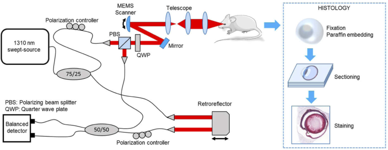
The system layout for mouse eye imaging is shown in Fig. 1 and is based on a modification of our previous imaging setup described in detail elsewhere [24]. A conversion to a purely single-mode fiber system was performed by replacing the previously used few-mode fiber with SMF28 fiber (Corning SMF-28e+). Further, instead of a free-space optical interference layout, a fused fiber coupler was used to interfere reference and sample arm signals instead of a free-space optical interference layout. A swept-source laser (Axsun Technologies, Inc., 1310 nm SS-OCT Laser Engine) operating at 1310 nm with a tuning range of 140 nm and 100 kHz repetition rate was used as the light source and provided a measured axial resolution of 6.5 µm in air. The imaging range achieved using the internal k-clock of the laser was 5 mm in air, spanned over 768 pixels in the OCT image. The beam power incident on the eye was 2 mW, yielding a sensitivity of 100 dB. A 50 mm focal length lens provided a lateral resolution of 8.8 µm as measured from a USAF resolution test target and the depth of focus of the scanning beam was calculated to be 1.04 mm in free space. For each eye, a raster scan consisting of pixels (repeating 5 B-scans per position to increase the signal-to-noise ratio (SNR)) was acquired corresponding to a field of view of mm. Prior to the imaging session, the polarization state in the sample arm was adjusted to provide maximum throughput through the circulator. Then the reference arm polarization state was matched using the polarization controller in the reference arm. After calibration, the polarization state was kept constant during the entire experiment for all the mouse eyes. In addition, two sets of 128 repeated B-scans were acquired along the horizontal meridian, setting the focus at two different positions: one at the cornea and one at the posterior surface of the lens. From these datasets, 10 consecutive B-scans were averaged to create the images in the results.
Fig. 1.
OCT imaging system and experimental protocol. The anesthetized mice were imaged in vivo with the OCT device depicted on the left. The eyes were extracted post mortem for histological processing as described on the right. MEMS: Microelectromechanical scanner.
2.2. Animals
APP/PS1 mice provided by Professor M. Jucker (Hertie Institute of Clinical Brain Research, University of Tübingen, Germany) [25] were used to establish a colony at the Division of Biomedical Research at the Medical University of Vienna. Both male and female transgenic mice with their respective aged-matched wildtype littermates as controls were housed under controlled lighting conditions (12 hours light, 12 hours dark) with food and water ad libitum. Genotyping was performed independently for human amyloid precursor protein (APP) and mutant human presenilin (PS1) [25]. A total of 15 mice (7 transgenic and 8 controls) were imaged in both eyes aged in the range between 17 and 24 months. In order to immobilize the mice for OCT imaging, the animals were anesthetized using a mixture of 0.75 µl/g ketamine and 0.15 µl/g xylazine diluted in 6.6 µl/g phosphate-buffered saline. Artificial tear drops were used to keep the eye moisturized and heating pads were placed underneath the mice to maintain the body temperature since lower temperatures also induce cataracts [26]. The measurement time was limited to 20 minutes in order to avoid the formation of cataracts due to a long period of anesthesia [27]. The experiment was conducted as indicated by the ARVO Statement for the Use of Animals in Ophthalmic and Vision Research and Directive 2010/63/EU. The study protocol was approved by the an ethics committee and by the Austrian Federal Ministry of Education, Science and Research (BMBWF/66.009/0272-V/3b/2019).
2.3. Histological preparation
After OCT imaging, mice were euthanized by cervical dislocation. The eyes were marked at the dorsal edge of the cornea with a permanent marking pen to maintain the orientation during microtomy, before being enucleated and immersed unopened in Davidson’s fixative for 24 hours at C. Whole eyes were dehydrated in graded alcohols and processed through xylene into paraffin. Three-micron-thick sections were cut parallel to the dorsoventral axis of the eye, mounted onto slides, deparaffinized, rehydrated, and stained with Hematoxylin and Eosin or Azan after Heidenhain. Sections were examined on a Zeiss Axio Imager.Z2 and scanned using a 3DHistech Pannoramic SCAN II slide scanner.
2.4. OCT data processing
The raw data were acquired with a custom-made LabView program (LabVIEW 2015, National Instruments) that was also used for controlling the imaging setup. For the image analysis, a postprocessing pipeline was implemented in Matlab (MATLAB, R2015b, MathWorks) [28]. First, in order to remove the spectral sampling shifts caused by A-line trigger jitter, the spectrum of every single A-line was aligned with a unique spectrum using cross-correlation. Spectral shaping and background removal were then performed to improve the image quality [29]. After computing the B-scan images by Fourier transforming the spectral data, motion artifacts between the five repeated B-scans were corrected using cross-correlation. The aligned sets of repeated B-scans were then averaged to improve the SNR. Although the laser power remained constant, slight intensity differences between the data sets occurred due to eventual shifts of focal plane between the measurements.
2.5. Statistical analysis
Blind grading of lens severity was performed independently by three experts. For this purpose, all the volumetric datasets of each individual eye were randomized and independently graded under visual inspection by each observer between 0 (weak scattering) and 3 (severe opacification with strong scattering signal). The grades were then grouped for wildtype and transgenic mice, respectively, and plotted as histograms. A Wilcoxon signed rank test was performed to test for the equality of the grade distributions between wildtype and transgenic mice using a significance level of . To analyze the severity of opacifications, the Weber contrast (WC) [30] was used as a quantitative measure. A circular region of interest (ROI) spanning a diameter of 10 pixels was manually annotated in the B-scan in the opacification core and then numerically compared to an equivalent ROI selected within the anterior chamber where only background signal was present. The WC was then calculated using the mean intensity values of the respective ROIs as:
| (1) |
where is the averaged intensity (logarithmic scale) in the opacity ROI and the averaged intensity (logarithmic scale) in the background ROI. The WCs were plotted as box-and-whisker diagrams in order to compare transgenic and wildtype mice. The WC values were also compared between age groups. A paired t-test assuming a significance level of p 0.05 was used to analyze the WC contrast correlation between wildtype and transgenic mice and also for adult (17/18 months) and old (23/24 months) mice groups.
3. Results
3.1. Mouse lens imaging
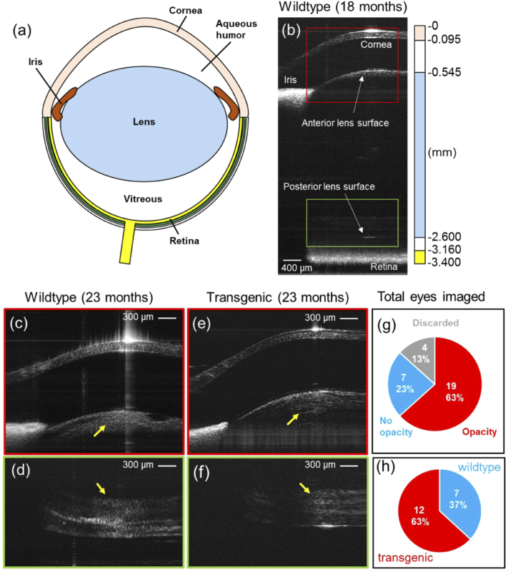
OCT revealed the presence of hyperscattering lesions in the lenses of both wildtype mice and their transgenic litter mates. While no loss of transparency was observed in the lens nucleus, several mice exhibited hyperreflective features in the lens cortex both at the anterior and posterior lens pole, as shown in Fig. 2(c-f). From a total of 30 eyes measured, 7 showed no opacity lesions in the lens, 19 exhibited hyperreflective opacifications in the lens and 4 had to be discarded due to poor image quality, as represented in Fig. 2(g). From the 19 eyes with opacifications, 7 were from wildtype mice (5 different individuals) and 12 from transgenic mice (7 different individuals), as represented in Fig. 2(h). Focusing on the posterior lens pole, hyperreflective signal appeared diffuse within the cortex adjacent to the lens capsule as shown in Fig. 2(d,f). Whenever an opacity was identified in the anterior surface a corresponding cloudy formation in the posterior part was also observed. However, not every imaged eye exhibiting hyperreflective features at the posterior pole also showed similar opacities at the anterior pole of the lens (N = 4).
Fig. 2.
Crystalline lens imaging with OCT. a) Illustration of the mouse eye. b) Whole OCT section of a wildtype mouse eye where the two focus locations selected for this experiment are indicated with a red box (anterior lens surface) and a green box (posterior lens surface). The scale bar on the right indicates the anatomical axial lengths of the different ocular components [31]. Note that the images were not corrected for light refraction by the individual structures. The retina appears flipped over the zero delay in this image due to the available imaging range such that the vitreous length appears reduced. c) Zoom-in on the anterior lens pole of a wildtype mouse (age 23 months) where an opacity has formed as indicated by the yellow arrow. The bright artifact at the corneal epithelium is due to a strong backreflection at the air-tissue interface. d) Zoom-in to the posterior part of the lens of the corresponding mouse. e) Zoom-in to the anterior lens part of a transgenic mouse of the same age where an opaque region can be observed posterior to the lens surface. f) Posterior lens surface of the transgenic mouse. Images were not corrected for light refraction. g) Pie chart representing the presence of lesions for the total number of eyes imaged. Four datasets were discarded due to poor image quality or eye condition. h) Pie chart representing the number of eyes with opacity lesions observed for wildtype and transgenic mice.
3.2. Cataract manifestations in aged and AD mice
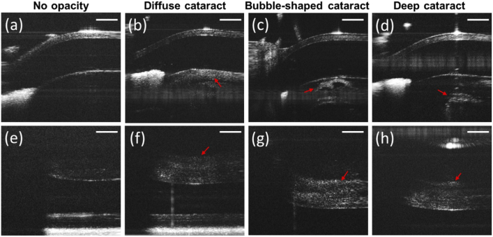
When analyzing the individual results, it was possible to appreciate different shapes and cataract morphologies, in particular close to the anterior lens surface. In the cases where no opacification was visually observed (N = 7), the lens appeared as a semi-translucent tissue devoid of a substantial OCT signal (with the exception of the lens surface interface) as shown in Fig. 3(a,e). A commonly observed cataractous lesion (N = 11) was a diffuse cloudy formation extended underneath the lens surface as shown in Fig. 3(b). Another particular cataract formation observed in some mouse eyes (N = 6) was a bubble-like shape as shown in Fig. 3(c). These lesions were very compact and localized, usually along the central axis and close to the anterior lenticular cortex. However, when analyzing the posterior lens surface of the eyes exhibiting these bubble-shaped lesions, the same diffuse cloudy opacity as in the other hyperscattering cases was observed, as shown in Fig. 3(g). In other cases (N = 2), the cataractous lesions were formed deeper in the lens and far from the surface, as it can be seen in Fig. 3(d). As in the cases described above, the posterior lens opacity developed in these eyes had a diffuse shape spread adjacent to the cortex, see Fig. 3(h).
Fig. 3.
Examples of different cataract manifestations in OCT. Details of the anterior lens (a-d) and the corresponding counterparts in the posterior lens (e-h) are shown. a) Representative B-scan of an anterior lens devoid of opacities. b) Diffuse cataract in the anterior part of the lens. Increased scattering can be observed across the anterior lens capsule. c) Bubble-shaped opacity formation close to the anterior lens surface. d) Cataract located in the lens nucleus. No opacity can be observed at the lens surface. e-h) Corresponding B-scans focusing on the posterior part of the lens. e) Only a weak scattering signal can be observed in the posterior lens. The retina is located at the bottom of the image. f-g) Diffuse opacifications with increased scattering signal formed in the lens cortex. Red arrows indicate opacification. Images were not corrected for light refraction. The horizontal lines in panels (a-d) are artifacts caused by a reflection within the system optics that could not be entirely removed by post processing. Scale bars correspond to 0.5 mm.
3.3. Correlation to histology
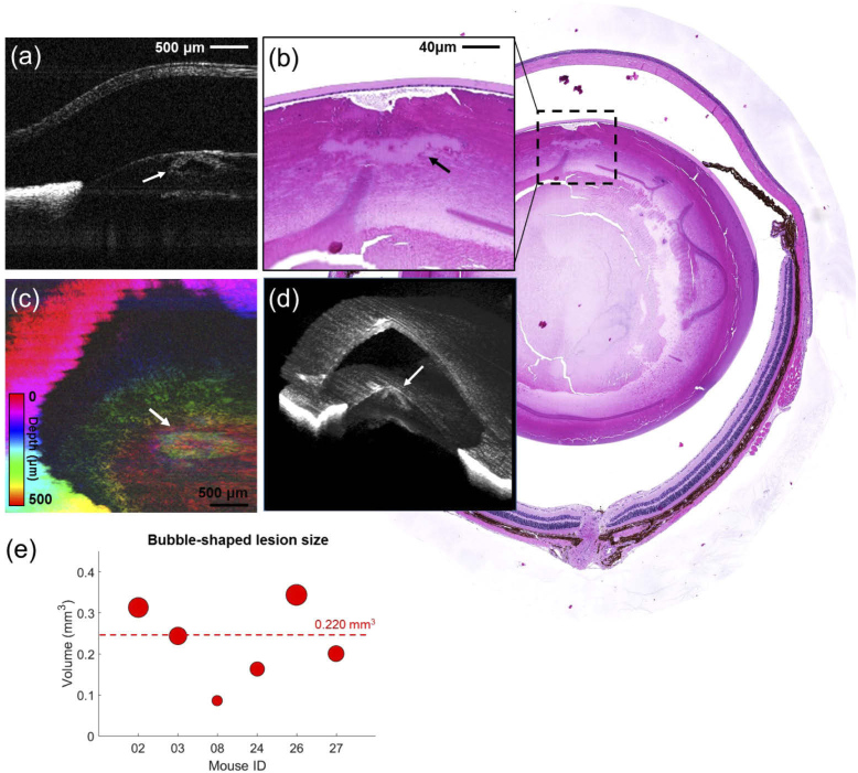
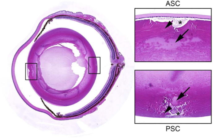
The 3D SS-OCT scans provided access to volumetric information and a quantitative analysis of the ocular lesions. The bubble-shaped lesions observed in 4 wildtype and 2 transgenic mouse eyes were investigated in more depth and compared to histological sections obtained from the same eyes. The dimensions of these bubble lesions were manually measured using ITK-Snap [32], giving an average volume of 0.220 0.100 (assuming a average lens refractive index of 1.44 [33]). Unlike the bubble formations, the other opacities found were diffuse, with a non-uniform distribution and an undefined edge which made it difficult to obtain a specific volume. The results are shown in Fig. 4. These lesions appeared as a hyperscattering area surrounding a hyposcattering void in OCT (Fig. 4(a)). All six mice presented with the typical histopathology of subcapsular cataract including sub-epithelial vacuolisation, extracellular cleft formation, disintregation of lens fibers, and the presence of eosinophilic (Morgagnian) globules of variable size, as shown in Fig. 5. In two eyes of two wildtype mice, a massive clod of homogenous subcapsular material (Fig. 4(b)) appeared to precisely match the size and relative position of the hyporeflective foci in the centre of the bubble-shaped lesions observed by OCT (Fig. 4(a)). There is a possibility that that focal cataracts present were missed during tissue preparation for microscopical analysis because the precise alignment of whole eyes for sectioning is challenging and series of sections through entire lenses cannot easily be obtained. A color-encoded depth projection over 500 µm shows the location of the lesion in Fig. 4(c). The capsular form of the lesion (in greenish color) is observed at a depth where no hyperscattering signal would be expected in normal eyes. The lesion location in relation to the anatomical structures can be clearly seen in the 3D reconstruction of Fig. 4(d). In the cutout portion, the bubble-shaped lesion with a hyperscattering body surrounding the hyposcattering center can be observed close to the lens capsule. The measured sizes of the 6 bubble lesions observed in the OCT images are shown in Fig. 4(e). The entire dataset can be seen in Visualization 1 (11.2MB, avi) .
Fig. 4.
Lenticular lesion in a 23-month old wildtype mouse investigated using the SS-OCT system. a) B-scan of a mouse anterior lens which exhibits a bubble-shaped lesion. b) Corresponding histological section of the imaged area. The lesion is indicated by an arrow. c) Depth intensity projection over 500 µm in the lesion region. d) 3D reconstruction of the anterior segment. The investigated bubble-shaped lesion can be observed right beneath the lens surface. OCT images were not corrected for light refraction. e) Measured sizes of the 6 bubble lesions observed in the 3D datasets.
Fig. 5.
The figure below shows representative lesions we observed in the lens of an mouse. Both anterior subcapsular cataract (ASC) and posterior subcapsular cataract (PSC) were observed. Anterior and posterior sutures were composed of enlarged fiber cells around the suture, fragmenting into eosinic globules (Morgagnian globules, arrowheads) and subcapsular vacuoles filled with debris (asterisks). Clods of cortical material are indicated with arrows.
3.4. Quantification of mouse lens opacifications
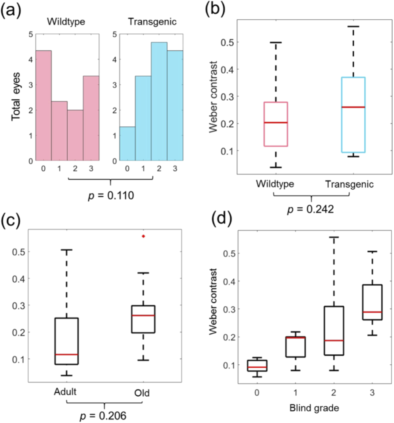
The experts performed the dataset grading individually, giving a total of 330 different grades which were averaged among the four sets of scores to provide the result shown in Fig. 6(a). Hyperscattering lesions were observed in both transgenic mice and their wildtype littermates. The opacity classification revealed a tendency towards more severe opacifications for transgenic mice. In wildtype mice, a lower number of high grade lesions and more cases with clear lens were observed. However, the lesions scored in wildtype and transgenic mice were found not to have significantly different distributions (p = 0.110). In order to assess the scattering lesions quantitatively, we calculated the Weber contrast between the averaged intensity in the opaque ROI and the averaged intensity in the background, as described in the methods section. However, no statistically significant difference (p = 0.242) was found between the WC in wildtype and transgenic animals, as it is shown in of Fig. 6(b). We also analyzed if the mouse age was correlated to the Weber contrast by plotting WC as a function of age for adult (17 and 18 months) and old (23 and 24 months) mice as shown in Fig. 6(c). No significant difference (p = 0.206) was found for WC between the two age groups, although a slight tendency toward higher values in older mice could be observed. A comparison between the lens severity scale and the Weber contrast is shown in Fig. 6(d).
Fig. 6.
Analysis of grade and contrast of the lens opacifications. a) Histogram distributions of the opacification gradings given for wildtype and transgenic mice. b) Weber contrast for wildtype and transgenic mouse groups. c) Weber contrast for adult (17/18 months) and old (23/24 months) mouse groups. d) Comparison of the Weber contrast values retrieved from the data and the blind visual grades. A tendency to increased WC values in high grades can be observed.
4. Discussion
The benefit of using OCT for ocular imaging has been widely demonstrated over the past years. The application of OCT for imaging the ocular lens, however, has only been studied scarcely in humans and even less in rodents [16–21]. In this work, we have demonstrated the use of a custom-made OCT system for studying the murine crystalline lens in vivo. Volumetric OCT enabled the visualization of different types of opacities and provided information about their shape and location in the murine lens.
In order to evaluate the imaging performance in the mouse eye, both wildtype mice and a mouse model of Alzheimer’s disease were investigated. OCT images revealed differently shaped hyperscattering lesions in the crystalline lens, both in transgenic mice and their wildtype litter mates. Grading by three readers resulted in different distributions of the severity scores for lens opacifications observed in transgenic versus wildtype animals. A numerical quantification of the opacification intensities using the WC did not reveal a significant difference for the signal intensities between transgenic and wildtype. This indicates that a mere analysis of the SNR in cataractous lesions may not suffice for numerically distinguishing differences between lesions in healthy and transgenic animals. However, an tendency towards increased WC values was observed for high grades, suggesting that it could be a good quantitative indicator of cataract severity. More sophisticated numerical approaches also incorporating the shape and texture of the lesions may open the door to dedicated segmentation and objective classification of cataractous lesions.
Alzheimer’s disease related pathology has already been investigated using OCT both in the brain and in the eye [34–38]. Retinal diseases such as glaucoma, age-related macular degeneration and diabetic retinopathy have been associated with dementia and AD [39], however the relation of AD and cataract formation in the crystalline lens is still unclear. Goldstein et al. postulated the existence of a relationship between supranuclear cataracts co-localized with microaggregates of amyloid-beta (A) peptides in the human lens [22]. Jun et al. suggested the connection of the delta-catenin protein to a high contingency of cataracts in Alzheimer’s patients [40]. In a generational study running since 1948 investigating more than 1000 subjects, a very strong correlation between AD and cataracts was found [40]. Recently, Lee et al. reported in their study that no association was found between cataracts and risk of developing AD [39]. Given the fact that we observed lens opacifications also in wildtype mice, it is very likely that those are due to old age since all the investigated mice were already 17 months old or older. A slight tendency toward higher WC was observed for older mice but more data points covering a wider age range (in particular also younger mice) would be necessary for a reliable estimate between mouse age and opacification development. For future studies, it would be interesting to increase the number of mice and perform a longitudinal investigation over an extended age range, analyzing also the (early) development and formation of the lens opacifications. Since we were not able to investigate the mice awake, measurements could be influenced by the anesthesia which may also have induced opacifications in the mouse lenses, even though we kept the animals warm and their eyes constantly moist during the experiment to avoid such artifacts [27].
There are several aspects that could be implemented in the future for improving our system capabilities and the data analysis when investigating the rodent lens. The use of a light source operating at a shorter wavelength such as 1060 nm would improve the scattering contrast of the opacities in the lens [41]. The use of shorter wavelengths (such as visible light) would be more accurate in reflecting the visual effects of the eye in terms of scattering, but at the cost of greater restriction of power levels and potentially reduced light penetration. Another challenging aspect of murine lens imaging is the shallow depth of focus offered by common OCT devices, making simultaneous imaging of the entire lens difficult. This could be solved by using extended-focus techniques or dynamic tunable focusing [42–44]. The transparency of the crystalline lens further complicates the visualization with techniques such as OCT. However, the SNR can be improved by signal averaging as performed in this study. As averaging of rather extensive image sequences can be challenging for in vivo imaging settings due to motion artifacts, a good animal fixation and/or motion compensation approaches may be employed [45,46] .
The OCT images presented here were not corrected for light refraction by the cornea and other ocular structures. In consequence, the observed ocular dimensions such as the shape of the lens are affected by the preceding optical structures and appear distorted. A rough conversion to the eyes’ actual geometry can be performed using ray tracing based on refractive index data for each respective ocular section reported in the literature [47]. However, the crystalline lens is known to have a non-homogeneous refractive index with a gradient distribution [48,49]. Computational methods have been proposed to retrieve the produced optical distortions and reconstruct the crystalline lens using OCT [50–52]. While the results presented in this article already demonstrate the benefits of OCT for the visualization of structures in the murine lens in vivo, the implementation of ray tracing algorithms to recover the true ocular geometry may, in a future study, provide further access to biometrical parameters such as anterior chamber depth, lens thickness or the actual dimensions of cataractous lesions. For the data shown in this manuscript, we also did not compensate for shadowing posterior to cataracts, a fact that should be taken into account in future studies.
The transparency of the lens is related to the arrangement of the lenticular fiber cells [3,53]. The disruption of these fibrillar structures and the aggregation of cells are the main cause of cataracts and opacifications [53]. The extension of our device to a polarization sensitive OCT system might enable the visualization of depolarization produced by cataractous lesions [54,55]. Since the basic principle of many imaging methods currently used for cataracts investigations are based on light scattering, our recently proposed bright and dark-field (BRAD) OCT technique might also be an interesting candidate for investigating scattering properties and cataractous lesions in the crystalline lens [24,28].
In conclusion, we have demonstrated SS-OCT for visualizing opacifications in the murine lens in vivo. The access to volumetric information enabled a more complete analysis of the crystalline lens as well as the selection of specific cross-sections, advantages over the subjective methods clinically used such as slit-lamp photography. Imaging and quantification of lens lesions based on OCT images may facilitate diagnosis and suppose a breakthrough in the investigation of crystalline-related diseases. However, more studies need to be performed to strengthen the case for using OCT for cataract evaluation and to compare OCT to established diagnostic techniques .
Acknowledgement
This work was funded by the European Research Council (ERC StG 640396 OPTIMALZ). The authors would like to thank the staff at the animal care facility of the Medical University of Vienna. We would like to acknowledge Andreas Hodul for technical support and Adelheid Woehrer at the neuropathology department. Special thanks to Christoph K. Hitzenberger for his support and expertise during the study.
Funding
European Research Council10.13039/501100000781 (ERC StG 640396 OPTIMALZ).
Disclosures
The authors declare that there are no conflicts of interest related to this article.
References
- 1.Flaxman S. R., Bourne R. R., Resnikoff S., Ackland P., Braithwaite T., Cicinelli M. V., Das A., Jonas J. B., Keeffe J., Kempen J., Leasher H., Limburg H., Naidoo K., Pesudovs K., Silvester A., Stevens G., Tahhan N., Wong T. Y., Taylor H. R., “Global causes of blindness and distance vision impairment 1990–2020: a systematic review and meta-analysis,” The Lancet Glob. Heal. 5(12), e1221–e1234 (2017). 10.1016/S2214-109X(17)30393-5 [DOI] [PubMed] [Google Scholar]
- 2.Remington L. A., Goodwin D., Clinical anatomy of the visual system E-Book (Elsevier Health Sciences, 2011). [Google Scholar]
- 3.Michael R., Bron A., “The ageing lens and cataract: a model of normal and pathological ageing,” Philos. Trans. R. Soc., B 366(1568), 1278–1292 (2011). 10.1098/rstb.2010.0300 [DOI] [PMC free article] [PubMed] [Google Scholar]
- 4.Maggs D. J., Miller P., Mrcpsych M. D., Ofri R., Slatter’s fundamentals of veterinary ophthalmology (Elsevier Health Sciences, 2013). [Google Scholar]
- 5.Okano T., Uga S., Ishikawa S., Shumiya S., “Histopathological study of hereditary cataractous lenses in SCR strain rat,” Exp. Eye Res. 57(5), 567–576 (1993). 10.1006/exer.1993.1161 [DOI] [PubMed] [Google Scholar]
- 6.Chylack L. T., Wolfe J. K., Singer D. M., Leske M. C., Bullimore M. A., Bailey I. L., Friend J., McCarthy D., Wu S.-Y., “The lens opacities classification system III,” Arch. Ophthalmol. 111(6), 831–836 (1993). 10.1001/archopht.1993.01090060119035 [DOI] [PubMed] [Google Scholar]
- 7.Grewal D. S., Brar G. S., Grewal S. P. S., “Correlation of nuclear cataract lens density using Scheimpflug images with Lens Opacities Classification System III and visual function,” Ophthalmology 116(8), 1436–1443 (2009). 10.1016/j.ophtha.2009.03.002 [DOI] [PubMed] [Google Scholar]
- 8.Rosales P., Marcos S., “Pentacam Scheimpflug quantitative imaging of the crystalline lens and intraocular lens,” J. Cataract Refractive Surg. 25(5), 421–428 (2009). 10.3928/1081597X-20090422-04 [DOI] [PubMed] [Google Scholar]
- 9.Weiner X., Baumeister M., Kohnen T., Bühren J., “Repeatability of lens densitometry using Scheimpflug imaging,” J. Cataract Refractive Surg. 40(5), 756–763 (2014). 10.1016/j.jcrs.2013.10.039 [DOI] [PubMed] [Google Scholar]
- 10.Lim S. A., Hwang J., Hwang K.-Y., Chung S.-H., “Objective assessment of nuclear cataract: comparison of double-pass and Scheimpflug systems,” J. Cataract Refractive Surg. 40(5), 716–721 (2014). 10.1016/j.jcrs.2013.10.032 [DOI] [PubMed] [Google Scholar]
- 11.Saad A., Saab M., Gatinel D., “Repeatability of measurements with a double-pass system,” J. Cataract Refractive Surg. 36(1), 28–33 (2010). 10.1016/j.jcrs.2009.07.033 [DOI] [PubMed] [Google Scholar]
- 12.Fercher A. F., Drexler W., Hitzenberger C. K., Lasser T., “Optical coherence tomography-principles and applications,” Rep. Prog. Phys. 66(2), 239–303 (2003). 10.1088/0034-4885/66/2/204 [DOI] [Google Scholar]
- 13.Adhi M., Duker J. S., “Optical coherence tomography–current and future applications,” Curr. opinion ophthalmology 24(3), 213–221 (2013). 10.1097/ICU.0b013e32835f8bf8 [DOI] [PMC free article] [PubMed] [Google Scholar]
- 14.Ramos J. L. B., Li Y., Huang D., “Clinical and research applications of anterior segment optical coherence tomography–a review,” Clin. & Exp. Ophthalmol. 37(1), 81–89 (2009). 10.1111/j.1442-9071.2008.01823.x [DOI] [PMC free article] [PubMed] [Google Scholar]
- 15.Ang M., Baskaran M., Werkmeister R. M., Chua J., Schmidl D., dos Santos V. A., Garhoefer G., Mehta J. S., Schmetterer L., “Anterior segment optical coherence tomography,” Prog. Retinal Eye Res. 66, 132–156 (2018). 10.1016/j.preteyeres.2018.04.002 [DOI] [PubMed] [Google Scholar]
- 16.DiCarlo C. D., Roach W. P., Gagliano D. A., Boppart S. A., Hammer D. X., Cox A. B., Fujimoto J. G., “Comparison of optical coherence tomography imaging of cataracts with histopathology,” J. Biomed. Opt. 4(4), 450–459 (1999). 10.1117/1.429951 [DOI] [PubMed] [Google Scholar]
- 17.Chan T. C., Li E. Y., Yau J. C., “Application of anterior segment optical coherence tomography to identify eyes with posterior polar cataract at high risk for posterior capsule rupture,” J. Cataract Refractive Surg. 40(12), 2076–2081 (2014). 10.1016/j.jcrs.2014.03.033 [DOI] [PubMed] [Google Scholar]
- 18.Pérez-Merino P., Velasco-Ocana M., Martinez-Enriquez E., Marcos S., “OCT-based crystalline lens topography in accommodating eyes,” Biomed. Opt. Express 6(12), 5039–5054 (2015). 10.1364/BOE.6.005039 [DOI] [PMC free article] [PubMed] [Google Scholar]
- 19.Panthier C., Burgos J., Rouger H., Saad A., Gatinel D., “New objective lens density quantification method using swept-source optical coherence tomography technology: Comparison with existing methods,” J. Cataract Refractive Surg. 43(12), 1575–1581 (2017). 10.1016/j.jcrs.2017.09.028 [DOI] [PubMed] [Google Scholar]
- 20.de Castro A., Benito A., Manzanera S., Mompeán J., Canizares B., Martínez D., Marín J. M., Grulkowski I., Artal P., “Three-dimensional cataract crystalline lens imaging with swept-source optical coherence tomography,” Invest. Ophthalmol. Visual Sci. 59(2), 897–903 (2018). 10.1167/iovs.17-23596 [DOI] [PubMed] [Google Scholar]
- 21.Grulkowski I., Manzanera S., Cwiklinski L., Mompeán J., de Castro A., Marin J. M., Artal P., “Volumetric macro-and micro-scale assessment of crystalline lens opacities in cataract patients using long-depth-range swept source optical coherence tomography,” Biomed. Opt. Express 9(8), 3821–3833 (2018). 10.1364/BOE.9.003821 [DOI] [PMC free article] [PubMed] [Google Scholar]
- 22.Goldstein L. E., Muffat J. A., Cherny R. A., Moir R. D., Ericsson M. H., Huang X., Mavros C., Coccia J. A., Faget K. Y., Fitch K. A., Masters C., Tanzi R. E., Chylack L. T., Bush A. I., “Cytosolic β-amyloid deposition and supranuclear cataracts in lenses from people with Alzheimer’s disease,” Lancet 361(9365), 1258–1265 (2003). 10.1016/S0140-6736(03)12981-9 [DOI] [PubMed] [Google Scholar]
- 23.Michael R., Otto C., Lenferink A., Gelpi E., Montenegro G. A., Rosandić J., Tresserra F., Barraquer R. I., Vrensen G. F., “Absence of amyloid-beta in lenses of Alzheimer patients: a confocal Raman microspectroscopic study,” Exp. Eye Res. 119, 44–53 (2014). 10.1016/j.exer.2013.11.016 [DOI] [PubMed] [Google Scholar]
- 24.Eugui P., Lichtenegger A., Augustin M., Harper D. J., Muck M., Roetzer T., Wartak A., Konegger T., Widhalm G., Hitzenberger C. K., Woehrer A., Baumann B., “Beyond backscattering: optical neuroimaging by BRAD,” Biomed. Opt. Express 9(6), 2476–2494 (2018). 10.1364/BOE.9.002476 [DOI] [PMC free article] [PubMed] [Google Scholar]
- 25.Radde R., Bolmont T., Kaeser S. A., Coomaraswamy J., Lindau D., Stoltze L., Calhoun M. E., Jäggi F., Wolburg H., Gengler S., Haass C., Ghetti B., Czech C., Holscher C., Matthews P. M., Jucker M., “Aβ42-driven cerebral amyloidosis in transgenic mice reveals early and robust pathology,” EMBO Rep. 7(9), 940–946 (2006). 10.1038/sj.embor.7400784 [DOI] [PMC free article] [PubMed] [Google Scholar]
- 26.Bermudez M. A., Vicente A. F., Romero M. C., Arcos M. D., Abalo J. M., Gonzalez F., “Time course of cold cataract development in anesthetized mice,” Curr. Eye Res. 36(3), 278–284 (2011). 10.3109/02713683.2010.542868 [DOI] [PubMed] [Google Scholar]
- 27.Calderone L., Grimes P., Shalev M., “Acute reversible cataract induced by xylazine and by ketamine-xylazine anesthesia in rats and mice,” Exp. Eye Res. 42(4), 331–337 (1986). 10.1016/0014-4835(86)90026-6 [DOI] [PubMed] [Google Scholar]
- 28.Eugui P., Harper D. J., Lichtenegger A., Augustin M., Merkle C. W., Woehrer A., Hitzenberger C. K., Baumann B., “Polarization-sensitive imaging with simultaneous bright-and dark-field optical coherence tomography,” Opt. Lett. 44(16), 4040–4043 (2019). 10.1364/OL.44.004040 [DOI] [PubMed] [Google Scholar]
- 29.Tripathi R., Nassif N., Nelson J. S., Park B. H., de Boer J. F., “Spectral shaping for non-gaussian source spectra in optical coherence tomography,” Opt. Lett. 27(6), 406–408 (2002). 10.1364/OL.27.000406 [DOI] [PubMed] [Google Scholar]
- 30.Lozzi A., Agrawal A., Boretsky A., Welle C. G., Hammer D. X., “Image quality metrics for optical coherence angiography,” Biomed. Opt. Express 6(7), 2435–2447 (2015). 10.1364/BOE.6.002435 [DOI] [PMC free article] [PubMed] [Google Scholar]
- 31.Remtulla S., Hallett P., “A schematic eye for the mouse, and comparisons with the rat,” Vision Res. 25(1), 21–31 (1985). 10.1016/0042-6989(85)90076-8 [DOI] [PubMed] [Google Scholar]
- 32.Yushkevich P. A., Piven J., Hazlett H. C., Smith R. G., Ho S., Gee J. C., Gerig G., “User-guided 3D active contour segmentation of anatomical structures: significantly improved efficiency and reliability,” NeuroImage 31(3), 1116–1128 (2006). 10.1016/j.neuroimage.2006.01.015 [DOI] [PubMed] [Google Scholar]
- 33.Chakraborty R., Lacy K. D., Tan C. C., na Park H., Pardue M. T., “Refractive index measurement of the mouse crystalline lens using optical coherence tomography,” Exp. Eye Res. 125, 62–70 (2014). 10.1016/j.exer.2014.05.015 [DOI] [PMC free article] [PubMed] [Google Scholar]
- 34.Coppola G., Di Renzo A., Ziccardi L., Martelli F., Fadda A., Manni G., Barboni P., Pierelli F., Sadun A. A., Parisi V., “Optical coherence tomography in Alzheimer’s disease: a meta-analysis,” PLoS One 10(8), e0134750 (2015). 10.1371/journal.pone.0134750 [DOI] [PMC free article] [PubMed] [Google Scholar]
- 35.Cunha J. P., Moura-Coelho N., Proença R. P., Dias-Santos A., Ferreira J., Louro C., Castanheira-Dinis A., “Alzheimer’s disease: A review of its visual system neuropathology. Optical coherence tomography—a potential role as a study tool in vivo,” Graefe’s Arch. Clin. Exp. Ophthalmol. 254(11), 2079–2092 (2016). 10.1007/s00417-016-3430-y [DOI] [PubMed] [Google Scholar]
- 36.Poroy C., Yücel A. A., “Optical coherence tomography: Is really a new biomarker for Alzheimer’s disease?” Annals Indian Acad. Neurol. 21(2), 119 (2018). 10.4103/aian.AIAN_368_17 [DOI] [PMC free article] [PubMed] [Google Scholar]
- 37.Lichtenegger A., Muck M., Eugui P., Harper D. J., Augustin M., Leskovar K., Hitzenberger C. K., Woehrer A., Baumann B., “Assessment of pathological features in Alzheimer’s disease brain tissue with a large field-of-view visible-light optical coherence microscope,” Neurophotonics 5(03), 1 (2018). 10.1117/1.NPh.5.3.035002 [DOI] [PMC free article] [PubMed] [Google Scholar]
- 38.Gesperger J., Lichtenegger A., Roetzer T., Augustin M., Harper D. J., Eugui P., Merkle C. W., Hitzenberger C. K., Woehrer A., Baumann B., “Comparison of intensity-and polarization-based contrast in amyloid-beta plaques as observed by optical coherence tomography,” Appl. Sci. 9(10), 2100 (2019). 10.3390/app9102100 [DOI] [Google Scholar]
- 39.Lee C. S., Larson E. B., Gibbons L. E., Lee A. Y., McCurry S. M., Bowen J. D., McCormick W. C., Crane P. K., “Associations between recent and established ophthalmic conditions and risk of Alzheimer’s disease,” Alzheimers Dement. 15(1), 34–41 (2019). 10.1016/j.jalz.2018.06.2856 [DOI] [PMC free article] [PubMed] [Google Scholar]
- 40.Jun G., Moncaster J. A., Koutras C., Seshadri S., Buros J., McKee A. C., Levesque G., Wolf P. A., George-Hyslop P. S., Goldstein L. E., Farrar L. A., “δ-catenin is genetically and biologically associated with cortical cataract and future Alzheimer-related structural and functional brain changes,” PLoS One 7(9), e43728 (2012). 10.1371/journal.pone.0043728 [DOI] [PMC free article] [PubMed] [Google Scholar]
- 41.Wang Y., Nelson J. S., Chen Z., Reiser B. J., Chuck R. S., Windeler R. S., “Optimal wavelength for ultrahigh-resolution optical coherence tomography,” Opt. Express 11(12), 1411–1417 (2003). 10.1364/OE.11.001411 [DOI] [PubMed] [Google Scholar]
- 42.Leitgeb R., Villiger M., Bachmann A., Steinmann L., Lasser T., “Extended focus depth for fourier domain optical coherence microscopy,” Opt. Lett. 31(16), 2450–2452 (2006). 10.1364/OL.31.002450 [DOI] [PubMed] [Google Scholar]
- 43.Su J. P., Li Y., Tang M., Liu L., Pechauer A. D., Huang D., Liu G., “Imaging the anterior eye with dynamic-focus swept-source optical coherence tomography,” J. Biomed. Opt. 20(12), 126002 (2015). 10.1117/1.JBO.20.12.126002 [DOI] [PMC free article] [PubMed] [Google Scholar]
- 44.Grulkowski I., Manzanera S., Cwiklinski L., Sobczuk F., Karnowski K., Artal P., “Swept source optical coherence tomography and tunable lens technology for comprehensive imaging and biometry of the whole eye,” Optica 5(1), 52–59 (2018). 10.1364/OPTICA.5.000052 [DOI] [Google Scholar]
- 45.Swanson E. A., Izatt J. A., Hee M. R., Huang D., Lin C., Schuman J., Puliafito C., Fujimoto J. G., “In vivo retinal imaging by optical coherence tomography,” Opt. Lett. 18(21), 1864–1866 (1993). 10.1364/OL.18.001864 [DOI] [PubMed] [Google Scholar]
- 46.Kraus M. F., Potsaid B., Mayer M. A., Bock R., Baumann B., Liu J. J., Hornegger J., Fujimoto J. G., “Motion correction in optical coherence tomography volumes on a per a-scan basis using orthogonal scan patterns,” Biomed. Opt. Express 3(6), 1182–1199 (2012). 10.1364/BOE.3.001182 [DOI] [PMC free article] [PubMed] [Google Scholar]
- 47.Chou T.-H., Kocaoglu O. P., Borja D., Ruggeri M., Uhlhorn S. R., Manns F., Porciatti V., “Postnatal elongation of eye size in DBA/2J mice compared with C57BL/6J mice: in vivo analysis with whole-eye OCT,” Invest. Ophthalmol. Visual Sci. 52(6), 3604–3612 (2011). 10.1167/iovs.10-6340 [DOI] [PMC free article] [PubMed] [Google Scholar]
- 48.Campbell M., Hughes A., “An analytic, gradient index schematic lens and eye for the rat which predicts aberrations for finite pupils,” Vision Res. 21(7), 1129–1148 (1981). 10.1016/0042-6989(81)90016-X [DOI] [PubMed] [Google Scholar]
- 49.Philipson B., “Distribution of protein within the normal rat lens,” Invest. Ophthalmol. Visual Sci. 8, 258–270 (1969). [PubMed] [Google Scholar]
- 50.Ortiz S., Siedlecki D., Grulkowski I., Remon L., Pascual D., Wojtkowski M., Marcos S., “Optical distortion correction in optical coherence tomography for quantitative ocular anterior segment by three-dimensional imaging,” Opt. Express 18(3), 2782–2796 (2010). 10.1364/OE.18.002782 [DOI] [PubMed] [Google Scholar]
- 51.Verma Y., Rao K., Suresh M., Patel H., Gupta P., “Measurement of gradient refractive index profile of crystalline lens of fisheye in vivo using optical coherence tomography,” Appl. Phys. B 87(4), 607–610 (2007). 10.1007/s00340-007-2689-4 [DOI] [Google Scholar]
- 52.de Castro A., Ortiz S., Gambra E., Siedlecki D., Marcos S., “Three-dimensional reconstruction of the crystalline lens gradient index distribution from OCT imaging,” Opt. Express 18(21), 21905–21917 (2010). 10.1364/OE.18.021905 [DOI] [PubMed] [Google Scholar]
- 53.Bassnett S., Shi Y., Vrensen G. F., “Biological glass: structural determinants of eye lens transparency,” Philos. Trans. R. Soc., B 366(1568), 1250–1264 (2011). 10.1098/rstb.2010.0302 [DOI] [PMC free article] [PubMed] [Google Scholar]
- 54.Baumann B., Augustin M., Lichtenegger A., Harper D. J., Muck M., Eugui P., Wartak A., Pircher M., Hitzenberger C. K., “Polarization-sensitive optical coherence tomography imaging of the anterior mouse eye,” J. Biomed. Opt. 23(08), 1 (2018). 10.1117/1.JBO.23.8.086005 [DOI] [PubMed] [Google Scholar]
- 55.Götzinger E., Bolz M., Zotter S., Torzicky T., Pircher M., Roberts P., Schlanitz F., Schmidt-Erfurth U., Hitzenberger C. K., “Polarization sensitive spectral domain optical coherence tomography of cataract lenses,” in Biomedical Optics and 3-D Imaging, OSA Technical Digest, (Optical Society of America, 2012), pp. BTu3A–91. [Google Scholar]


