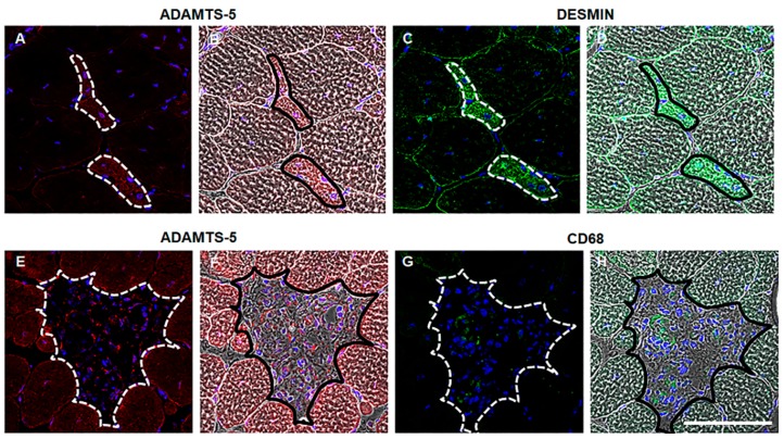
Figure 2.
ADAMTS-5 is highly expressed in regions of regeneration and inflammation in dystrophic muscles. ADAMTS-5 immunoreactivity (red), desmin or CD68 immunoreactivity (green) and nuclei (blue) in serial cross-sections. Phase contrast images overlaid with the fluorescent signal provide morphological evidence of ADAMTS-5 and desmin or CD68 co-localization. ADAMTS-5 immunoreactivity in TA muscle cross-sections from mdx mice (A and E), with corresponding phase contrast image (B and F, respectively). Desmin immunoreactivity in TA muscle cross-sections from mdx mice (C), with corresponding phase contract (D). CD68 immunoreactivity in TA muscle cross-sections from mdx mice (G), with corresponding phase contrast image (H). Regions of co-localization are indicated by the outline on the fluorescent and the phase image. N = 3 mdx mice. Scale bar = 100 µm.

