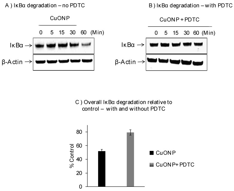
Figure 2.
Effect of CuONPs on IĸB-α degradation. SH-SY5Y cells were plated at 2 × 10⁶ cells/well (6 well plate) and exposed to CuONPs (10 μM) in the presence or absence of the potent NFκB inhibitor—pyrrolidine dithiocarbamate (PDTC, 50 nM) at the indicated time points. Cells were lysed, and lysates were Western blotted for the presence of IκB-α—a protein inhibitor for NFκB activation. Blots were collected, digitized, and quantified using a Bio-Rad VersaDoc™ Digital Imaging System (MP4000). Experiments were performed at n = 3 independent trials and representative Western blots were presented. (A) Western blot from cells exposed to CuONPs but not PDTC; (B) western blot from cells exposed to CuONPs and PDTC; (C) summary graph of relative degradation (compared to controls) in cells exposed to CuONPs and CuONPs and PDTC.

