Coronavirus disease 2019 (COVID-19) is caused by the single-stranded enveloped RNA virus SARS-CoV‑2. It has become a worldwide pandemic since it appeared in Wuhan in December 2019. SARS-CoV‑2 is the seventh human coronavirus, which affects primarily the respiratory tract. Severely ill patients suffer from pneumonia. They might need intensive care and artificial ventilation after developing acute respiratory syndrome (ARDS) or multiple organ dysfunction syndrome (MODS). The main human cellular receptor for the binding of the viral spike proteins is angiotensin-converting enzyme 2 (ACE2), following activation by transmembrane protease serine 2 (TMPRSS2). Angiotensin-converting enzyme 2 is expressed by type II alveolar cells, the predominant portal of entry in the lungs; ACE2 is also expressed in the heart and vasculature. It is upregulated by ACE inhibitors, which are often prescribed for cardiac patients. Their influence on SARS-Cov‑2 infectivity is unclear.
Myocarditis in SARS 2002
SARS-CoV viral RNA was detected in 35% (7⁄20) of human heart samples obtained at autopsy during the SARS outbreak in Toronto [1]. The positive samples showed an increase in macrophage infiltration together with myocyte necrosis and SARS-CoV RNA expression by polymerase chain rection (PCR). It was associated with a reduction in ACE2 protein expression. In situ hybridization was not available, so that direct evidence of viral RNA in the myocytes is still missing.
Cardiac inflammation in SARS-CoV-2
Hu et al. reported a 37-year-old male patient who was admitted to hospital in January 2020 with chest pain, dyspnea, and diarrhea [2]. His sputum was positive for SARS-CoV‑2. Chest radiography demonstrated pneumonia, pleural effusion, and enlargement of the heart. The troponin T level was >10,000 ng/l, creatine kinase MB (CK-MB) 112.9 ng/l, and brain natriuretic peptide (BNP) 21.025 ng/l. Echocardiography revealed an enlarged left ventricle with an end-diastolic dimension of 58 mm and an ejection fraction of 28%. Computed tomography coronary angiography excluded coronary artery stenosis. The patient developed cardiogenic shock and was diagnosed with fulminant myocarditis. He was ventilated and received a combination of methylprednisolone (200 mg/day), immunoglobulins (20 g/day), each for 4 days, norepinephrine and milrinone to stabilize blood pressure, and piperacillin sulbactam against bacterial superinfection. One week later, his heart size and cardiac marker enzymes had normalized.
Chen C et al. reported on 120 SARS-CoV-2-infected patients, 33% showed elevated NT-proBNP levels, and 10% elevated troponin T levels. Plasma interleukin (IL)-6 was dramatically increased. They consider a cytokine storm as the underlying fatal pathophysiology and classified it as “fulminant myocarditis” [3]. High levels of IL-1-beta, interferon (IFN)-gamma, and monocyte chemoattractant protein (MCP)-1 might have activated the T‑helper‑1 cell response [4]. The balance of pro- and anti-inflammatory cytokines apparently controls the clinical phenotype. An excessive immune response and a cytokine storm may lead to MODS.
Phases and faces of myocarditis
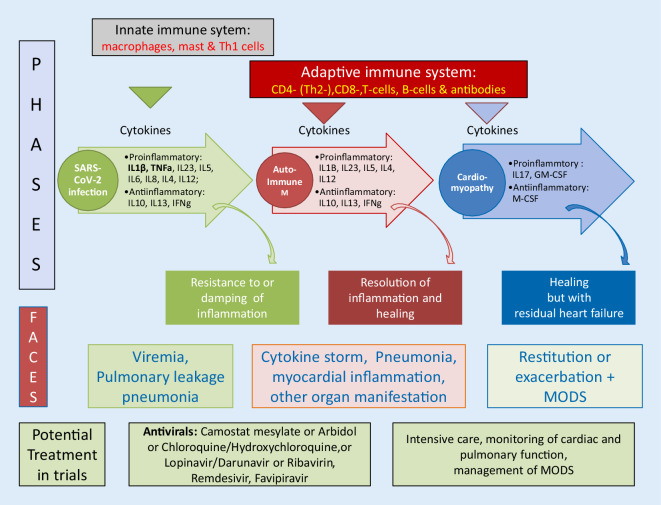
As with other forms of viral myocarditis, SARS-CoV‑2 runs through different phases of the disease (Fig. 1): (1) viremia and direct infection of lungs and myocardium, (2) recruitment of the innate immune system by macrophages and cytokine release, (3) response of the adaptive immune system, (4) resulting in death or recovery with lasting immunity [5].
Fig. 1.
SARS-CoV‑2 infection: phases of immune response with cytokine patterns and associated clinical phenotypes (faces). See text for abbreviations. (Modified from Maisch 2019 [5])
The clinical phenotype (= face) in phase 1 features a broad spectrum from mild throat infection to pneumonia and pleural effusion, in phases 2 and 3 the adaptive immune system may lead to exacerbation with hyperinflammation by a cytokine storm. Then the phenotype resembles MODS. Phase 4 can be characterized by death or aggravation of pre-existing cardiovascular disease or complete recovery of organ function including the heart. Determinants of the outcome are genetic predisposition, the immune status of the individual, the management of the disease and its complications, and the availability of the appropriate medication in different phases and faces of the COVID-19 disease.
Treatment strategies
In infected individuals with no or few symptoms only, watchful waiting and symptomatic treatment are appropriate. In patients with pneumonia and severe cardiac disease the full spectrum of intensive medical care including ventilation and veno-venous extracorporeal membrane oxygenation (vvECMO) should be applied. Approved antiviral treatment against COVID-19 is not yet available. Antivirals such as camostat mesylate (inhibitor of TMPRSS2), chloroquine/hydroxychloroquine (inhibitor of endocytosis), lopinavir/darunavir (inhibitor of 3‑chymotrypsin-like protease) or ribavirin, remdesivir, favipiravir (inhibitor of RNA-dependent RNA polymerase), or prednisolone should be restricted to controlled or randomized trials such as the worldwide WHO-cosponsored Solidarity Trial (https://www.who.int/emergencies/diseases/novel-coronavirus-2019/global-research-on-novel-coronavirus-2019-ncov/solidarity-clinical-trial-for-covid-19-treatments). Particular attention also deserve studies on the application of ivIg or with IL‑6 inhibitors (tocilizumab) to reduce hyperinflammation.
Conflict of interest
B. Maisch declares that he has no competing interests.
References
- 1.Oudit GY, Kassiri Z, Jiang C, et al. SARS-coronavirus modulaton of myocadial ace2 expression and inflammation in patients with sars. Eur J Clin Invest. 2009;39(7):618–625. doi: 10.1111/j.1365-2362.2009.02153.x. [DOI] [PMC free article] [PubMed] [Google Scholar]
- 2.Hu H, Ma F, Wie X, Fang Y. Coronavirus fulminant myocarditis saved with glucocorticoid and human immunoglobulin. Cardiovascular flashlight. Eur Heart J. 2020 doi: 10.1093/eurheartj/ehaa190. [DOI] [PMC free article] [PubMed] [Google Scholar]
- 3.Chen C, Zhou Y, Wang DW. SARS-CoV-2: a potential novel etiology of fulinant myocarditis(2020) Herz. 2020 doi: 10.1007/s00059-020-04909-z. [DOI] [PMC free article] [PubMed] [Google Scholar]
- 4.Huang C, Wang Y, Li X, et al. Clinical features of patients infected with 2019 novel coronavirus in Wuhan, China. Lancet. 2020 doi: 10.1016/1050140-6736(20)30183-5. [DOI] [PMC free article] [PubMed] [Google Scholar]
- 5.Maisch B. Cardio-immunology of myocarditis: focus on immune mechanisms and treatment options. Front Cardiovasc Med. 2019;6:48. doi: 10.3389/fcvm.2019.00048. [DOI] [PMC free article] [PubMed] [Google Scholar]


