Fig. 4.

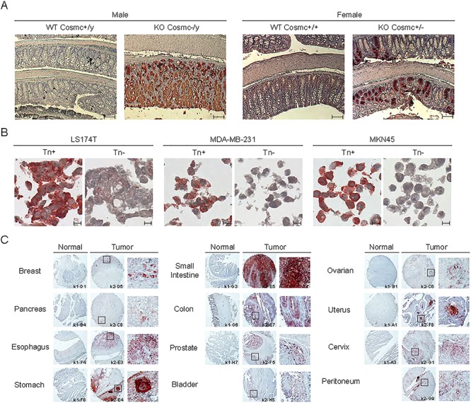
Immunohistochemical staining in IEC-Cosmc KO mice and human cancer cell block sections, and human cancer tissue array. (A) IHC staining with Remab6 using small intestine-colon-rectum sections in villi-specific Cosmc KO mice (male; KO Cosmc−/y, female; KO Cosmc+/−) compared to WT. Scale bar represents 100 μm. (B) Cell block section staining with Remab6 of Tn-positive or Tn-negative populations of human carcinoma cell lines (LS174T, MDA-MB-231 and MKN-45). Scale bar represents 10 μm. (C) Human cancer tissue array (FDA808k-1/2, US Biomax Inc.), including normal and tumor tissues as indicated, with Remab6-Fab-HRP reagent. Squares represent Tn-positive staining sites with high magnification. Brownish-red indicates Tn staining with antibody, and blue indicates nuclear staining.
