Abstract
A method, quantitative analysis of multicomponents by single marker (QAMS), was established and fully verified based on high-performance liquid chromatography (HPLC) for simultaneous determination of six chromone indicators of Saposhnikoviae Radix (SR). In the present study, cimifugin (C), 5-O-methylvisamminol (V), hamaudol (H), and their corresponding glycosides, prim-O-glucosylcimifugin (GC), 4′-O-β-D-glucosyl-5-O-methylvisamminol (GV), and sec-O-glucosylhamaudol (GH), were selected as bioactive constituents and indicators for the quality evaluation of SR. GV was chosen as the unique reference standard, and relative correction factors (RCF) between GV and the other five chromones were calculated. The feasibility of QAMS for the analysis of chromones was investigated by comparing with the traditional external standard method (ESM). Furthermore, the method was proven to have accuracy (96.98%–102.50%), repeatability (RSD <3%), stability (RSD <3%), precision (RSD <3%), and desirable linearity (R2 ≧0.9999). Subsequently, 55 batches of commercial SR from different regions were determined by QAMS, and their contents were analyzed by principal component analysis (PCA), correlation analysis, and hierarchical cluster analysis (HCA), respectively. Based on the results, a more refined quality standard of commercial SR was proposed: SR was qualified when the total contents of six chromones were greater than 3 mg·g−1. Furthermore, SR could initially be regarded as a superior medicine when it satisfied both conditions at the same time: the total content of GC, C, GV, V, GH, and H was greater than 8 mg·g−1, and the proportion of the total content of C, V, and H was greater than 10%. This study demonstrated that the quality of SR could be successfully evaluated by the developed QAMS method; meanwhile, valuable information was provided for improving the quality standard of SR.
1. Introduction
Saposhnikoviae Radix (SR), known as “FangFeng” in China, is derived from the dried root of the plant Saposhnikovia divaricate (Turcz.) Schischk, belonging to the family Umbelliferae. This herb is pungent in flavour, sweet, and lukewarm, and enters the bladder, liver, and spleen meridian. According to the theory of Traditional Chinese Medicine (TCM), SR has significant effects on dispelling wind to relieve exogenous syndrome, removing dampness to kill pain, and stopping spasms [1]. As an important ingredient in many traditional Chinese prescriptions such as Yu-Ping-Feng-San, Fang-Feng-Tong-Sheng pill, and Tong-Xie-Yao-Fang [2, 3], SR has great application value. Furthermore, many pharmacological studies have indicated that a number of curative effects, including antipyretic, analgesic, anti-inflammatory, antibacterial, antitumor, antiallergic, and antioxidation, are existed in SR [4–8].
SR has several components: chromones, polysaccharides, coumarins, volatile oils, and other components [9–12]. It is worth mentioning that chromones are the most representative components in SR. On the one hand, there are extremely abundant content [13]. On the other hand, it was closely related to the pharmacological efficacy of SR, for example, anti-inflammatory, analgesic, and antioxidation [14–16]. With the development of SR market, increasing importance has been given to the quality control of SR. According to the record of Chinese Pharmacopoeia (2015 edition), prim-O-glucosylcimifugin (GC) and 4′-O-β-D-glucosyl-5-O-methylvisamminol (GV) were selected as quality-control indicators. However, each Chinese herbal medicine is an integrated complex with diversity of active components, contributing to particular efficacy through synergy and mutual effect based on the theory of TCM [17]. Thus, using a few components to control the quality is insufficient for the complex botanical products and traditional Chinese medicines. In line with the importance status of the chromones mentioned above, multiple representative chromone components could be selected as indicators to evaluate the quality of SR. According to phytochemical studies, GV, GC, sec-O-glucosylhamaudol (GH), cimifugin (C), 5-O-methylvisamminol (V), and hamaudol (H) are widely existing in SR [18, 19], and their pharmacological effects are significant and most related to the efficacy of SR [20, 21]. Therefore, when G, GC, V, GV, H, and GH are selected as new quality-control indicators, the chemical characterization, medicinal function, and inner quality of SR could be represented comprehensively. Some studies have shown that the methods of refluxing and ultrasonic were always used for the extraction of chromones [22, 23]. Considering the polarity and solubility of the six chromone indicators, the heating refluxing method could be applied for sample preparation, and the methanol could be used as the extraction solvent based on an optimization of sample preparation process in this work.
In many studies, the contents of multicomponents are usually determined by the external standard method (ESM). In this method, the reference standards are necessary that we need to spend more and more time and economic cost to separate and purify [24]. As an alternative method, quantitative analysis of multicomponents by single marker (QAMS) is only requiring a single reference standard to simultaneously determine the contents of multicomponents, which is more effective and appropriate for the quality control (QC) [25]. When some reference standards are unstable, low in abundance, or hard to extract from the plant, QAMS could not only reduce the cost but also reduce the difficulty in preparation [26, 27]. Besides, this method could improve the practicability of QC and expand the application for herbal or botanical products effectively. In QAMS, the content of internal standard could be obtained directly by HPLC and the other components could be calculated by using multiple conversion factors. Hence, the relative correction factor (RCF) is a critical parameter in the content computational formula about analytes. As the result of molar absorptivity of different analytes are often different, RFC plays a role of calibration when a single reference is used to determine multicomponents [28, 29]. In ESM, the concentration of analyte (Ck) can be calculated by the ratio of the peak areas of analytes in sample solution (Ak) to the peak area of its corresponding standard solution in a unit concentration (As/Cs), as shown in the following equation:
| (1) |
In QAMS, Ck should be calibrated by RCF of each analyte (fk) based on the calculation of ESM. The formula is as follows:
| (2) |
Importantly, the value of fk is calculated by the ratio of peak areas in a unit concentration of standard substance (As/Cs) to analyte (Ak/Ck) as follows:
| (3) |
It is worth mentioning that the final value of RCF is usually the average value of multiple RCFs by a series of determining under different concentration levels of internal referring substance [30]. Since six control components, i.e., C, GC, V, GV, H, and GH, are used, ESM will cause high costs and complicated operations. Therefore, the QAMS method is used to compute the contents of six chromones, and GV is used as the internal standard for its strong representative nature, high stability, high content, and significant pharmacological activities [31, 32].
In the present study, a new substitute method named QAMS was applied for simultaneous determination of six chromones in SR. As the reference substance, the content of GV was determined by HPLC, and the contents of C, GC, V, H, and GH were calculated with RCF based on the intrinsic function and the proportional relationship between GV and these five chromones. The feasibility could be verified by comparing the results with the ESM, and this method was validated in terms of linearity, accuracy, precision (instruction precision and intermediate precision), and stability, referring to some reliable references [33]. Subsequently, 55 batches of commercial SR were determined and a more comprehensive and reliable quality evaluation standard of SR was preliminary inferred by principle component analysis (PCA), correlation analysis, and hierarchical cluster analysis (HCA), respectively.
2. Materials and Methods
2.1. Apparatus and Chromatographic Analysis
Analyses were primarily performed by using a Waters HPLC System (Waters Crop, Milford, MA, USA) equipped with a 1525 binary pump solvent management system, 2998 PDA detector, 2707 automatic sampling device, and Breeze 2 workstation. Two additional different HPLC instruments were used: One was high performance liquid chromatography (Waters Crop, Milford, MA, USA) equipped with 1525 binary pump solvent management system, 2489 UV detector, and Breeze 2 workstation. Another was a Waters Alliance e2695-2998 HPLC system (Empower workstation, Waters Crop, Milford, MA, USA). HPLC separation was carried out on a CAPCELL PAK C18 column (4.6 mm × 150 mm, 5 μm). Column temperature was set at 25°C, and inject volume was 10 μL. The mobile phase consisted of methanol (A) and 0.3% formic acid aqueous solution (B). The gradient elution was programmed at a flow rate of 1.0 mL·min−1 as follows: 0–12 min, 32% A; 12–40 min, 32%–50% A; 40–50 min, 50%–70% A; 50–52 min, 70% A. The detection wavelength was set at 254 nm.
2.2. Chemicals and Materials
Fifty-five batches of SR were collected from different regions in China, which were identified by Professor Zhang Yuan from the Beijing University of Traditional Chinese Medicine and proved to be the dried root of Saposhnikovia divaricate (Turcz.) Schischk following the method described in Chinese Pharmacopoeia (2015 edition) [1]. GC, C, GH, and GV were obtained from Chengdu Mansite Biotechnology Co., Ltd. (Chengdu, China). The purity (≧98%) of these reference standards was assumed as provided by the suppliers. The other two compounds, V and H, were separated and purified in our lab and the purity was identified to be of not less than 98% (determined by HPLC). The structures were determined on the basis of UV, MS, and NMR data and confirmed by comparison with data from the literature. The chemical structures of all standards are shown in Figure 1.
Figure 1.
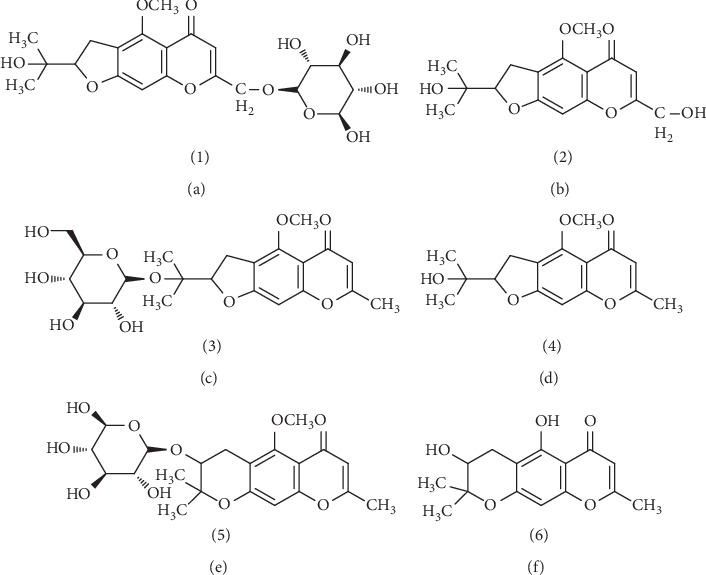
Chemical structures of prim-O-glucosylcimifugin (a), cimifugin (b), 5-O-methylvisammioside (c), 4′-O-D-glucosyl-5-O-methylvisamminol (d), sec-O-glucosylhamaudol (e), and hamaudol (f).
Methanol, acetonitrile, and formic acid of HPLC grade were purchased from Thermo Fisher Scientific Inc. (Waltham, MA, USA), and other reagents (Beijing Chemical Industry Factory) were of analytical grade. HPLC grade water was prepared using a Pall Cascada IX system (Pall, USA). All other reagents were of analytical grade.
2.3. Preparation of Mixed Standard Solutions
Substances of C, GC, H, GH, V, and GV were weighed precisely and dissolved into methanol to prepare the mixed stock solutions of reference standards with the concentrations of 0.1220 mg·ml−1, 0.2096 mg·ml−1, 0.0369 mg·ml−1, 0.1636 mg·ml−1, 0.0808 mg·ml−1, and 0.3340 mg·ml−1, respectively. Then, a series of concentrations of calibration standard solutions were produced by diluting the mixed stock solution (dilution factor = 1, 2, 5, 10, 20, 50, and 100) with the same methanol. The solutions were stored at 4°C in a refrigerator and filtered through a 0.45 μm membrane filter before injection. All samples being injected into HPLC system were prepared right before analysis.
2.4. Preparation of Sample Solutions
An appropriate amount of the samples to be tested was crushed and passed through an 80-mesh screen. Next, 0.25 g of the sample powder was precisely weighed and placed in a stuffed flask with accurate addition of 10 ml of methanol, subjected to heating reflux for 120 min. It is worth noting that the stuffed flask was weighed before and after refluxing, then added the solvent to keep the weight equivalent at room temperature if required. By filtering through the 0.45 μm filter and discarding the first 2 ml, the remaining filtrate was used as the test solution of samples.
2.5. Method Validation
2.5.1. Specificity
The mixed reference standard solution and test solution were separately injected into HPLC under the optimized chromatographic conditions (Section 2.1).
2.5.2. Linearity
The prepared stock standard solutions mentioned above (Section 2.3) with a series of appropriate concentration levels were used for HPLC based on the chromatographic conditions (Section 2.1), respectively. The limits of detection (LOD) and quantification (LOQ) were measured based on a signal-to-noise ratio (S/N) at about 3 and 10, respectively.
2.5.3. Precision
To ensure the validity of this newly developed method, the tests of instrument precision and intermediate precision were performed. For instrument precision, test solutions prepared (Section 2.4) were examined by HPLC for six replicates within one day, and on the purpose of detecting intermediate precision, the prepared test solutions were injected into HPLC by different operators with different instruments on different dates.
2.5.4. Stability
The same tested solutions, which were prepared (Section 2.4) and placed at room temperature, were injected into HPLC at different time points (0, 2, 4, 8, 12, and 24 h).
2.5.5. Repeatability
Six parallel sample solutions with the same batch were prepared following the method (Section 2.4) individually and determined by HPLC according to the chromatographic conditions (Section 2.1).
2.5.6. Accuracy
Six copies of the same batch of SR powder (0.125 g) with known content were weighed, respectively. Then, a certain amount of control standard solution was added into the samples according to the proportion of sample content to reference substance, about 1 : 1. Preparation and determination of six sample solutions were conducted in parallel referring to the method as described in Sections 2.1 and 2.4. Recoveries were computed.
2.6. QAMS Method
2.6.1. Calculation of RCF
Seven concentration levels of mixed standard solution were prepared (Section 2.3) and injected into HPLC under the chromatographic conditions (Section 2.1), respectively. Besides, the chromatographic peak areas of each component were recorded.
2.6.2. Durability Test of RCF
Three different instruments (as listed in Section 2.1), three kinds of chromatographic columns (Capcell Pak C18, Water SunFire C18, and Water Symmetry C18) (4.6 mm × 150, 5 μm), different flow rates (0.9, 1.0, and 1.1 ml·min−1), and different column temperatures (25, 30, and 35°C) were used to investigated the influence of different conditions on RCF.
2.6.3. Location of the Chromatographic Peak of Measured Component
For better authentication as well as convenience to quality control of the commercial SR, the chromatographic peak positions of GC, C, GV, V, GH, and H were investigated using different instruments and different columns.
2.6.4. Comparison of the Results between QAMS and ESM
In order to assess and validate QAMS feasibility of multicompounds in SR, the contents of C, GC, V, GV, H, and GV were determined by ESM and QAMS in 15 batches, respectively. For ESM, the determination of the six chromones was carried out with six reference standards (GC, C, GV, V, GH, and H), whereas for QAMS, the results were based on the nature of the calculation of fx, the intrinsic function, and the proportional relation between the selected reference analyte and other analytes. The content of the selected internal substance (GV) was determined like ESM, and then the contents of the other five chromones were calculated in accordance with relative conversion factors between analytes and the internal substance [34].
2.7. Application and Data Analysis
The developed QAMS method was applied for the quantitative assessment of 55 batches of commercial SR from different regions. The contents of GC, C, GV, V, GH, and H were determined and then analyzed by PCA, correlation analysis and HCA, respectively. Meanwhile, the further analysis of three chromone glycosides (C, H, and V) was carried out to make clarification of their importance for the overall quality of SR. The figures presented were developed by exploration of the analysis function using SPSS 22.0 software package.
3. Results and Discussion
3.1. Method Validation
3.1.1. Specificity
As shown in Figure 2, the analytes had good separation because it has no interference in the corresponding position of the six components, and the target peaks of the test solution corresponded to the peaks of reference standard solution according to retention time in the chromatogram. So, it indicated that this method had specificity.
Figure 2.
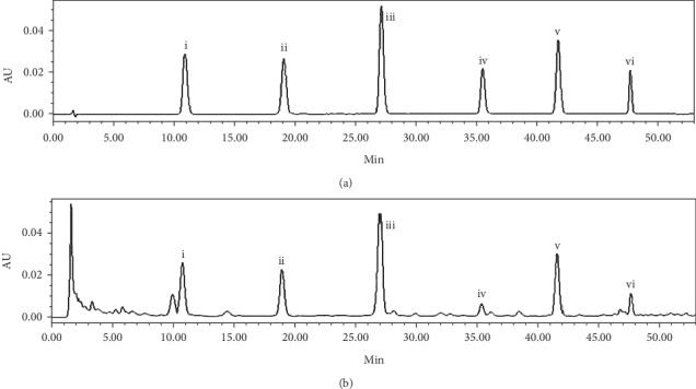
The chromatograms of the mixed reference standard solution (a) and test solution (b). The peaks represent (i) prim-(O)-glucosylcimifugin, (ii) cimifugin, (iii) 4′-(O)-β-D-glucosyl-5-(O)-methylvisammino, (iv) 5-(O)-methylvisamminol, (v) sec-(O)-glucosylhamaudol, and (vi) hamaudol.
3.1.2. Linearity
The standard curves of six reference substances were established by using the chromatographic peak area (y) as the vertical axis and the concentration of the reference solution (x) as abscissa, respectively. There was a good linearity as the result of all the correlation coefficients (R) which were not less than 0.999 over the concentration range. Furthermore, LOD and LOQ of six substances were also calculated as shown in Table 1.
Table 1.
Regression equations, correlation coefficients, linearity ranges, limits of detection, and limits of quantification for six indicators.
| Indicator | Regression equation1 | R | Linearity range (μg·ml−1) | LOD (μg·ml−1) | LOQ (μg·ml−1) |
|---|---|---|---|---|---|
| Prim-O-glucosylcimifugin | y = 1815300x + 8028 | 0.9999 | 2.10–209.60 | 0.29 | 0.96 |
| Cimifugin | y = 2928940x + 7329 | 0.9999 | 1.22–122.00 | 0.28 | 0.92 |
| 4′-O-β-D-glucosyl-5-O-methylvisammino | y = 1894482x + 14466 | 0.9999 | 3.34–334.00 | 0.28 | 0.94 |
| 5-O-methylvisamminol | y = 3222987x + 4879 | 0.9999 | 0.81–80.80 | 0.18 | 0.61 |
| Sec-O-glucosylhamaudol | y = 2660022x + 6702 | 0.9999 | 1.64–163.60 | 0.22 | 0.73 |
| Hamaudol | y = 4418335x + 1757 | 0.9999 | 0.37–36.90 | 0.09 | 0.31 |
1In the regression equation y = ax + b, where y refers to the peak area and x refers to the concentration of the indicator (μg·mL−1).
3.1.3. Precision
The areas of chromatographic peak and RSD of six compounds were recorded and calculated, respectively. In Table 2, the RSD results of six compounds for the instrument precision were in the range of 0.42%–0.75%. Apart from this, the RSDs which were calculated by intermediate precision were all lower than 3%. It indicated that this method has a good precision.
Table 2.
Instrument precision, intermediate precision, stability, repeatability, and recovery of six analytes.
| Indicator component | Instrument precision (n = 6) | Intermediate precision (n = 6) | Stability (n = 6) | Repeatability (n = 6) | Accuracy (n = 6) | |
|---|---|---|---|---|---|---|
| RSD (%) | RSD (%) | RSD (%) | RSD (%) | Mean recovery (%) | RSD (%) | |
| Prim-O-glucosylcimifugin | 0.75 | 2.4 | 0.84 | 1.03 | 99.87 | 1.66 |
| Cimifugin | 0.56 | 1.69 | 0.56 | 0.73 | 97.34 | 0.77 |
| 4′-O-β-D-glucosyl-5-O-methylvisamminol | 0.43 | 1.23 | 0.54 | 0.49 | 96.98 | 1.31 |
| 5-O-methylvisamminol | 0.42 | 0.36 | 0.57 | 0.8 | 102.5 | 1.03 |
| Sec-O-glucosylhamaudol | 0.37 | 1.06 | 0.34 | 0.96 | 98.13 | 0.71 |
| Hamaudol | 0.67 | 1.45 | 0.69 | 1.35 | 99.48 | 0.68 |
3.1.4. Stability
The peak areas of GC, C, GV, V, GH, and H were recorded, and the values of RSD were 0.84%, 0.56%, 054%, 0.57%, 0.34%, and 0.69%, respectively. Hence, the sample solution was stable at room temperature within 24 h (Table 2).
3.1.5. Repeatability
The results showed that average mass fractions of GC, C, GV, V, GH, and H were 1.478, 0.808, 2.510, 0.173, 1.053, and 0.151 mg·g−1, respectively. The RSD of corresponding average mass fraction was 1.03%, 0.73%, 0.49%, 0.80%, 0.96%, and 1.35% which proved that this quantitative method was of good repeatability.
3.1.6. Accuracy
There was favorable accuracy because the average recovery rates of six marker compounds were varied in the range of 96.98%–102.5%. Meanwhile, RSD values of recovery rates for each compound were lower than 3% totally.
3.2. QAMS Method
3.2.1. Calculation of RCF
GV was selected as the internal standard, and the values of RCF (fx) for other five indicators were computed in different concentrations according to the equation (3) mentioned above. The average RCF of each compounds was shown as follows: fGC = 1.047 (RSD% = 2.32), fC = 0.6489 (RSD% = 0.24), fv = 0.5909 (RSD% = 0.30), fGH = 0.7223 (RSD% = 0.68), and fH = 0.7223 (RSD% = 0.28).
3.2.2. Durability Test of RCF
The RCF of five chromones in different conditions (instruments, chromatographic columns, flow rates, and column temperatures) were obtained, and the RSDs were all less than 5%, which could clearly demonstrate that the RCF calculated by the proposed method has good durability and system suitability for routine testing.
3.2.3. Location of the Chromatographic Peaks of Measured Components
The chromatographic peak position was identified by the relative retention time (RRT), which was calculated according to the following equation:
| (4) |
where tRk is the retention time of measured components, tRS is the retention time of internal reference, and △tRks is the difference of retention time in both. Among them, the chromatographic peak position of GV was explicitly identified and designated as the reference peak in the SR samples. RRT was measured between GV and the other five components by different instruments and columns at the same time, and their RSDs were all less than 5%. It indicated that the calculation of RRT was stable and could be used for identifying the chromatographic peaks location of measured components.
3.2.4. Comparison of the Results between QAMS and ESM
The contents of 15 batches of commercial SR were determined by the QAMS method and ESM. The results are shown in Table 3. Relative error (RE) was built between the two component variables to examine the deviations between QAMS and ESM. By comparing two sets of contents of five components between QAMS and ESM, respectively, their content variations were found to be within the range of 5%. It met the requirement of Chinese Pharmacopoeia. To assess the consistency of the results, correlation analysis was used to evaluate the similarity between the QAMS method and ESM. Correlation coefficient value is a commonly used parameter in the similarity evaluation. The larger the values, the higher the similarity of the target sample will be. When they are equal to 1, the targets are identical. In this work, the data, as shown in Table 4, were above 0.900, which indicated that there were no significant differences between the QAMS and ESM, and the identified RCF and parameters of chromatographic peak location for measured chromones of SR were reliable. In conclusion, QAMS can be applied in the determination of six chromones.
Table 3.
Comparison of contents by QAMS and ESM for CV as internal standard (mg·g−1).
| No. | GV | GC | C | V | GH | H | ||||||||||
|---|---|---|---|---|---|---|---|---|---|---|---|---|---|---|---|---|
| ESM | ESM | QAMS | Relative error (%) | ESM | QAMS | Relative error (%) | ESM | QAMS | Relative error (%) | ESM | QAMS | Relative error (%) | ESM | QAMS | Relative error (%) | |
| 1 | 2.896 | 4.295 | 4.289 | −0.14 | 1.048 | 1.047 | −0.10 | 0.09829 | 0.09793 | −0.37 | 1.069 | 1.070 | 0.09 | 0.06459 | 0.06460 | 0.02 |
| 2 | 0.8574 | 2.646 | 2.642 | −0.15 | 0.3916 | 0.3914 | −0.05 | 0.04527 | 0.04511 | −0.35 | 0.2362 | 0.2364 | 0.08 | 0.1061 | 0.1061 | 0.00 |
| 3 | 1.889 | 3.783 | 3.778 | −0.13 | 0.7479 | 0.7475 | −0.05 | 0.06943 | 0.06918 | −0.36 | 0.9545 | 0.9553 | 0.08 | 0.1507 | 0.1507 | 0.00 |
| 4 | 2.480 | 2.885 | 2.881 | −0.14 | 0.2100 | 0.2098 | −0.10 | 0.05922 | 0.05901 | −0.35 | 0.2845 | 0.2847 | 0.07 | 0.020008 | 0.02009 | 0.05 |
| 5 | 2.848 | 3.510 | 3.505 | −0.14 | 0.9254 | 0.9248 | −0.06 | 0.08080 | 0.08051 | −0.36 | 0.7045 | 0.7051 | 0.09 | 0.06158 | 0.06159 | 0.02 |
| 6 | 1.832 | 2.019 | 2.016 | −0.15 | 0.6268 | 0.6264 | −0.06 | 0.05237 | 0.05219 | −0.34 | 0.2341 | 0.2343 | 0.09 | 0.02235 | 0.02236 | 0.04 |
| 7 | 1.961 | 2.241 | 2.239 | −0.09 | 0.5400 | 0.5396 | −0.07 | 0.05959 | 0.05937 | −0.37 | 0.2508 | 0.2510 | 0.08 | 0.07276 | 0.07278 | 0.03 |
| 8 | 2.062 | 2.249 | 2.246 | −0.13 | 0.2459 | 0.2458 | −0.04 | 0.06527 | 0.06503 | −0.37 | 0.2155 | 0.2157 | 0.09 | 0.0256 | 0.02565 | 0.00 |
| 9 | 1.390 | 4.122 | 4.123 | 0.02 | 0.3810 | 0.3809 | −0.03 | 0.0398 | 0.03918 | −0.51 | 0.4580 | 0.4593 | 0.28 | 0.08772 | 0.08785 | 0.15 |
| 10 | 3.961 | 2.466 | 2.466 | 0.00 | 1.469 | 1.469 | 0.00 | 0.2263 | 0.2251 | −0.53 | 1.264 | 1.267 | 0.24 | 0.3047 | 0.3051 | 0.13 |
| 11 | 2.004 | 3.054 | 3.054 | 0.00 | 1.491 | 1.491 | 0.00 | 0.1301 | 0.1294 | −0.54 | 0.7997 | 0.8018 | 0.26 | 0.1760 | 0.1763 | 0.17 |
| 12 | 2.519 | 2.363 | 2.363 | 0.00 | 1.029 | 1.030 | 0.10 | 0.08996 | 0.08953 | −0.48 | 0.6982 | 0.6997 | 0.21 | 0.06253 | 0.06263 | 0.16 |
| 13 | 2.848 | 4.417 | 4.416 | −0.02 | 1.754 | 1.756 | 0.11 | 0.1763 | 0.1754 | −0.51 | 0.9507 | 0.9528 | 0.22 | 0.1461 | 0.1463 | 0.14 |
| 14 | 2.233 | 4.983 | 4.983 | 0.00 | 0.799 | 0.800 | 0.09 | 0.07737 | 0.07700 | −0.48 | 0.8065 | 0.8082 | 0.21 | 0.07475 | 0.07487 | 0.16 |
| 15 | 4.038 | 3.972 | 3.973 | 0.03 | 1.239 | 1.238 | −0.08 | 0.1380 | 0.1372 | −0.58 | 0.6367 | 0.6384 | 0.27 | 0.05454 | 0.05462 | 0.15 |
|
| ||||||||||||||||
| Correlation coefficient | 0.999996894 | 0.999998781 | 0.999996565 | 0.99999876 | 0.999999518 | |||||||||||
Table 4.
Content of six chromone compounds in 55 batches of commercial SR.
| No. | Compound content (mg·g−1) | ||||||
|---|---|---|---|---|---|---|---|
| GC | C | GV | V | GH | H | Total | |
| 1 | 1.317 | 0.4275 | 1.261 | 0.02920 | 0.2946 | 0.03354 | 3.363 |
| 2 | 2.418 | 0.4976 | 2.382 | 0.04779 | 0.3659 | 0.03554 | 5.747 |
| 3 | 2.651 | 0.5458 | 2.698 | 0.06159 | 0.3781 | 0.02972 | 6.364 |
| 4 | 1.337 | 0.6792 | 1.654 | 0.04434 | 0.2859 | 0.06364 | 4.064 |
| 5 | 2.689 | 0.2197 | 2.876 | 0.03020 | 0.3112 | 0.01964 | 6.146 |
| 6 | 4.573 | 1.107 | 1.955 | 0.03020 | 0.5887 | 0.08134 | 8.335 |
| 7 | 1.729 | 0.4001 | 1.835 | 0.03092 | 0.1675 | 0.02509 | 4.188 |
| 8 | 2.564 | 0.3944 | 3.718 | 0.04341 | 0.2723 | 0.02125 | 7.013 |
| 9 | 2.430 | 0.1014 | 2.255 | 0.03667 | 0.2496 | 0.01529 | 5.088 |
| 10 | 2.258 | 0.6732 | 2.216 | 0.00000 | 0.3463 | 0.05838 | 5.552 |
| 11 | 1.304 | 0.344 | 1.449 | 0.03023 | 0.3543 | 0.06303 | 3.545 |
| 12 | 2.452 | 0.1243 | 2.755 | 0.03687 | 0.1534 | 0.05423 | 5.576 |
| 13 | 1.346 | 0.9504 | 1.465 | 0.06466 | 0.3278 | 0.11580 | 4.270 |
| 14 | 1.777 | 0.5765 | 1.127 | 0.02988 | 0.2396 | 0.08358 | 3.834 |
| 15 | 1.786 | 0.6144 | 1.364 | 0.03312 | 0.1714 | 0.05189 | 4.021 |
| 16 | 2.896 | 0.422 | 2.682 | 0.06387 | 0.5827 | 0.09963 | 6.746 |
| 17 | 4.69 | 1.02 | 2.551 | 0.10200 | 1.1490 | 0.15070 | 9.663 |
| 18 | 1.043 | 0.2811 | 1.573 | 0.02553 | 0.1242 | 0.00000 | 3.047 |
| 19 | 1.717 | 0.6368 | 1.962 | 0.08296 | 0.2502 | 0.15210 | 4.801 |
| 20 | 2.28 | 0.983 | 2.533 | 0.09785 | 0.3417 | 0.07771 | 6.313 |
| 21 | 2.729 | 0.3448 | 2.464 | 0.04038 | 0.2646 | 0.02275 | 5.866 |
| 22 | 4.2 | 1.147 | 3.442 | 0.14280 | 0.6112 | 0.06122 | 9.604 |
| 23 | 2.139 | 0.1695 | 1.929 | 0.00000 | 0.3146 | 0.01560 | 4.568 |
| 24 | 2.744 | 0.8955 | 2.324 | 0.08142 | 0.7392 | 0.1081 | 6.892 |
| 25 | 4.45 | 1.843 | 2.509 | 0.09471 | 0.6873 | 0.07324 | 9.657 |
| 26 | 2.817 | 1.401 | 2.608 | 0.09266 | 0.9524 | 0.07638 | 7.947 |
| 27 | 2.368 | 0.5529 | 2.202 | 0.05162 | 0.3305 | 0.07492 | 5.580 |
| 28 | 1.487 | 0.1982 | 1.880 | 0.03231 | 0.1906 | 0.01684 | 3.805 |
| 29 | 3.599 | 0.6820 | 2.030 | 0.04863 | 0.5045 | 0.06149 | 6.926 |
| 30 | 2.116 | 1.4090 | 3.498 | 0.11720 | 0.3130 | 0.04773 | 7.501 |
| 31 | 4.237 | 1.2030 | 1.818 | 0.03805 | 0.7812 | 0.06076 | 8.138 |
| 32 | 2.077 | 0.9935 | 2.653 | 0.08411 | 0.3187 | 0.07907 | 6.205 |
| 33 | 2.221 | 1.1080 | 2.931 | 0.10130 | 0.2647 | 0.06694 | 6.693 |
| 34 | 1.863 | 0.3630 | 0.941 | 0.03012 | 0.3419 | 0.07997 | 3.619 |
| 35 | 1.818 | 0.3075 | 1.096 | 0.00000 | 0.3717 | 0.12460 | 3.718 |
| 36 | 2.134 | 0.3686 | 1.076 | 0.00000 | 0.1803 | 0.06089 | 3.820 |
| 37 | 2.173 | 1.3010 | 1.232 | 0.04985 | 0.5113 | 0.12210 | 5.389 |
| 38 | 3.773 | 0.3078 | 3.220 | 0.06403 | 0.3651 | 0.02726 | 7.757 |
| 39 | 2.234 | 0.4056 | 1.289 | 0.00000 | 0.1985 | 0.03652 | 4.164 |
| 40 | 2.168 | 0.4823 | 1.776 | 0.00000 | 0.2772 | 0.05842 | 4.762 |
| 41 | 1.490 | 0.4874 | 1.240 | 0.00000 | 0.3820 | 0.12980 | 3.729 |
| 42 | 1.708 | 0.3880 | 1.587 | 0.00000 | 0.3404 | 0.13020 | 4.154 |
| 43 | 1.834 | 0.2949 | 2.261 | 0.03667 | 0.1705 | 0.02114 | 4.618 |
| 44 | 2.199 | 0.5615 | 2.569 | 0.06130 | 0.2619 | 0.13020 | 5.783 |
| 45 | 3.667 | 1.3310 | 3.572 | 0.12560 | 0.8210 | 0.08447 | 9.601 |
| 46 | 1.985 | 0.1671 | 1.610 | 0.04060 | 0.1129 | 0.03844 | 3.954 |
| 47 | 1.765 | 0.2047 | 1.508 | 0.03583 | 0.1699 | 0.00000 | 3.683 |
| 48 | 2.363 | 1.0300 | 2.519 | 0.08953 | 0.6997 | 0.06263 | 6.764 |
| 49 | 3.670 | 1.3180 | 1.900 | 0.11270 | 0.8467 | 0.18620 | 8.034 |
| 50 | 2.447 | 0.5516 | 1.778 | 0.07714 | 0.4362 | 0.07921 | 5.369 |
| 51 | 2.466 | 1.4690 | 3.961 | 0.22510 | 1.2670 | 0.30510 | 9.693 |
| 52 | 4.416 | 1.7560 | 2.848 | 0.17540 | 0.9528 | 0.14630 | 10.300 |
| 53 | 5.136 | 1.2960 | 3.769 | 0.17010 | 0.7804 | 0.07168 | 11.220 |
| 54 | 1.901 | 0.5797 | 0.953 | 0.03516 | 0.2994 | 0.10780 | 3.876 |
| 55 | 1.825 | 0.3481 | 0.867 | 0.03686 | 0.3172 | 0.09194 | 3.486 |
3.3. Application and Data Analysis
3.3.1. Sample Analysis and Characteristics of Six Chromone Compounds in 55 Batches of Commercial SR
In 55 batches of commercial SR from different regions, the contents of six chromones, GC, C, GV, V, GH, and H, were determined by QAMS. Results are listed in Table 4. It was observed that the maximum total content of six compounds was 11.22 mg·g−1 in no. S53, while the minimum total content of those was 3.047 mg·g−1 in no. S18. Such a wide concentration variance of these 55 batches of commercial SR may be attributed to a variety of factors, including plant sources, genetic variation, and geography differences. To further verify the relationships among the samples and evaluate the variation of six compounds, PCA, coefficient analysis, and HCA were performed using the SPSS 22.0 software (IBM, USA).
3.3.2. PCA and Correlation Analysis
The contents of GC (X1), C (X2), GV (X3), V (X4), GH (X5), and H (X6) of 55 batches of commercial samples were subjected to PCA. The results are shown in Figure 3. A two-component PCA model was established accounting for the accumulated variation of 80.260%, where the first principal component (Z1) was 62.614% and the second (Z2) was 17.646%. According to the component score coefficient matrix (Table 5; Figure 4), every coefficient between Z1 and six indicators was significant, which could indicate that Z1 represented the total content of six components. That is to say, each component was indispensable for the quality evaluation of SR. Furthermore, Z2 was mainly reflecting X6 as the result of their coefficient which was the largest (0.733). The comprehensive score (Z) for each batch sample could be obtained as follows:
| (5) |
Figure 3.
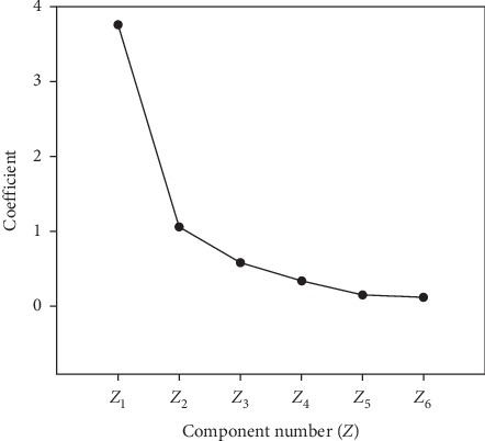
The coefficient of principal components.
Table 5.
The component score coefficient matrix.
| Principal component | ||
|---|---|---|
| Z 1 | Z 2 | |
| X 1 | 0.749 | −0.365 |
| X 2 | 0.866 | 0.122 |
| X 3 | 0.679 | −0.580 |
| X 4 | 0.900 | −0.049 |
| X 5 | 0.903 | 0.184 |
| X 6 | 0.599 | 0.733 |
Figure 4.
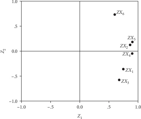
The coefficient between the content of 6 chromone compounds (X1, X2, X3, X4, X5, and X6) and Z1 and Z2.
The details of 55 batche samples are shown in Table 6. Through correlation analysis of results from every batch sample, a good correlation between Z and the total content of six components could be included in accordance with the correlation coefficient which was 0.875. Thus, it was feasible to evaluate the quality of SR comprehensively if the total content of six components was used as indicator. Meanwhile, we preliminary set that the total content of six chromones should not be less than 3 mg·g−1 for qualified SR based on the above determined content range (3.047–11.22 mg·g−1).
Table 6.
Comprehensive evaluation of 55 batches of commercial SR.
| No. | Z |
|---|---|
| 1 | −2.2088 |
| 2 | −0.8149 |
| 3 | −0.3972 |
| 4 | −1.2788 |
| 5 | −1.3328 |
| 6 | −1.4037 |
| 7 | −2.187 |
| 8 | −0.7398 |
| 9 | −1.9465 |
| 10 | −1.1080 |
| 11 | −1.8149 |
| 12 | −1.5472 |
| 13 | −0.1805 |
| 14 | −1.5233 |
| 15 | −1.7940 |
| 16 | 0.6962 |
| 17 | 4.3433 |
| 18 | −3.1265 |
| 19 | 0.1636 |
| 20 | 0.7331 |
| 21 | −1.2827 |
| 22 | 3.0727 |
| 23 | −2.3900 |
| 24 | 1.7230 |
| 25 | 3.4021 |
| 26 | 2.8143 |
| 27 | −0.4977 |
| 28 | −2.5341 |
| 29 | 0.3312 |
| 30 | 1.4697 |
| 31 | 1.6586 |
| 32 | 0.5130 |
| 33 | 0.7921 |
| 34 | −1.6345 |
| 35 | −1.5166 |
| 36 | −2.3571 |
| 37 | 0.8094 |
| 38 | −0.0475 |
| 39 | −2.3745 |
| 40 | −1.7203 |
| 41 | −1.2931 |
| 42 | −1.2774 |
| 43 | −2.0675 |
| 44 | 0.0793 |
| 45 | 3.6328 |
| 46 | −2.3500 |
| 47 | −2.7293 |
| 48 | 1.3859 |
| 49 | 3.8177 |
| 50 | −0.0636 |
| 51 | 7.7397 |
| 52 | 5.6501 |
| 53 | 4.5777 |
| 54 | −1.1113 |
| 55 | −1.5619 |
3.3.3. HCA
The HCA was applied to analyze the concentrations of GC, G, GV, V, GH, and H. The result indicated that 54 batches of commercial SRs (the 51st batch of SR was self-contained, not considered) were divided into two categories as shown in Figure 5. On the one hand, Cluster 1 consisted of 45 batch samples (34, 55, 14, 54, 1, 11, 4, 15, 36, 39, 10, 40, 41, 42, 35, 18, 47, 7, 28, 43, 46, 23, 27, 50, 16, 29, 2, 3, 5, 21, 9, 12, 8, 38, 19, 44, 13, 37, 24, 48, 26, 20, 32, 33, 30), in which their total content of six components was not more than 8 mg·g−1. On the other hand, the remaining 9 batch samples (6, 31, 25, 22, 45, 53, 17, 49, 52) were classified for second category, which were greater than 8 mg·g−1 of the indicator. Contacting with the results of Section 3.1.1, it could strongly prove that taking the total content of GC, G, GV, V, GH, and H as an indicator can reasonably, comprehensively, and objectively evaluate the quality of SR.
Figure 5.
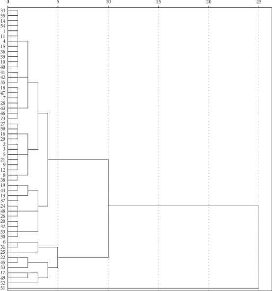
The cluster analysis tree of 55 batches of commercial SR (the indicator is the total content of six chromone compounds).
For the quality evaluation of SR, the chromone aglycone should not be ignored because this plays an important role in the pharmacological activity of SR. In addition, as shown in Table 5, the correlation coefficients of C, V, and H were significant (0.866, 0.900, and 0.0599), respectively. Using the ratio of the total content of C, V, and H to the total content of six chromones as an indicator, 53 batches of commercial SRs were divided into two categories by HCA (the 13th and 37th batches were self-contained, not considered). The details are shown in Table 7 and Figure 6. Cluster 1 included 41 batches (32, 54, 14, 19, 20, 4, 33, 15, 48, 25, 30, 51, 49, 52, 26, 1, 6, 31, 45, 24, 41, 11, 42, 27, 10, 17, 50, 34, 44, 53, 55, 22, 2, 18, 3, 7, 39, 29, 40, 36, 35), in which the ratio was greater than 10%. Meanwhile, the remaining 12 batch samples (12, 23, 5, 38, 9, 8, 47, 28, 46, 21, 16, 43) were attributed to Cluster 2, where the ratio was less than 10%. It was indicated that these three chromone aglycones were so important that they could be used as quality indicators for the quality evaluation of SR.
Table 7.
Ratio of the total content of three chromone aglycones to the total content of six chromones.
| No. | Ratio |
|---|---|
| 1 | 14.58 |
| 2 | 10.11 |
| 3 | 10.01 |
| 4 | 19.37 |
| 5 | 4.39 |
| 6 | 14.62 |
| 7 | 10.89 |
| 8 | 6.55 |
| 9 | 3.01 |
| 10 | 13.18 |
| 11 | 12.34 |
| 12 | 3.86 |
| 13 | 26.49 |
| 14 | 18.00 |
| 15 | 17.39 |
| 16 | 8.68 |
| 17 | 13.17 |
| 18 | 10.06 |
| 19 | 18.16 |
| 20 | 18.35 |
| 21 | 6.95 |
| 22 | 14.07 |
| 23 | 4.05 |
| 24 | 15.74 |
| 25 | 20.82 |
| 26 | 19.76 |
| 27 | 12.18 |
| 28 | 6.50 |
| 29 | 11.44 |
| 30 | 20.98 |
| 31 | 16.00 |
| 32 | 18.64 |
| 33 | 19.07 |
| 34 | 13.07 |
| 35 | 11.62 |
| 36 | 11.24 |
| 37 | 27.33 |
| 38 | 5.14 |
| 39 | 10.62 |
| 40 | 11.36 |
| 41 | 16.55 |
| 42 | 12.48 |
| 43 | 7.64 |
| 44 | 13.02 |
| 45 | 16.05 |
| 46 | 6.23 |
| 47 | 6.53 |
| 48 | 17.48 |
| 49 | 20.13 |
| 50 | 13.19 |
| 51 | 20.62 |
| 52 | 20.18 |
| 53 | 13.70 |
| 54 | 18.64 |
| 55 | 13.68 |
Figure 6.
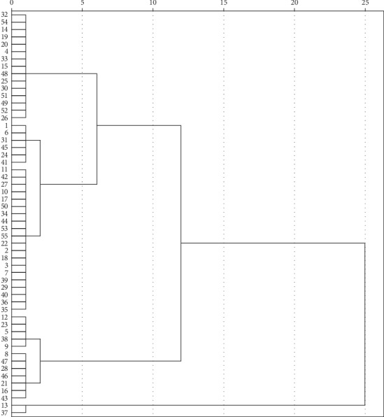
The cluster analysis tree of 55 batches of commercial SR (the indicator is the ratio of the total content of three chromone aglycones to the total content of six chromone compounds).
Last but not least, based on the results of the twice cluster analysis, samples of the Cluster 2 (6, 31, 25, 22, 45, 53, 17, 49, 52) in the first HCA were all contained to the Cluster 1 (32, 54, 14, 19, 20, 4, 33, 15, 48, 25, 30, 51, 49, 52, 26, 1, 6, 31, 45, 24, 41, 11, 42, 27, 10, 17, 50, 34, 44, 53, 55, 22, 2, 18, 3, 7, 39, 29, 40, 36, 35) in the second HCA. Thus, we could initially infer a conclusion that the SR could be regarded as a superior medicine when the total concentration of GC, C, GV, V, GH, and H was greater than 8 mg·g−1. Meanwhile, the proportion of aglycons (C, V, and H) was greater than 10%. In this study, 9 batch samples (6, 31, 25, 22, 45, 53, 17, 49, 52) were the superior medicine with the six chromone content which was greater than 8 mg·g−1, and meanwhile, the total content of C, V, and H was greater than 0.8 mg·g−1.
3.4. Optimization of Sample Preparation
Optimization of extraction methods, solvents, solvent volume, and extraction time were investigated by single-factor test to obtain the best extraction efficiency. The results revealed that extraction efficiency of refluxing was more efficient than ultrasonic extraction for analytes, so the remaining experiment was carried out by refluxing, and methanol was chosen as the best solvent by comparing with various solvents including methanol, 70% methanol, and 50% methanol. In addition, the extraction volume (5, 10, and 15 ml) and the extraction times (60, 120, and 240 min) were tested subsequently. Consequently, the optimal sample preparation parameter was refluxing with 10 mL methanol for 120 min as for 0.25 g powder of SR.
4. Conclusion
A method named QAMS was established to evaluate the quality of SR based on routine HPLC apparatus. In this method, GV was chosen as the internal standard to determine the RCF between GV and other five chromones (GC, C, V, GH, and H) of SR. QAMS was accurate and feasible for the quality evaluation according to the results of method validation, and no significant difference existed in the content results obtained by QAMS and ESM. Using this developed method, 55 batches of commercial SR from different regions were determined, and the results were analyzed by PCA, correlation analysis, and HCA, respectively. It could include that chromones played an important role for the quality of SR; meanwhile, the total content of GC, C, GV, V, GH, and H was used as the evaluation indicator that was comprehensive, objective, and reliable. Other than this, the SR could be regarded as qualified medicine if the total content of six chromones was not less than 3 mg·g−1. Moreover, the importance of chromone aglycones for the quality evaluation of SR was further demonstrated. It could initially infer that it was a superior SR medicine if the total content of GC, C, GV, V, GH and H was greater than 8 mg·g−1; meanwhile, the proportion of C, V, and H was greater than 10%.
All in all, these results provided useful information for the development of commercial SR, and the quality of SR from different origins or different purchase locations was confusing and unstable. Therefore, a compulsive processing standard for SR should be established and standardized. Last but not least, the abovementioned series of analyses will play a positive role in the improvement of the quality evaluation system of SR.
Acknowledgments
This research was supported by the Beijing Municipal Education Commission Common Construction Project (no. 2016022).
Contributor Information
Yanyan Jiang, Email: jyyjm1129@163.com.
Bin Liu, Email: liubinyn67@163.com.
Data Availability
All chromatographic data used to support the findings of this study are available from the corresponding author upon request.
Conflicts of Interest
The authors declare that there are no conflicts of interest regarding the publication of this paper.
References
- 1.Chinese Pharmacopoeia Commission. Pharmacopoeia of the People’s Republic of China. Beijing, China: China Medical Science and Technology Press; 2015. [Google Scholar]
- 2.Du C. Y. Q., Choi R. C. Y., Zheng K. Y. Z., et al. Yu Ping Feng San, an ancient Chinese herbal decoction containing astragali radix, atractylodis macrocephalae rhizoma and Saposhnikoviae radix, regulates the release of cytokines in murine macrophages. PLoS One. 2018;11(8) doi: 10.1371/journal.pone.0078622.e78622 [DOI] [PMC free article] [PubMed] [Google Scholar]
- 3.Zhang L. K., Sheng L., Guo W. F. Significance of “Saposhnikoviae radix” in complex prescription compatibility of traditional Chinese medicine. Journal of Basic Chinese Medicine. 2016;8(22):1107–1108. [Google Scholar]
- 4.Kong X. Y., Liu C. F., Zhang C., et al. The suppressive effects of Saposhnikovia divaricata (Fangfeng) chromone extract on rheumatoid arthritis via inhibition of nuclear factor-κB and mitogen activated proteinkinases activation on collagen-induced arthritis model. Journal of Ethnopharmacology. 2013;148(3):842–850. doi: 10.1016/j.jep.2013.05.023. [DOI] [PubMed] [Google Scholar]
- 5.Wu X. B., Jin S. R., Li S. M. Effect of saposhnikoviae radix extract on PAR-2 expression and related cytokine secretion of mast cells. Chinese Journal of Experimental Traditional Medical Formulae. 2016;5(22):123–126. [Google Scholar]
- 6.Zhang Z. Q., Tian Y. J., Zhang J. Studies on the antioxidative activity of polysaccharides from radix saposhnikoviae. Journal of Chinese Medicinal Materials. 2008;2(31):268–272. [PubMed] [Google Scholar]
- 7.Wang Z. W., Yang J. M., Jiang H. Influence of polysaccharides on pharmacodynamics and pharmacokinetics of bolting Saposhnikoviae radix. Chinese Traditional Patent Medicine. 2015;11(37):2392–2397. [Google Scholar]
- 8.Liu S. L., Jiang C. X., Zhao Y. Advance in study on chemical constituents of Saposhnikoviae divaricate and their pharmacological effects. Chinese Traditional and Herbal Drugs. 2017;10(48):2146–2152. [Google Scholar]
- 9.Yu L.-F., Li X.-R., Liu S.-Y., Xu G.-W., Liang Y.-Z. Comparative analysis of essential components between the herbal pair radix saposhnikoviae-rhizoma seu radix notopterygii and its single herbs by GC-MS combined with a chemometric resolution method. Analytical Methods. 2009;1(1):45–51. doi: 10.1039/b9ay00044e. [DOI] [PubMed] [Google Scholar]
- 10.Kreiner J., Pang E., Lenon G. B., Yang A. W. H. Saposhnikoviae divaricate: a phytochemical, pharmacological, and pharmacokinetic review. Chinese Journal of Natural Medicines. 2017;15(4):255–264. doi: 10.1016/s1875-5364(17)30042-0. [DOI] [PMC free article] [PubMed] [Google Scholar]
- 11.Wang Y., Wu T., Liu C. M. Isolation and identification of chemical components of chromones in Saposhnikoviae radix. Lishizhen Medicine and Materia Medica Research. 2018;7(29):1558–1561. [Google Scholar]
- 12.Chen L. X., Chen X. Y., Su L., Jiang Y., Liu B. Rapid characterisation and identification of compounds in Saposhnikoviae radix by high-performance liquid chromatography coupled with electrospray ionisation quadrupole time-of-flight mass spectrometry. Natural Product Research. 2018;32(8):898–901. doi: 10.1080/14786419.2017.1366482. [DOI] [PubMed] [Google Scholar]
- 13.Li W., Wang Z., Chen L., et al. Pressurized liquid extraction followed by LC-ESI/MS for analysis of four chromones in RADIX Saposhnikoviae. Journal of Separation Science. 2010;33(17-18):2881–2887. doi: 10.1002/jssc.201000336. [DOI] [PubMed] [Google Scholar]
- 14.Jiang H., Yang J. M., Cao L., Jia G., Dai H., Meng X. Quality evaluation of heat stress treated Radix saposhnikoviae using pharmacokinetic and pharmacologic methods. Journal of Chinese Pharmaceutical Sciences. 2018;27(2):109–115. doi: 10.5246/jcps.2018.02.012. [DOI] [Google Scholar]
- 15.Li W., Wang Z., Sun Y. S., Chen L., Han L.-K., Zheng Y.-N. Application of response surface methodology to optimise ultrasonic-assisted extraction of four chromones in radix saposhnikoviae. Phytochemical Analysis. 2011;22(4):313–321. doi: 10.1002/pca.1282. [DOI] [PubMed] [Google Scholar]
- 16.Yadav P., Sharma S. K., Manchanda P., Parshad B. Chromones and their derivatives as radical scavengers: a remedy for cell impairment. Current Topics in Medicinal Chemistry. 2014;14(22):2552–2575. doi: 10.2174/1568026614666141203141317. [DOI] [PubMed] [Google Scholar]
- 17.Chen A. Z., Su L., Yuan H., Wu A., Lu J., Ma S. Simultaneous qualitative and quantitative analysis of 11 active compounds in rhubarb using two reference substances by UHPLC. Journal of Separation Science. 2018;41(19):3686–3696. doi: 10.1002/jssc.201800479. [DOI] [PubMed] [Google Scholar]
- 18.Liao H., Li Q., Liu R., Liu J., Bi K. Fingerprint analysis and multi-ingredient determination using a single reference standard for Saposhnikoviae radix. Analytical Sciences. 2014;30(12):1157–1163. doi: 10.2116/analsci.30.1157. [DOI] [PubMed] [Google Scholar]
- 19.Li Y.-Y., Wang H., Chen J., et al. RRLC-TOF/MS in identification of constituents and metabolites of radix saposhnikoviae in rat plasma and urine. Academic Journal of Second Military Medical University. 2010;30(7):760–763. doi: 10.3724/sp.j.1008.2010.00760. [DOI] [Google Scholar]
- 20.Sun S., Xu L. L., Kong L. Y. Chromones from angelica morri hayata. Journal of China Pharmaceutical University. 2003;34(2):125–127. [Google Scholar]
- 21.Chin Y. W., Jung Y. H., Chae H. S., Yoon K. D., Kim J. W. Anti-inflammatory constituents from the roots of Saposhnikovia divaricate. Korean Chemical Society. 2011;32(6):2123–2134. doi: 10.5012/bkcs.2011.32.6.2132. [DOI] [Google Scholar]
- 22.Han Z. M., Yang R. Y., Wang Y. H. Extraction of chromone from saposhnikovia divaricata by ultrasonic wave. Lishizhen Medicine and Materia Medica Research. 2008;19(12):3035–3037. [Google Scholar]
- 23.Zhang T. L., Yu C. Y., Wei X. B. Study on the extraction technology of active constituents from saposhnikovia divaricata (Turcz.) Heilongjiang Medicine and Pharmacy. 2012;35(3):26–27. [Google Scholar]
- 24.Zhang Y. B., Juan D. A., Zhang J. X., et al. A feasible, economical, and accurate analytical method for simultaneous determination of six alkaloid markers in aconiti lateralis radix praeparata from different manufacturing sources and processing ways. Chinese Journal of Natural Medicines. 2017;15(4):301–309. doi: 10.1016/s1875-5364(17)30048-1. [DOI] [PubMed] [Google Scholar]
- 25.Xie J., Li J., Liang J., Luo P., Qing L.-S., Ding L.-S. Determination of contents of catechins in oolong teas by quantitative analysis of multi-components via a single marker (QAMS) method. Food Analytical Methods. 2017;10(2):363–368. doi: 10.1007/s12161-016-0592-5. [DOI] [Google Scholar]
- 26.Luo D. Q., Jia P., Zhao S. S., et al. Identification and differentiation of polygonum multiflorum Radix and polygoni multiflori Radix preaparata through the quantitative analysis of multicomponents by the single-marker method. Journal of Analytical Methods in Chemistry. 2019;2019:13. doi: 10.1155/2019/7430717.7430717 [DOI] [PMC free article] [PubMed] [Google Scholar]
- 27.Wang S. H., Xu Y., Wang Y. W., et al. Simultaneous determination of six active components in Oviductus ranae via quantitative analysis of multicomponents by single marker. Journal of Analytical Methods in Chemistry. 2017;2017:9. doi: 10.1155/2017/9194847.9194847 [DOI] [PMC free article] [PubMed] [Google Scholar]
- 28.Dong Y. H., Guo Q., Liu J. J., Ma X. Simultaneous determination of seven phenylethanoid glycosides in cistanches herba by a single marker using a new calculation of relative correction factor. Journal of Separation Science. 2018;41(9):1913–1922. doi: 10.1002/jssc.201701219. [DOI] [PubMed] [Google Scholar]
- 29.Hou J. J., Wu W. Y., Da J., et al. Ruggedness and robustness of conversion factors in method of simultaneous determination of multi-components with single reference standard. Journal of Chromatography A. 2011;1218(33):5618–5627. doi: 10.1016/j.chroma.2011.06.058. [DOI] [PubMed] [Google Scholar]
- 30.Wang C.-Q., Jia X.-H., Zhu S., Komatsu K., Wang X., Cai S.-Q. A systematic study on the influencing parameters and improvement of quantitative analysis of multi-component with single marker method using notoginseng as research subject. Talanta. 2015;134:587–595. doi: 10.1016/j.talanta.2014.11.028. [DOI] [PubMed] [Google Scholar]
- 31.Jang Y. Y., Liu B., Shi R. B. Isolation and structure identification of chemical constituents from Saposhnikovia divaricate (Turcz) Schischk. Acta Pharmaceutica Sinica. 2007;42(5):505–510. [PubMed] [Google Scholar]
- 32.Wang L., Liang R. X., Cao Y. Effect of prim-O-glucosylcimifugin and 4′-O-β-D-glucosyl-5-O-methylvisa-mminol con on proliferation of smooth muscle cell stimulated by TNF-a. China Journal of Chinese Materia Medica. 2008;33(17):2157–2160. [PubMed] [Google Scholar]
- 33.Cui L., Zhang Y., Shao W., Gao D. Analysis of the HPLC fingerprint and QAMS from Pyrrosia species. Industrial Crops and Products. 2016;85:29–37. doi: 10.1016/j.indcrop.2016.02.043. [DOI] [Google Scholar]
- 34.Li D.-W., Zhu M., Shao Y.-D., Shen Z., Weng C.-C., Yan W.-D. Determination and quality evaluation of green tea extracts through qualitative and quantitative analysis of multi-components by single marker (QAMS) Food Chemistry. 2016;197:1112–1120. doi: 10.1016/j.foodchem.2015.11.101. [DOI] [PubMed] [Google Scholar]
Associated Data
This section collects any data citations, data availability statements, or supplementary materials included in this article.
Data Availability Statement
All chromatographic data used to support the findings of this study are available from the corresponding author upon request.


