Abstract
Background
Environmental organic dust exposures enriched in Toll-like receptor (TLR) agonists can reduce allergic asthma development but are associated with occupational asthma and chronic bronchitis. The TLR adaptor protein myeloid differentiation factor88 (MyD88) is fundamental in regulating acute inflammatory responses to organic dust extract (ODE), yet its role in repetitive exposures is unknown and could inform future strategies.
Methods
Wild-type (WT) and MyD88 knockout (KO) mice were exposed intranasally to ODE or saline daily for 3 weeks (repetitive exposure). Repetitively exposed animals were also subsequently rested with no treatments for 4 weeks followed by single rechallenge with saline/ODE.
Results
Repetitive ODE exposure induced neutrophil influx and release of pro-inflammatory cytokines and chemokines were profoundly reduced in MyD88 KO mice. In comparison, ODE-induced cellular aggregates, B cells, mast cell infiltrates and serum IgE levels remained elevated in KO mice and mucous cell metaplasia was increased. Expression of ODE-induced tight junction protein(s) was also MyD88-dependent. Following recovery and then rechallenge with ODE, inflammatory mediators, but not neutrophil influx, was reduced in WT mice pretreated with ODE coincident with increased expression of IL-33 and IL-10, suggesting an adaptation response. Repetitively exposed MyD88 KO mice lacked inflammatory responsiveness upon ODE rechallenge.
Conclusions
MyD88 is essential in mediating the classic airway inflammatory response to repetitive ODE, but targeting MyD88 does not reduce mucous cell metaplasia, lymphocyte influx, or IgE responsiveness. TLR-enriched dust exposures induce a prolonged adaptation response that is largely MyD88-independent. These findings demonstrate the complex role of MyD88-dependent signaling during acute vs. chronic organic dust exposures.
Keywords: Environmental respiratory and skin disease, Agriculture, Occupational, Organic dust, Airway inflammation, Adaptation, MyD88
Background
Environmental agriculture organic dust exposures are rich in Toll-like receptor (TLR) agonists that are associated with regulating airway allergic and non-allergic inflammatory diseases [1]. Early life exposures to these TLR-enriched environments appears to be protective against the development of IgE-mediated diseases, including eosinophilic asthma [2–4]. However, workplace exposure to these organic dusts is associated with increased occupational asthma, workplace exacerbated asthma, neutrophil-predominant pulmonary inflammation, chronic bronchitis, and chronic obstructive pulmonary disease (COPD) [2, 5–7]. This dichotomy implies that persistent exposures to these environmental inflammatory-inducing agent(s) modulate the lung inflammatory response with potential long-term consequences.
The complexity and biodiversity of agriculture dust exposures is increasingly recognized, and animal and human studies have defined roles for TLR2/TLR4/TLR9-signaling pathways [8–12]. The TLR/IL-1R/IL-18R adaptor protein myeloid differentiation factor 88 (MyD88) that is used by all TLRs except for TLR3 [13] has been shown to have a fundamental role in the acute inflammatory response to organic dust [10, 14, 15] as well as other inflammatory exposures [16, 17]. MyD88 signaling mediates bleomycin-induced IL-17B expression in alveolar macrophages to promote pulmonary fibrosis [16]. In silica-induced fibrosis, silica dust increases MyD88 expression in macrophages [17]. In experimental asthma, MyD88 expression in epithelial cells mediates eosinophilia whereas MyD88 expression in conventional dendritic cells controls the neutrophilic response [18]. In response to agriculture organic dust extract (ODE), MyD88 knockout (KO) mice demonstrate reduced airway hyper-responsiveness and near absence of neutrophil influx and inflammatory cytokine release following acute ODE exposure [10]. However, these animals demonstrate an elevated, as opposed to reduced, mucus metaplasia response to acute ODE challenges [14]. Despite the well-characterized role for MyD88 in mediating acute responses to ODE, its impact on airway inflammatory responses to repetitive, prolonged exposures is unknown. This information could be important in future targeted approaches.
Repetitive exposures with ODE from large animal farming facilities induces perivascular lymphocytic aggregates that slowly, but not completely, resolve in size and number by 4 weeks after the inflammatory insult is removed [19]. It is not known if there is a heightened or dampened airway inflammatory response upon re-exposure. In other settings, endotoxin exposure can induce a refractory state of hypo-responsiveness for several days/weeks [20]. Alternatively, the lung microenvironment could be poised to be hyper-responsive to rechallenge, which collectively would have implications for workers returning to the job site after prolonged absences. It is unclear whether MyD88 signaling is involved in rechallenge responses.
In this study, we sought to investigate the lung cellular and microenvironment response to repeated ODE exposure for 3 weeks and to determine whether prolonged recovery (i.e. 4 weeks) following repetitive exposures would result in a heightened or refractory response to subsequent ODE rechallenge. Moreover, we hypothesized that the MyD88 signaling pathway would be central to governing lung responses to ODE. Collectively, our findings demonstrate a prominent role for MyD88 in mediating repetitive ODE-induced airway inflammatory responses, including an adaptive role for regulating the expression of epithelial barrier integral proteins. Moreover, our results also suggest that repetitive ODE treatment heightens neutrophil influx but decreases inflammatory cytokine release, while simultaneously upregulating an anti-inflammatory and macrophage response following a later ODE insult. These studies highlight a contextual role of MyD88-dependent signaling in orchestrating the pulmonary inflammatory response to acute and repetitive organic dust exposures.
Methods
Animals
MyD88 gene knock-out (KO) mice on a C57BL/6 J background were provided by S. Akira (Okasa, Japan). C57BL/6 J wild-type (WT) mice from The Jackson Laboratory (Bar Harbor, ME) were used as controls. Mice were fed alfalfa-free chow ad libitum (Harlan Laboratories) as recommended by The Jackson Laboratory. Mice were group housed in an SPF (specific-pathogen-free) facility under 12- h light and dark cycles. All animal procedures were approved by the Institutional Animal Care and Use Committee at the University of Nebraska Medical Center and conducted according to the U.S. National Institutes of Health guidelines for the use of rodents. Male and female mice aged 6–12 weeks were utilized in experimental procedures.
Organic dust extract
Aqueous organic dust extract (ODE) was prepared from settled dust collected from horizontal surfaces 3 ft. above the floor in swine confinement feeding operations. Extracts were batch prepared and filter (0.22 μm) sterilized as previously described [21, 22]. Stock ODE was diluted to a concentration of 12.5% (vol/vol) in sterile phosphate buffered saline (PBS, pH 7.4, diluent), a concentration previously shown to elicit optimal lung inflammation in mice [21, 22]. ODE 12.5% contains approximately 3–4 mg/ml total protein as measured by Nanodrop spectrophotometry (NanoDrop Technologies, Wilmington, DE) and endotoxin levels range from 22.1 to 91.1 EU/ml as measured by the limulus amebocyte lysate assay using manufacturer instructions (Sigma). Specific microbial biomarkers inferred from shotgun metagenomics of DNA pyrosequencing reads of the dust samples have been previously detailed [23].
Murine ODE exposure model
Under light sedation with isoflurane inhalation, mice were administered 50 μl of 12.5% ODE or sterile saline (PBS) by intranasal inhalation every day for 3 weeks with weekends excluded as previously described [21, 22]. Mice were euthanized 5 h after final exposure for experimental endpoints (Fig. 1). In separate studies, mice were treated with saline or ODE daily for 3 weeks (“repetitive exposure”) and then allowed to rest for 4 weeks without any treatments (“rest/recovery phase”) and subsequently rechallenged once with either saline or ODE and euthanized 5 h following this final rechallenge exposure for experimental endpoints (Fig. 1).
Fig. 1.
Experimental design protocol. MyD88 WT and knockout (KO) mice were intranasally treated with saline or ODE daily for 3 weeks and then were euthanized (“repetitive exposure”) or allowed to rest for 4 weeks withouat any treatments (“rest/recovery period”) and subsequently rechallenged once with either saline or ODE and euthanized (“rechallenged following rest”)
Bronchoalveolar lavage fluid studies
Bronchoalveolar lavage fluid (BALF) was collected by instilling 3 × 1 ml PBS into the airway. Cytokines implicated in agriculture respiratory disease including IL-6, neutrophil chemoattractants CXCL1 and CXCL2, and TNF-α [1] were measured in the cell-free supernatant of the first lavage by ELISA (R&D Systems, Minneapolis, MN) with limits of detectability of 1.8, 2.0, 1.5, and 7.2 pg/ml, respectively. Total number of cells recovered from pooled lavages were enumerated and differential cell counts were performed on cytospin-prepared slides (Cytopro Cytocentrifuge; Wescor, Logan, UT) stained with DiffQuick (Siemens, Neward, DE).
Immunostaining and microscopy of mouse lungs
After BALF collection, whole lungs were harvested and inflated with 1 ml 10% formalin and hung under pressure of 20 cm H2O for 24 h for optimal preservation of lung parenchymal architecture. Lung tissue was formalin-fixed, embedded in paraffin, and cut into 5 μm sections. Lung sections were then stained for detection of neutrophils and B-cells using anti-Ly-6B.2 and anti-CD45.R antibodies respectively, according to antibody-specific staining techniques as previously described [24]. Lung lesions (histopathology) in tissue sections were semi-quantitatively scored for severity of alveolitis (infiltration of neutrophils in alveolar parenchyma) and perivascular/peribronchiolar lymphoid cell infiltrates (ectopic lymphoid aggregates) by a veterinary pathologist blinded to treatment conditions (JH), following defined histopathological criteria previously described in detail [24]. Mucous cell in bronchiolar airway epithelium were microscopically detected by periodic acid Schiff (PAS) histochemical staining for intracellular mucosubstances. Severity of mucous cell metaplasia in bronchiolar epithelium was likewise semi-quantitatively scored in PAS-stained lung sections from each animal [24]. In brief, the severity of lung lesions were scored according to the percentage of total lung tissue affected, i.e. severity score 0 for no microscopic findings, 1 for < 10% lung tissue involvement, 2 for 10–25%, 3 for 26–50%, 4 for 51–75%, and 5 for > 75% [24].
To identify mast cells, lung sections were stained for tryptase, and quantification of tryptase+ cells per entire lung section was performed using Definiens software with the evaluator blinded to treatment conditions. To assess collagen, Masson’s Trichome staining was performed. Following staining, slides were scanned with iScan Coreo Au (Ventana, Tucson, AZ) slide scanner by the UNMC Tissue Sciences Facility and converted into digital format. Quantification of collagen was performed using ImageJ software after deconvolution and thresholding (Rasband, W.S., Image J, U.S. National Institutes of Health, Bethesda, MD, https://imagej.nih.gov/ij/, 1997–2016) using methods previously described [25, 26].
In the separate studies where animals received repetitive ODEs, rested for 4 weeks and then rechallenged, lung sections were stained for H&E and confocal imaging. For confocal imaging, sections were stained for B cells using ALEXA FLUOR™ 488 conjugated Rat anti-Mouse CD45R (BD Biosciences, San Jose, CA) and macrophages using ALEXA FLUOR™ 594 conjugated rabbit anti-CD68 polyclonal antibody (Bioss Antibodies, Woburn, MA). ALEXA FLUOR™ 488 conjugated Rat IgG and ALEXA FLUOR™ 594 conjugated rabbit IgG (Bioss) were used as isotype controls as previously described [22]. Images were obtained using a Zeiss 510 Meta Confocal Laser Scanning Microscope and analyzed using Image J software (NIH).
Serum IgE levels
Whole blood was collected from the axillary artery at the time of euthanasia in BD Microtainer tubes (Becton, Dickinson, and Company, Franklin Lakes, NJ), centrifuged for 2 min at 6000 x g and supernatant collected. Serum IgE levels were quantified according to manufacturer’s instructions using ELISA (R&D Systems, Minneapolis, MN).
Lung homogenate mediator and tight junction analysis
Following BALF collection and vascular perfusion, half of the lung was homogenized in 500 μl of sterile PBS with a gentle MACS dissociator (Miltenyi Biotec, Bergisch Gladbach, Germany) and cell-free supernatant stored at − 80 °C. Levels of the alarmin IL-33, the anti-inflammatory cytokine IL-10, and epidermal growth factor receptor agonist amphiregulin (AREG; involved in lung repair/recovery processes [27]) were quantified by ELISA (Quantikine kit from R&D Systems, Minneapolis, MN) with lower limit of detection of 6.85, 12 and 20 pg/ml. To broadly explore an array of cytokines/chemokines potentially involved in mediating environmental ODE-induced inflammatory lung disease, a custom 18-plex Mouse Magnetic Luminex kit was utilized, which measured CCL2, CCL3, CCL4, CCL5, CCL7, CCL8, CCL11, CCL19, CCL22, CXCL1, CXCL10, IL-4, IL-5, IL-13, periostin, MMP-12, S100A8, and CH13-LI. Protein levels were determined by Bradford assay.
Expression of the tight junction integral proteins claudin-1, − 2, − 3, − 4, − 7, and occludin was investigated by real-time PCR, as previously described, [28] with forward and reverse primer sequences detailed in Additional File 1. Immunoblot analysis was performed using antibodies against claudin-3 (#PA5–16867; Invitrogen), claudin-4 (#PA5–32354, Invitrogen), and occludin (#40–4700; Invitrogen) based on their differential expression in qRT-PCR. Immunoblot analysis of pSTAT3 (#9145S; Cell signaling technology) was included as a control as previously described [29]. Signals were detected using an enhanced chemiluminescence detection kit (Amersham Biosciences). Equal protein loading was determined by re-probing with an anti-β actin antibody (#A2228-100UL; Sigma) after stripping each membrane.
Statistical methods
Data are presented as means and SEM. A sample-size calculation was estimated from a previous single ODE exposure in WT and MyD88 KO murine study [10], whereby we calculated a sample size of N = 3 in each group, to achieve 80% power at the 0.05 level of significance to detect a difference in airway neutrophil influx, assuming a mean (SD) of 11.15 × 10⁵ (4.5 × 10⁵) for WT + ODE and a mean of 0.02 × 10⁵ (0.0065 × 10⁵) for KO + ODE. The experimental groups were run with sex- and age-matched litter mates as available, generally over-sampled, and subsequently pooled. Sample size for each outcome listed in figure legends reflects sample quantity and quality available from the pooled studies. To detect significant differences among groups, a one-way ANOVA was performed, and in the event p < 0.05 by ANOVA across groups, a non-parametric Mann-Whitney test was used to determine statistical difference between two groups. Prism software (version 7.0c; GraphPad Software, La Jolla, CA) was used. Significance was accepted at p-values < 0.05.
Results
MyD88-deficient mice demonstrate a significantly reduced airway inflammatory response following repetitive ODE exposure
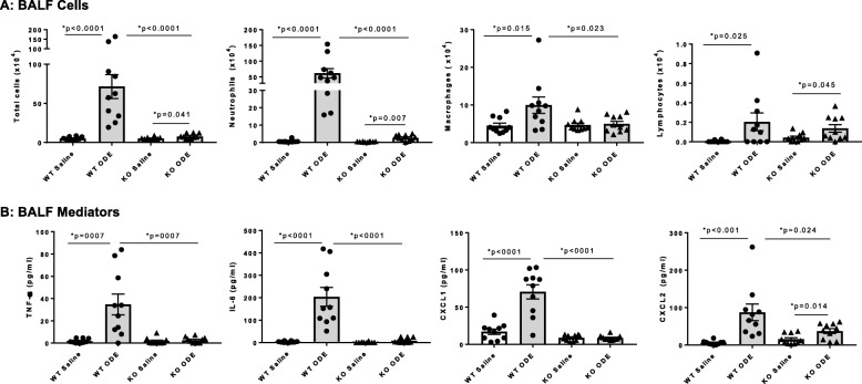
The airway inflammatory response to repetitive daily exposure to ODE for 3 weeks was strikingly reduced in MyD88 KO mice as compared to WT animals. BALF total cells, neutrophils, macrophages, and lymphocytes were increased in ODE-challenged WT mice, and there were significant reductions in total cells, neutrophils, but not lymphocytes, in MyD88 KO animals (Fig. 2a). There was no significant influx of eosinophils detected among the groups (data not shown). Additionally, ODE-induced TNF-α, IL-6, and murine neutrophil chemoattractants (CXCL1 and CXCL2) were either reduced or completely abrogated in MyD88 KO mice (Fig. 2b).
Fig. 2.
Decreased airway inflammatory response in MyD88 KO mice following repetitive exposure to ODE for 3 weeks. Wild-type (WT) and MyD88 knock-out (KO) mice were treated i.n. with saline or ODE daily for 3 weeks, whereupon bronchoalveolar lavage fluid (BALF) was collected five hours following final exposure. Panel a, Total cells, neutrophils, macrophages, and lymphocytes were increased in ODE-treated WT mice, and this response was significantly reduced in MyD88 KO animals. Panel b, ODE-induced TNF-α, IL-6, and murine neutrophil chemoattractants (CXCL1 and CXCL2) were reduced or abrogated in MyD88 KO mice. Scatter plots show mean with SEM; N = 10 mice/group (N = 6 male, N = 4 female) from 3 independent experiments
MyD88-deficient mice demonstrate reduced ODE-induced neutrophilic alveolitis without a reduction in perivascular lymphoid infiltrates
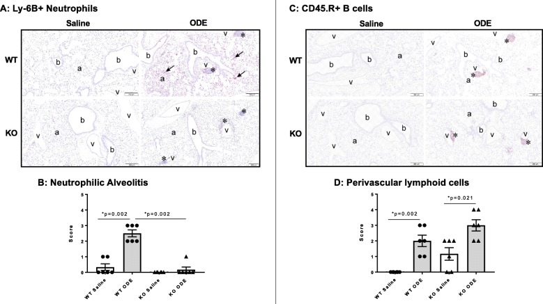
Lung tissue sections from exposure groups were stained for neutrophil (Ly-6B.2) and B-cell (CD45.R) infiltrates and semi-quantitatively scored as described in the Methods section. Repetitive ODE exposure was found to induce neutrophilic alveolitis and perivascular B cell infiltrates in WT mice (Fig. 3a-d). This alveolitis response to repetitive ODE exposure was reduced in MyD88 KO mice, but the perivascular lymphoid cell response (ectopic lymphoid aggregates) was not affected (Fig. 3a-d).
Fig. 3.
Repetitive ODE exposure-induced neutrophilic alveolitis, but not perivascular lymphoid cell aggregates, are reduced in MyD88 KO mice. Wild-type (WT) and MyD88 knock-out (KO) mice were treated i.n. with saline or ODE daily for 3 weeks, whereupon lung tissues were collected, formalin-fixed, and paraffin embedded. Lung sections (5 μm) were stained for neutrophils (a, Ly-6B.2+) and B cells (c, CD45.R+) with representative images for each treatment group shown. Key: b, bronchiolar airway; a, alveolar parenchyma; v = blood vessel; asterisk, lymphoid aggregates, and arrow Ly-6B.2+ neutrophils (scale bar is 200 μm). Lung sections were semi-quantitatively scored from 0 to 5 (see Methods section) for neutrophilic alveolitis (b) and perivascular lymphoid cells (d). Scatter plots (b, d) depict mean with SEM; N = 6 mice/group (N = 4 male, N = 2 female)
MyD88-deficient mice have increased ODE-induced mucus cell metaplasia
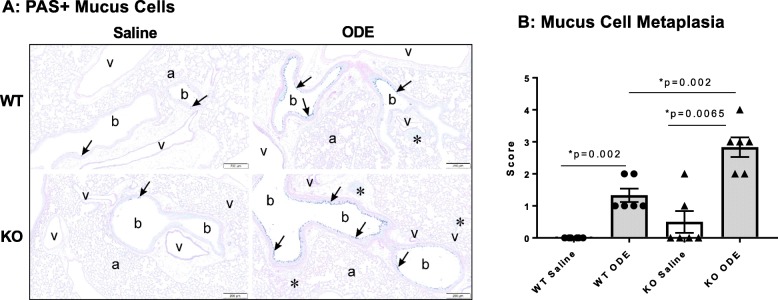
It has been previously shown that repetitive ODE exposure for 1 week induces mucus cell metaplasia that is further augmented in MyD88 KO animals [14]. In the present study, mucous cell metaplasia as evident by PAS+ staining was increased after repetitive ODE exposure for 3 weeks in both MyD88 WT and KO mice as compared to saline control, and this response was significantly augmented in the ODE-treated MyD88 KO as compared to the ODE-treated WT mice (Fig. 4a-b).
Fig. 4.
Mucous cell metaplasia is increased in MyD88 KO mice repetitively exposed to inhalant ODE. Wild-type (WT) and MyD88 knock-out (KO) mice were treated i.n. with saline or ODE daily for 3 weeks, whereupon lung tissues were collected, formalin-fixed, and paraffin embedded. Lung sections (5 μm) were PAS-stained for mucus cell determination with representative images for each treatment group shown (a). Key: b, bronchiolar airway; a, alveolar parenchyma; v = blood vessel; asterisk, lymphoid aggregates, and arrow denotes positive PAS stained mucus cells (scale bar is 200 μm). Lung sections were semi-quantitatively scored from 0 to 5 for mucus cell metaplasia with scatter plot (b) depicting means with SEM; N = 6 mice/group (N = 4 male, N = 2 female)
Lung mast cells, lung IL-33 and serum IgE levels are increased following repetitive ODE exposure and differentially modulated by MyD88
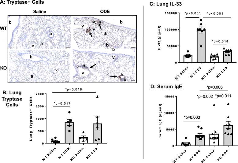
Repetitive ODE exposure increased tryptase+ lung mast cells in both WT and MyD88 KO mice, predominately observed within the perivascular aggregates (Fig. 5a-b). There was no difference between ODE-treated WT and KO animals. We then sought to quantitate levels of IL-33, an alarmin cytokine involved in mast cell activation and mucin expression [30]; and moreover, IL-33R signals via MyD88 to potentially reflect an autocrine/paracrine action [31]. ODE exposure significantly induced lung IL-33 levels in WT and KO animals as compared to saline, but this response was significantly reduced in ODE-treated MyD88 KO mice as compared to WT mice (Fig. 5c). It has been previously demonstrated that ODE exposure slightly, but significantly increases serum IgE levels [32]. In these studies, serum IgE levels were increased in both ODE-treated WT and MyD88 KO mice in comparison to their respective saline-treated controls (Fig. 5d). Serum IgE levels were significantly increased in saline control MyD88 KO mice as compared to saline control WT mice, which is consistent with a prior report showing elevated levels of serum IgE in saline control treated MyD88 KO animals [33]. However, a separate study showed decreased IgE levels in MyD88 KO animals [34, 35].
Fig. 5.
Tryptase+ lung mast cells, serum IgE and lung IL-33 levels are increased following repetitive ODE exposure and variably modulated by MyD88. WT and MyD88 KO mice were treated i.n. daily for 3 weeks with saline or ODE whereupon lung sections (5 μm thick) were stained for tryptase with representative images for each treatment group shown a. Key: b, bronchiolar airway; a, alveolar parenchyma; v = blood vessel; asterisk, cellular aggregates; and arrow denotes positive tryptase stained cells in cellular aggregates (scale bar is 100 μm). b, Scatter plot depicts means with SEM of tryptase positive cells per entire lung section as quantified by Definiens software with N = 5–6 mice/group. c, Serum IgE levels quantified by ELISA with N = 8–9 mice group. d, ODE-induced lung homogenate IL-33 levels were reduced in MyD88 KO mice with N = 6–7 mice/group
Repetitive ODE exposure increases mediators involved in neutrophil, lymphocyte, monocyte, and mast cell recruitment as well as factors involved in tissue remodeling, which are largely attenuated in MyD88 deficient mice
To further delineate the immunophenotype induced by ODE, we investigated a number of inflammatory, allergy, and non-allergy mediators by cytokine array analysis (Table 1; significant differences across treatment groups are in bold). Repetitive exposure to ODE did not induce an increase in the Th2 cytokines IL-4, IL-5, and IL-13 which are known to be involved in the classical allergy response and agrees with the lack of MyD88 involvement in regulating serum IgE levels. However, in both WT and MyD88 KO mice repetitively exposed to ODE there were significant increases in CCL2, CCL3, CCL4, CCL5, and CCL7. These mediators are known to be involved with recruitment of neutrophils, lymphocytes, monocytes, and mast cells. Moreover, these responses were attenuated in MyD88 KO mice as compared to WT animals, confirming the reduced recruitment of these effector cells in the lungs of MyD88 KO mice following ODE. Additionally, ODE exposure increased levels of neutrophilic inflammatory mediator S100A8 and tissue remodeling factors including periostin, MMP-12, CHI3-L1, and amphiregulin (AREG) in WT mice. These mediators were not induced in ODE-challenged MyD88 KO mice. There was no significant difference in total protein levels in lung homogenates across treatment groups.
Table 1.
Levels of mediators in lung tissue homogenates screened by treatment group
| Mediator | Action | WT Saline | WT ODE | KO Saline | KO ODE |
|---|---|---|---|---|---|
| CCL2 | Attracts monocytes, memory T cells, DCs | 181.5 ± 27.7 | 625.2 ± 18.4 ** | 371.1 ± 87.3 | 557.0 ± 96.0* |
| CCL3 | Attracts neutrophils | 21.5 ± 4.5 | 874.5 ± 198.3***,### | 43.3 ± 11.9 | 100.4 ± 36.4* |
| CCL4 | Attracts NK cells, monocytes | 37.5 ± 5.3 | 140.6 ± 23.8***, ### | 37.5 ± 3.7 | 48.8 ± 7.5 |
| CCL5 | Attracts T cells, eosinophils, basophils | 252.1 ± 67.5 | 1410 ± 450.5** | 545.1 ± 98.9 | 2519 ± 600.2* |
| CCL7 | Attracts monocytes, activates MΦ | 34.4 ± 9.2 | 552.5 ± 117.3***,## | 27.5 ± 5.7 | 150.5 ± 53.7* |
| CCL8 | Attracts mast cells, eosinophils and basophils, monocytes, T cells, NK cells | 356.3 ± 163.3 | 1620 ± 620.6 | 591.8 ± 112.4 | 1181 ± 215.2 |
| CCL11 | Attracts eosinophils | 94.2 ± 39.7 | 177.4 ± 52.3 | 77.8 ± 9.3 | 97.7 ± 10.7 |
| CCL19 | Attracts T and B cells | 14.6 ± 2.5 | 18.7 ± 1.1 | 15.9 ± 1.4 | 22.2 ± 6.5 |
| CCL22 | Attracts Th2 and Treg | 117.5 ± 38.3 | 432.1 ± 52.6** | 208.5 ± 62.4 | 335.4 ± 88.3 |
| CXCL1 | Attracts neutrophils | 62.3 ± 20.6 | 713.1 ± 211.3**,## | 30.2 ± 6.2 | 47.8 ± 11.0 |
| CXCL10 | Attracts monocytes/macrophages, T cells, NK cells, DCs | 44.2 ± 12.1 | 131.6 ± 29.3* | 40.8 ± 3.1 | 107.2 ± 38.9 |
| IL-4 | Th2, allergy | 236.2 ± 25.1 | 173.5 ± 16.3 | 303.9 ± 24.5 | 273.8 ± 5.1 |
| IL-5 | Eosinophil recruitment | 3.4 ± 0.9 | 3.0 ± 0.2 | 2.6 ± 0.1 | 2.8 ± 0.1 |
| IL-13 | Regulates IgE, mucus | 246.2 ± 14 | 173.5 ± 16.3 | 303.9 ± 24.5 | 273.8 ± 5.1 |
| Periostina | Tissue remodeling | 9.99 ± 1.11 | 21.14 ± 2.71***, # | 8.91 ± 9.88 | 12.42 ± 1.40 |
| MMP-12 | Tissue remodeling | 757.7 ± 337.9 | 13,351 ± 5086**,## | 647.8 ± 133.5 | 854.5 ± 196.1 |
| S100A8a | Neutrophilic inflammation | 229.9 ± 61.1 | 652.7 ± 66.4***,### | 172.5 ± 33.8 | 215.4 ± 9.6 |
| CHI3-L1a | Inflammation and tissue remodeling | 291.5 ± 42.1 | 620.0 ± 9.5*, # | 339.4 ± 67.6 | 468.9 ± 70.0 |
| AREG | Tissue remodeling and repair | 102.3 ± 11.6 | 221.8 ± 23.5** | 122.4 ± 3.7 | 161.5 ± 19.0 |
| Total Proteina | 7.50 ± 0.47 | 8.81 ± 0.41 | 6.47 ± 0.83 | 5.61 ± 1.46 |
Mean ± SEM values shown in pg/ml; except avalues are ng/ml. N = 4 mice/ group (2 males/2 females). Statistical difference denoted (*p < 0.05 **p < 0.01, ***p < 0.001) vs. saline. Statistical difference between WT ODE and KO ODE denoted (#p < 0.05, ##p < 0.01, ###p < 0.001). Differences are in bold
Protein levels in the BALF increased in ODE treated WT mice, but not MyD88 KO animals, suggesting modest disruption in alveolar permeability [36]. Specifically, mean ± SEM of BALF protein levels were significantly different (p = 0.019) between WT saline: 140.1 ± 13.4 μg/ml vs. WT ODE: 204.8 ± 19.9 μg/ml, but were not significantly different (p > 0.05) between MyD88 KO saline: 142.8 ± 7.2 μg/ml vs. KO ODE: 158.3 ± 10.6 μg/ml.
Repetitive ODE exposure augments the expression of several tight junction proteins in the lung that are differentially regulated by MyD88
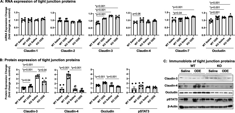
Studies have reported a potential adaptive role for specific tight junction proteins in the inflamed and injured lung, in particular claudin-4 [37, 38]. Our initial RT-qPCR analysis supported trends (p = 0.06) for increased gene expression of claudin-4 in ODE-challenged WT mice as well as claudin-3 and occludin (Fig. 6a). In the same samples, expression of claudin-1, − 2 and − 7 were unaltered supporting the specificity of the above outcomes. Immunoblot analysis further demonstrated robust and significant (p < 0.001) upregulation of claudin-4 in ODE-challenged WT mice as compared to WT saline (Fig. 6b-c). There were also significant increases in claudin-3 and occludin with ODE exposure as compared to saline in WT animals (Fig. 6b-c). In comparison, gene and protein expression of these tight junction proteins were not increased in ODE-treated MyD88 KO animals as compared to saline-treated KO mice. As compared to WT saline, gene expression of claudin-3, claudin-7, and occludin were increased in MyD88 KO animals (with/without ODE), and by protein measurements, only claudin-3 was increased in these KO animals. The significance of this basal difference in MyD88 KO animals is unknown.
Fig. 6.
Repetitive ODE exposure increases expression of several tight junction proteins known to be upregulated in inflamed/injured lung in WT mice but not in MyD88 KO mice. WT and MyD88 KO mice were treated i.n. daily for 3 weeks with saline or ODE. Panel a, Expression of tight junction mRNA was measured by real-time quantitative PCR and are reported as fold-changes normalized to control. Panel b, Quantification of tight junction protein expression in ODE-treated mice compared to control mice as determined by the immunoblot presented in Panel c. Scatter plots demonstrate mean with standard error bars of N = 3 animals per group with 2 replicates per sample
MyD88 is required for eliciting immune responses upon rechallenge after a period of rest following repetitive ODE exposure
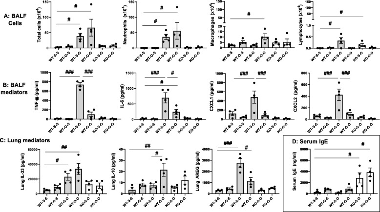
Total cells and neutrophils were increased in the BALF of WT mice pretreated with saline or ODE and then challenged once with ODE (WT-S-O and WT-O-O) as compared to saline control (WT-S-S) (Fig. 7a). There was no difference between WT mice rechallenged with ODE after repetitive saline (WT-S-O) and WT mice rechallenged with ODE after repetitive ODE (WT-O-O) (Fig. 7a). BALF macrophages were significantly increased only in WT mice pre-treated with ODE and then rechallenged with ODE (WT-O-O). BALF lymphocytes were significantly increased in WT mice pre-treated with saline and then rechallenge with ODE (WT-S-O). There were no significant increases in total cells, neutrophils, or macrophages in MyD88 KO mice pretreated with either saline or ODE and then rechallenged with ODE (KO-S-O or KO-O-O). However, lymphocytes were increased in MyD88 KO mice pretreated with saline and then rechallenged with ODE (KO-S-O) as compared to KO-O-O mice (mice pretreated with ODE and then rechallenged with ODE).
Fig. 7.
An airway inflammatory adaptive response following ODE rechallenge in WT mice after a 4 week recovery from repetitive exposures with minimal responsiveness observed in MyD88 KO mice. WT and MyD88 KO mice were repetitively exposed to saline or ODE for 3 weeks followed by a 4 week rest/recovery phase and then subsequently rechallenged once with saline or ODE. Scatter dot plots demonstrate mean with standard error bars with treatment groups denoted as S-S (saline repeated exposure-saline rechallenge), O-S (ODE repeated exposure-saline rechallenge), S-O (saline repeated exposure-ODE rechallenge), O-O (ODE repeated exposure-ODE rechallenge). Panel a: Total cells, neutrophils, macrophages, and lymphocytes in BALF. Panel b: Proinflammatory cytokines/chemokines quantified in cell-free BALF. Panel c: Mediator levels quantified in cell-free lung homogenates. Panel d: Serum IgE levels. Statistical significance (#p < 0.05; ##p < 0.01; ###p < 0.001) vs. saline or denoted by line. N = 4 mice group (2 female/2 male)
A different pattern of responses was observed with ODE-induced inflammatory mediators (i.e. TNF-α, IL-6, CXCL1 and CXCL2) (Fig. 7b). Namely, these inflammatory mediators were robustly increased in WT mice pretreated with saline and rechallenged with ODE (WT-S-O). However, these responses were strikingly attenuated in mice pretreated with ODE and rechallenged with ODE (WT-O-O). The MyD88 KO mice, regardless of pretreatment with saline or ODE, failed to mount an inflammatory mediator response to the ODE rechallenge.
Lung mediators and serum IgE were variably regulated in mice rechallenged with ODE following the prolonged 4-week recovery phase
Lung homogenates from the same treated WT and MyD88 KO mice were analyzed for several mediators. Lung IL-33 levels were increased in WT mice pretreated with saline or ODE and then challenged once with ODE (WT-S-O and WT-O-O) as compared to saline controls (Fig. 7c), and there was no significant difference between WT-S-O and WT-O-O. There was no detectable IL-33 response in MyD88 KO mice. Levels of the anti-inflammatory cytokine IL-10 in the lung were increased only in the WT mice pretreated with ODE and then rechallenged once with ODE (WT-O-O) (Fig. 7c). The repair/recovery mediator AREG was increased in WT mice pretreated with saline or ODE and rechallenged with ODE (WT-S-O or WT-O-O) as compared to mice rechallenged with saline (WT-S-S). There was a non-significant (p = 0.057) decrease in AREG release in WT mice pretreated with ODE and rechallenged with ODE (WT-O-O) as compared to WT saline pretreated and ODE rechallenged (WT-S-O) (Fig. 7c). Lung IL-10 and AREG levels were not significantly modulated in MyD88 KO mice. Lastly, we quantitated serum IgE levels in these same mice, and serum IgE levels were increased only in the MyD88 KO mice (regardless of pretreatment conditions) and were unchanged across the WT mice treatment groups (Fig. 7d).
Exacerbated lung histopathology occurs with ODE rechallenge following a 3-week exposure and 4-week rest/recovery phase
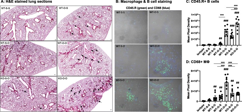
Microscopically, there was a persistence of perivascular lymphoid infiltrates after repetitive ODE in both WT and MyD88 KO animals following 4-week rest (Fig. 8a; isotype control antibody staining shown in Additional File 2). These perivascular ectopic lymphoid aggregates were more pronounced in the MyD88 KO animals repetitively treated with ODE and rechallenged with ODE (KO-O-O) as compared to the WT-O-O treated mice. CD45.R+ B cells were prominent in ODE-induced perivascular lymphocytic aggregates (Fig. 8b). There was an increase in B cell infiltrates in WT and KO mice pretreated with ODE and rechallenged with ODE (O-O) as compared to animals rechallenged with saline (O-S). B cell infiltrates in aggregates were increased in KO mice as compared to WT (Fig. 8c). CD68+ macrophages were increased with ODE treatment (Fig. 8b, d) and macrophage recruitment was the highest in the WT-O-O treatment group.
Fig. 8.
Lung histopathology accentuated with rechallenge following a 4 week recovery after repetitive ODE exposure. WT and MyD88 KO mice were repetitively exposed to saline or ODE for 3 weeks followed by a 4 week rest/recovery phase and then subsequently re-challenge once with saline or ODE. Panel a, Lung sections (5 μm) were H&E stained with representative images shown from each group of 4 animals/group (scale denotes 100 μm; 2x magnification). Arrows denote lymphocytic aggregates. Panel b, Confocal images of lung tissues stained for B cells (CD45.R; green) and macrophages (CD68; blue) shown at 40x magnification with selected images focused on aggregates where available. Scatter dot plots demonstrate mean with standard error bars of mean pixel density representing two regions of interest (ROIs) from each mouse (N = 8 ROI/group) depicting CD45.R+ B cells c and CD68+ Macrophages d. Treatment groups denoted as S-S (saline repeated exposure-saline rechallenge), O-S (ODE repeated exposure-saline rechallenge), S-O (saline repeated exposure-ODE rechallenge), and O-O (ODE repeated exposure-ODE rechallenge). Statistical significance denoted as ##p < 0.01, ###p < 0.001 vs. WT-S-S; and **p < 0.01, ***p < 0.001 denoted between groups (line)
Discussion
Organic dusts from agricultural environments contain an array of bacterial motifs recognized to play important roles in mediating airway inflammatory disease [1], and we have previously shown that MyD88 has a fundamental role in the acute inflammatory response to organic dust [10]. Here we demonstrate that MyD88 remains critical to mediating the airway inflammatory response following daily, repetitive exposures for 3 weeks as demonstrated by neutrophil influx and increases in a wide array of inflammatory mediators that were nearly abrogated in MyD88 KO mice. MyD88 was not responsible for lymphocyte recruitment as lymphocytes in the BALF and B cell infiltrates in the lung parenchyma were not reduced in ODE-challenged MyD88 KO mice. Mucus cell metaplasia was augmented in MyD88 KO animals, which cannot be explained by alterations in inflammatory mediators classically implicated in mucus cell biology, such as IL-6, IL-4, IL-13, and IL-33. Repetitive ODE exposure also induces a prolonged adaptation response. Despite a 4-week rest/recovery phase following repetitive ODE exposures, there was significant reduction in inflammatory mediator levels, but not neutrophil influx, in WT animals following a single rechallenge. These findings were also dependent upon MyD88. Upregulated expression of claudin-4, − 3 and occludin proteins in ODE-challenged mice further support this concept as these proteins are known to be upregulated in response to lung inflammation/injury and render adaptive roles [37, 38]. Together, these findings illustrate the complex role of the innate immune MyD88 signaling pathway with repetitive organic dust exposures and rechallenge.
Agriculture organic dust exposures are well-established to induce a neutrophilic as opposed to an eosinophilic influx with a corresponding release of pro-inflammatory mediators including TNF-α, IL-6, and neutrophil chemoattractants CXCL1 and CXCL2 [1]. Expanding on these findings, repetitive ODE exposures were also found to induce IL-33, the neutrophil mediator S100A8, and several factors involved with neutrophil, lymphocyte, monocyte, and mast cell recruitment (i.e. CCL2, CCL3, CCL4, CCL5, and CCL7). The classic Th2 cytokines implicated in allergic responses (i.e. IL-4, IL-5, and IL-13) were not induced. Further, tissue remodeling/pre-fibrosis/repair mediators including periostin, MMP-12, and CHI3-L1 as well as amphiregulin (AREG) were increased after repetitive ODE. Nearly all these responses were MyD88-dependent. Thus, not only is the innate MyD88 signaling pathway central to acute ODE induced inflammatory responses, these new studies suggest that there is not a compensatory response with repeated daily exposures over time regarding these multiple inflammatory attributes. Furthermore, MyD88 was central to mediating the airway inflammatory response following a prolonged rest period and subsequent rechallenge. Therefore, targeting MyD88 could represent a strategy in occupationally exposed workers to reduce airway inflammatory disease burden.
Various agriculture organic dust exposures in humans and mice also induce lymphocytic responses typified by a Th1 and/or Th17 microenvironment in the lung [22, 39, 40]. In the current study, we focused on B cells due to our recent report demonstrating their importance in experimental ODE-induced lung disease [22]. Current studies suggest that MyD88 may be important in resolution processes of environmentally triggered lung disease. Whereas there was no difference in B cell infiltrates with repetitive ODE exposure between WT and MyD88 KO animals, B cell infiltrates were increased in mice lacking MyD88 at 4 weeks post-recovery and a one-time rechallenge further augmented B cell infiltrates in the MyD88 KO mice. Others have also found that absence of MyD88 results in unresolved pulmonary infiltrates following long-term radiation-induced lung injury, [41] silica-induced fibrosis [42], and bacterial pneumonia [43]. The explanation for the persistence of lung infiltrates has been attributed to the generation of regulatory cells triggered through TLR/MyD88 signaling [41–43].
It is likely that macrophages and IL-10 contribute to the resolution process and response to ODE rechallenge. It is known that depletion of lung macrophages impairs lung recovery following repetitive ODE exposures [44]. In addition, mice lacking scavenger receptor A (CD204), a receptor responsible for enhancing IL-10 production under inflammatory states [45, 46], have impaired recovery [47]. Here, we report that macrophages and IL-10 levels were significantly increased in WT mice pretreated with ODE and rechallenged with ODE, which we interpret to explain the profoundly affected (i.e. reduced) airway inflammatory cytokine response to rechallenge. This reduced response is in contrast to a hypothesis that the lung could have a heightened inflammatory response upon rechallenge as observed in Byssinosis (i.e. endotoxin-mediated disease), whereby textile workers experience worsening dyspnea on Mondays or after vacations [48]. Agriculture organic dusts are complex and not just comprised of endotoxin, and it is likely that the wide diversity of bacterial motif exposures explain the prolonged adaptive/modulated response of the lung microenvironment. Interestingly, others have recognized the benefit of this type of immunostimulatory driver, as inhaled treatment of a synergistic combination of TLR agonists protect mice against a wide array of lethal pneumonias and are being explored for potential therapeutic applications [49–51].
Mucus production and barrier control are additional innate responses to ODE that were found to be governed by MyD88. In this regard, our studies found that ODE exposure specifically increased expression of the tight junctions claudin-4, − 3 and occludin, which were lost in MyD88-deficient mice. Increased claudin-4 has been reported in the inflamed and injured lung, and has been associated with adaptive (regulatory) responses, as inhibiting claudin-4 function in mice promoted the severity of lung injury [37, 38]. We attribute a similar adaptive role for occludin. These findings emphasize the potential communication between MyD88 signaling and barrier function in regulating the lung airway homeostasis and injury responses as these responses were absent in ODE-challenged MyD88 KO mice. Curiously, claudin-3 expression was dramatically increased in MyD88 KO mice. While we currently do not understand the implications of this finding, studies have reported independent roles for claudin-3 and claudin-4 in regulating lung epithelial barrier properties despite their high sequence similarity [52]. The functional consequence of these changes in tight junction protein expression remains unclear. There was evidence to support that alveolar permeability was impacted with ODE exposure as protein levels were modestly increased in the lavage fluid of ODE-treated WT, which was inhibited in MyD88 KO mice. Collectively, these studies would support new directions to investigate the functional role of tight junctions through potentially utilizing genetically modified mice or through pharmacologic directed approaches.
In addition, AREG can promote the lung repair process, but prolonged overexpression can also lead to fibrotic remodeling [27, 53, 54]. It was recently reported that ODE increases AREG levels, and importantly, a 3-day treatment period with lung-delivered recombinant AREG hastened lung resolution/recovery following dust exposure [55]. Here, we confirm increased AREG levels with repetitive ODE exposure and demonstrate that upon re-exposure to ODE, AREG levels are dampened. These responses were inhibited in MyD88 KO animals. It is possible that the absence of an AREG response could be potentially important in explaining the persistence of the lymphoid pathology demonstrated in the MyD88 KO animals. It is also recognized that intranasal inhalation delivery method has consistently resulted in a predominance of central airway involvement as evident by pathology being focused around the bronchopulmonary compartment. However, with repeated exposures, the lung parenchyma or interstitial space is increasingly involved, but to a lesser degree as marked by increase in neutrophilic alveolitis. This neutrophilic alveolitis was dependent upon MyD88.
The exaggerated mucous cell metaplasia in mice lacking MyD88 following repetitive ODE for 3 weeks is consistent with prior work demonstrating enhanced mucus cell metaplasia after 1 week of exposure [14]. In this previous work, there were no differences in mucous cell numbers, but negative regulation of Muc5ac transcription by MyD88 in secretory cells was demonstrated [14]. In the current report, we demonstrated that this response remains uncoupled from classic inflammatory mucus cell metaplasia mediators, including neutrophils, IL-33, Th2 cytokines, and various pro-inflammatory cytokines. It is recognized that full-length IL-33 has modest biological activity and that its activity can be rapidly enhanced by removal of N-terminus by serine proteases released from neutrophils and mast cells as well as rapidly inactivated by disulfide bonding of critical cysteine residues. We reported on full-length IL-33, and it may be warranted to investigate processed forms of IL-33 [56]. Increased numbers of airway mast cells are also associated with enhanced mucin [57], and although ODE increased mast cells, there was no difference in MyD88 KO and WT animals. Interestingly, IgE levels were inherently increased in MyD88 KO mice and further augmented with ODE. Therefore, it is possible to speculate that an IgE-dependent mechanism, presumably through local mast cell activation, could be playing a potential role in ODE-induced mucus cell metaplasia. However, using tryptase as a marker of mast cell activation, we were unable to detect any differences across treatment groups in serum or BALF tryptase levels (data not shown). It might be warranted in future studies to directly target IgE in regulating mucin in this non-eosinophilic, neutrophilic-associated airway disease, as targeting IgE is a clinically available therapeutic approach.
Conclusions
Collectively, our findings demonstrate that MyD88 remains essential to the airway inflammatory response following repetitive and rechallenges with ODE exposures. The MyD88 pathway strongly regulates neutrophilic and pro-inflammatory responses to ODE that predominately impacting the central airways, but also the alveolar compartment. However, despite these profound reductions with inflammatory mediators, lymphocytic infiltrates and mucin production remains and even to some degree, potentiated, in this MyD88 deficient state. There is a long-lasting adaptation response following ODE exposures with anti-inflammatory features demonstrated upon rechallenge. Exploiting the MyD88 signaling pathway in the lung could represent future preventative and/or therapeutic targets.
Supplementary information
Additional file 1. Real-time PCR forward and reverse sequence primers tight junction proteins expression of mice.
Additional file 2. Confocal images of isotype antibody staining of lung sections from WT and MyD88 KO mice repetitively exposed to ODE for 3 weeks, rested for 4 weeks, and then rechallenged once with ODE or saline. Treatment groups denoted as S-S (saline repeated exposure-saline rechallenge), O-S (ODE repeated exposure-saline rechallenge), SO (saline repeated exposure-ODE rechallenge), O-O (ODE repeated exposure-ODE rechallenge).
Acknowledgements
Data collections was also supported by the Fred & Pamela Buffett Cancer Center [Tissue Sciences Facility] Shared Resource, supported by the National Cancer Institute under award number P30CA36727.
Abbreviations
- TLR
Toll-like receptor
- COPD
Chronic obstructive pulmonary disease
- SPF
Specific-pathogen-free
- MyD88
Myeloid differentiation factor 88
- ODE
Organic dust extract
- KO
Knock-out
- WT
Wild-type
- PBS
Phosphate buffered saline
- BALF
Bronchoalveolar lavage fluid
- AREG
Amphiregulin
Authors’ contributions
ANJ – assisted with study design, generated data, analyzed data, drafted, edited and approved the manuscript; JRH – analyzed data, edited and approved the manuscript; AJN – generated data, analyzed data, edited and approved the manuscript; JDD – generated data, analyzed data, edited and approved the manuscript; JK – generated data, edited and approved the manuscript; MJD – generated data, edited and approved the manuscript; GMT – generated data, edited and approved the manuscript; BK – generated data, edited and approved the manuscript; ABS -- generated data, edited and approved the manuscript; RG – generated data, analyzed data, edited and approved the manuscript; SCG -- analyzed data, edited and approved the manuscript; YT – generated data, edited and approved the manuscript; DJR --- edited and approved the manuscript; TK -- edited and approved the manuscript; JAP -- assisted with study design, generated data, analyzed data, drafted, edited and approved the manuscript.
Funding
Design of the study and collection, analysis, and interpretation of data, and writing of the manuscript was supported by grants from the National Institute of Environmental Health Sciences (R01ES019325 to JAP) and National Institute of Occupational Safety and Health (U54OH010162 to JAP). Data collections was also supported by the Fred & Pamela Buffett Cancer Center [Tissue Sciences Facility] Shared Resource, supported by the National Cancer Institute under award number P30CA36727.
Availability of data and materials
The datasets used and/or analyzed during the current study are available from the corresponding author on reasonable request.
Ethics approval and consent to participate
Not applicable.
Consent for publication
Not applicable.
Competing interests
The authors declare that they have no competing interests.
Footnotes
Publisher’s Note
Springer Nature remains neutral with regard to jurisdictional claims in published maps and institutional affiliations.
Contributor Information
Amber N. Johnson, Email: ambern.johnson@unmc.edu
Jack R. Harkema, Email: harkemaj@msu.edu
Amy J. Nelson, Email: amyjnelson@unmc.edu
John D. Dickinson, Email: jdickins@unmc.edu
Julianna Kalil, Email: jkalil@uchicago.edu.
Michael J. Duryee, Email: mduryee@unmc.edu
Geoffrey M. Thiele, Email: gthiele@unmc.edu
Balawant Kumar, Email: balawant.kumar@unmc.edu.
Amar B. Singh, Email: amar.singh@unmc.edu
Rohit Gaurav, Email: rohit.gaurav@unmc.edu.
Sarah C. Glover, Email: scglover@umc.edu
Ying Tang, Email: tangy1122@ufl.edu.
Debra J. Romberger, Email: dromberg@unmc.edu
Tammy Kielian, Email: tkielian@unmc.edu.
Jill A. Poole, Email: japoole@unmc.edu
Supplementary information
Supplementary information accompanies this paper at 10.1186/s12931-020-01362-8.
References
- 1.Wunschel J, Poole JA. Occupational agriculture organic dust exposure and its relationship to asthma and airway inflammation in adults. J Asthma. 2016;53:471–477. doi: 10.3109/02770903.2015.1116089. [DOI] [PMC free article] [PubMed] [Google Scholar]
- 2.Kelly KJ, Poole JA. Pollutants in the workplace: effect on occupational asthma. J Allergy Clin Immunol. 2019;143:2014–2015. doi: 10.1016/j.jaci.2019.04.013. [DOI] [PMC free article] [PubMed] [Google Scholar]
- 3.Stein MM, Hrusch CL, Gozdz J, Igartua C, Pivniouk V, Murray SE, Ledford JG, Marques dos Santos M, Anderson RL, Metwali N, et al. Innate immunity and asthma risk in Amish and Hutterite farm children. N Engl J Med. 2016;375:411–421. doi: 10.1056/NEJMoa1508749. [DOI] [PMC free article] [PubMed] [Google Scholar]
- 4.Ege MJ, Mayer M, Normand AC, Genuneit J, Cookson WO, Braun-Fahrlander C, Heederik D, Piarroux R, von Mutius E, Group GTS Exposure to environmental microorganisms and childhood asthma. N Engl J Med. 2011;364:701–709. doi: 10.1056/NEJMoa1007302. [DOI] [PubMed] [Google Scholar]
- 5.Mazurek JM, White GE, Rodman C, Schleiff PL. Farm work-related asthma among US primary farm operators. J Agromed. 2015;20:31–42. doi: 10.1080/1059924X.2014.976729. [DOI] [PMC free article] [PubMed] [Google Scholar]
- 6.Eduard W, Pearce N, Douwes J. Chronic bronchitis, COPD, and lung function in farmers: the role of biological agents. Chest. 2009;136:716–725. doi: 10.1378/chest.08-2192. [DOI] [PubMed] [Google Scholar]
- 7.Raju S, Keet CA, Paulin LM, Matsui EC, Peng RD, Hansel NN, McCormack MC. Rural residence and poverty are independent risk factors for chronic obstructive pulmonary disease in the United States. Am J Respir Crit Care Med. 2019;199:961–969. doi: 10.1164/rccm.201807-1374OC. [DOI] [PMC free article] [PubMed] [Google Scholar]
- 8.Charavaryamath C, Juneau V, Suri SS, Janardhan KS, Townsend H, Singh B. Role of toll-like receptor 4 in lung inflammation following exposure to swine barn air. Exp Lung Res. 2008;34:19–35. doi: 10.1080/01902140701807779. [DOI] [PubMed] [Google Scholar]
- 9.Poole JA, Wyatt TA, Kielian T, Oldenburg P, Gleason AM, Bauer A, Golden G, West WW, Sisson JH, Romberger DJ. Toll-like receptor 2 regulates organic dust-induced airway inflammation. Am J Respir Cell Mol Biol. 2011;45:711–719. doi: 10.1165/rcmb.2010-0427OC. [DOI] [PMC free article] [PubMed] [Google Scholar]
- 10.Bauer C, Kielian T, Wyatt TA, Romberger DJ, West WW, Gleason AM, Poole JA. Myeloid differentiation factor 88-dependent signaling is critical for acute organic dust-induced airway inflammation in mice. Am J Respir Cell Mol Biol. 2013;48:781–789. doi: 10.1165/rcmb.2012-0479OC. [DOI] [PMC free article] [PubMed] [Google Scholar]
- 11.Gao Z, Dosman JA, Rennie DC, Schwartz DA, Yang IV, Beach J, Senthilselvan A. Association of Toll-like receptor 2 gene polymorphisms with lung function in workers in swine operations. Ann Allergy Asthma Immunol. 2013;110:44–50. doi: 10.1016/j.anai.2012.11.003. [DOI] [PubMed] [Google Scholar]
- 12.Eder W, Klimecki W, Yu L, von Mutius E, Riedler J, Braun-Fahrlander C, Nowak D, Holst O, Martinez FD, Team A Association between exposure to farming, allergies and genetic variation in CARD4/NOD1. Allergy. 2006;61:1117–1124. doi: 10.1111/j.1398-9995.2006.01128.x. [DOI] [PubMed] [Google Scholar]
- 13.Casanova JL, Abel L, Quintana-Murci L. Human TLRs and IL-1Rs in host defense: natural insights from evolutionary, epidemiological, and clinical genetics. Annu Rev Immunol. 2011;29:447–491. doi: 10.1146/annurev-immunol-030409-101335. [DOI] [PubMed] [Google Scholar]
- 14.Dickinson JD, Sweeter JM, Staab EB, Nelson AJ, Bailey KL, Warren KJ, Jaramillo AM, Dickey BF, Poole JA. MyD88 controls airway epithelial Muc5ac expression during TLR activation conditions from agricultural organic dust exposure. Am J Physiol Lung Cell Mol Physiol. 2019;316:L334–L347. doi: 10.1152/ajplung.00206.2018. [DOI] [PMC free article] [PubMed] [Google Scholar]
- 15.Poole JA, Wyatt TA, Romberger DJ, Staab E, Simet S, Reynolds SJ, Sisson JH, Kielian T. MyD88 in lung resident cells governs airway inflammatory and pulmonary function responses to organic dust treatment. Respir Res. 2015;16:111–015. doi: 10.1186/s12931-015-0272-9. [DOI] [PMC free article] [PubMed] [Google Scholar]
- 16.Yang D, Chen X, Wang J, Lou Q, Lou Y, Li L, Wang H, Chen J, Wu M, Song X, Qian Y. Dysregulated lung commensal Bacteria drive interleukin-17B production to promote pulmonary fibrosis through their outer membrane vesicles. Immunity. 2019;50:692–706. doi: 10.1016/j.immuni.2019.02.001. [DOI] [PubMed] [Google Scholar]
- 17.Chan JYW, Tsui JCC, Law PTW, So WKW, Leung DYP, Sham MMK, Tsui SKW, Chan CWH. Regulation of TLR4 in silica-induced inflammation: an underlying mechanism of silicosis. Int J Med Sci. 2018;15:986–991. doi: 10.7150/ijms.24715. [DOI] [PMC free article] [PubMed] [Google Scholar]
- 18.Thomas SY, Whitehead GS, Takaku M, Ward JM, Xu X, Nakano K, Lyons-Cohen MR, Nakano H, Gowdy KM, Wade PA, Cook DN. MyD88-dependent dendritic and epithelial cell crosstalk orchestrates immune responses to allergens. Mucosal Immunol. 2018;11:796–810. doi: 10.1038/mi.2017.84. [DOI] [PMC free article] [PubMed] [Google Scholar]
- 19.Warren KJ, Wyatt TA, Romberger DJ, Ailts I, West WW, Nelson AJ, Nordgren TM, Staab E, Heires AJ, Poole JA. Post-injury and resolution response to repetitive inhalation exposure to agricultural organic dust in mice. Safety. 2017;3:10. doi: 10.3390/safety3010010. [DOI] [PMC free article] [PubMed] [Google Scholar]
- 20.Neagos J, Standiford TJ, Newstead MW, Zeng X, Huang SK, Ballinger MN. Epigenetic regulation of tolerance to toll-like receptor ligands in alveolar epithelial cells. Am J Respir Cell Mol Biol. 2015;53:872–881. doi: 10.1165/rcmb.2015-0057OC. [DOI] [PMC free article] [PubMed] [Google Scholar]
- 21.Poole JA, Wyatt TA, Oldenburg PJ, Elliott MK, West WW, Sisson JH, Von Essen SG, Romberger DJ. Intranasal organic dust exposure-induced airway adaptation response marked by persistent lung inflammation and pathology in mice. Am J Physiol Lung Cell Mol Physiol. 2009;296:L1085–L1095. doi: 10.1152/ajplung.90622.2008. [DOI] [PMC free article] [PubMed] [Google Scholar]
- 22.Poole JA, Mikuls TR, Duryee MJ, Warren KJ, Wyatt TA, Nelson AJ, Romberger DJ, West WW, Thiele GM. A role for B cells in organic dust induced lung inflammation. Respir Res. 2017;18:214–017. doi: 10.1186/s12931-017-0703-x. [DOI] [PMC free article] [PubMed] [Google Scholar]
- 23.Boissy RJ, Romberger DJ, Roughead WA, Weissenburger-Moser L, Poole JA, Levan TD. Shotgun pyrosequencing metagenomic analyses of dusts from swine confinement and grain facilities. PLoS One. 2014;9:e95578. doi: 10.1371/journal.pone.0095578. [DOI] [PMC free article] [PubMed] [Google Scholar]
- 24.Bates MA, Akbari P, Gilley KN, Wagner JG, Li N, Kopec AK, Wierenga KA, Jackson-Humbles D, Brandenberger C, Holian A, et al. Dietary Docosahexaenoic acid prevents silica-induced development of pulmonary ectopic germinal centers and glomerulonephritis in the lupus-prone NZBWF1 mouse. Front Immunol. 2018;9:2002. doi: 10.3389/fimmu.2018.02002. [DOI] [PMC free article] [PubMed] [Google Scholar]
- 25.Dusad A, Duryee MJ, Shaw AT, Klassen LW, Anderson DR, Wang D, Ren K, Gravallese EM, O'Dell JR, Mikuls TR, Thiele GM. Induction of bone loss in DBA/1J mice immunized with citrullinated autologous mouse type II collagen in the absence of adjuvant. Immunol Res. 2014;58:51–60. doi: 10.1007/s12026-013-8479-7. [DOI] [PubMed] [Google Scholar]
- 26.Poole JA, Thiele GM, Janike K, Nelson AJ, Duryee MJ, Rentfro K, England BR, Romberger DJ, Carrington JM, Wang D, et al. Combined collagen-induced arthritis and organic dust-induced airway inflammation to model inflammatory lung disease in rheumatoid arthritis. J Bone Miner Res. 2019. [DOI] [PMC free article] [PubMed]
- 27.Berasain C, Avila MA. Amphiregulin. Semin Cell Dev Biol. 2014;28:31–41. doi: 10.1016/j.semcdb.2014.01.005. [DOI] [PubMed] [Google Scholar]
- 28.Pope JL, Ahmad R, Bhat AA, Washington MK, Singh AB, Dhawan P. Claudin-1 overexpression in intestinal epithelial cells enhances susceptibility to adenamatous polyposis coli-mediated colon tumorigenesis. Mol Cancer. 2014;13:167. doi: 10.1186/1476-4598-13-167. [DOI] [PMC free article] [PubMed] [Google Scholar]
- 29.Ahmad R, Rah B, Bastola D, Dhawan P, Singh AB. Obesity-induces organ and tissue specific tight junction restructuring and barrier deregulation by claudin switching. Sci Rep. 2017;7:5125. doi: 10.1038/s41598-017-04989-8. [DOI] [PMC free article] [PubMed] [Google Scholar]
- 30.Guo Z, Wu J, Zhao J, Liu F, Chen Y, Bi L, Liu S, Dong L. IL-33 promotes airway remodeling and is a marker of asthma disease severity. J Asthma. 2014;51:863–869. doi: 10.3109/02770903.2014.921196. [DOI] [PubMed] [Google Scholar]
- 31.Saluja R, Khan M, Church MK, Maurer M. The role of IL-33 and mast cells in allergy and inflammation. Clin Transl Allergy. 2015;5:33. doi: 10.1186/s13601-015-0076-5. [DOI] [PMC free article] [PubMed] [Google Scholar]
- 32.Poole JA, Mikuls TR, Duryee MJ, Warren KJ, Wyatt TA, Nelson AJ, Romberger DJ, West WW, Thiele GM. A role for B cells in organic dust induced lung inflammation. Respir Res. 2017;18:214. doi: 10.1186/s12931-017-0703-x. [DOI] [PMC free article] [PubMed] [Google Scholar]
- 33.Matsushita K, Yoshimoto T. B cell-intrinsic MyD88 signaling is essential for IgE responses in lungs exposed to pollen allergens. J Immunol. 2014;193:5791–5800. doi: 10.4049/jimmunol.1401768. [DOI] [PubMed] [Google Scholar]
- 34.Denis O, Vincent M, Havaux X, De Prins S, Treutens G, Huygen K. Induction of the specific allergic immune response is independent of proteases from the fungus Alternaria alternata. Eur J Immunol. 2013;43:907–917. doi: 10.1002/eji.201242630. [DOI] [PubMed] [Google Scholar]
- 35.Florsheim E, Yu S, Bragatto I, Faustino L, Gomes E, Ramos RN, Barbuto JA, Medzhitov R, Russo M. Integrated innate mechanisms involved in airway allergic inflammation to the serine protease subtilisin. J Immunol. 2015;194:4621–4630. doi: 10.4049/jimmunol.1402493. [DOI] [PMC free article] [PubMed] [Google Scholar]
- 36.Mouradian GC, Gaurav R, Pugliese S, El Kasmi K, Hartman B, Hernandez-Lagunas L, Stenmark KR, Bowler RP, Nozik-Grayck E. Superoxide dismutase 3 R213G single-nucleotide polymorphism blocks murine bleomycin-induced fibrosis and promotes resolution of inflammation. Am J Respir Cell Mol Biol. 2017;56:362–371. doi: 10.1165/rcmb.2016-0153OC. [DOI] [PMC free article] [PubMed] [Google Scholar]
- 37.Rokkam D, LaFemina MJ, Lee JW, Matthay MA, Frank JA. Claudin-4 levels are associated with intact alveolar fluid clearance in human lungs. Am J Pathol. 2011;179:1081–1087. doi: 10.1016/j.ajpath.2011.05.017. [DOI] [PMC free article] [PubMed] [Google Scholar]
- 38.Wray C, Mao Y, Pan J, Chandrasena A, Piasta F, Frank JA. Claudin-4 augments alveolar epithelial barrier function and is induced in acute lung injury. Am J Phys Lung Cell Mol Phys. 2009;297:L219–L227. doi: 10.1152/ajplung.00043.2009. [DOI] [PMC free article] [PubMed] [Google Scholar]
- 39.Robbe P, Spierenburg EA, Draijer C, Brandsma CA, Telenga E, van Oosterhout AJ, van den Berge M, Luinge M, Melgert BN, Heederik D, et al. Shifted T-cell polarisation after agricultural dust exposure in mice and men. Thorax. 2014;69:630–637. doi: 10.1136/thoraxjnl-2013-204295. [DOI] [PubMed] [Google Scholar]
- 40.Ivanov S, Palmberg L, Venge P, Larsson K, Linden A. Interleukin-17A mRNA and protein expression within cells from the human bronchoalveolar space after exposure to organic dust. Respir Res. 2005;6:44. doi: 10.1186/1465-9921-6-44. [DOI] [PMC free article] [PubMed] [Google Scholar]
- 41.Brickey WJ, Neuringer IP, Walton W, Hua X, Wang EY, Jha S, Sempowski GD, Yang X, Kirby SL, Tilley SL. MyD88 provides a protective role in long-term radiation-induced lung injury. Int J Radiat Biol. 2012;88:335–347. doi: 10.3109/09553002.2012.652723. [DOI] [PMC free article] [PubMed] [Google Scholar]
- 42.Re SL, Yakoub Y, Devosse R, Uwambayinema F, Couillin I, Ryffel B, Marbaix E, Lison D, Huaux F. Uncoupling between inflammatory and fibrotic responses to silica: evidence from MyD88 knockout mice. PLoS One. 2014;9:e99383. doi: 10.1371/journal.pone.0093532. [DOI] [PMC free article] [PubMed] [Google Scholar]
- 43.Ray A, Chakraborty K, Ray P. Immunosuppressive MDSCs induced by TLR signaling during infection and role in resolution of inflammation. Front Cell Infect Microbiol. 2013;3:52. doi: 10.3389/fcimb.2013.00052. [DOI] [PMC free article] [PubMed] [Google Scholar]
- 44.Poole JA, Gleason AM, Bauer C, West WW, Alexis N, van Rooijen N, Reynolds SJ, Romberger DJ, Kielian TL. CD11c+/CD11b+ cells are critical for organic dust-elicited murine lung inflammation. Am J Respir Cell Mol Biol. 2012;47:652–659. doi: 10.1165/rcmb.2012-0095OC. [DOI] [PMC free article] [PubMed] [Google Scholar]
- 45.Jozefowski S, Biedron R, Srottek M, Chadzinska M, Marcinkiewicz J. The class a scavenger receptor SR-A/CD204 and the class B scavenger receptor CD36 regulate immune functions of macrophages differently. Innate Immun. 2014;20:826–847. doi: 10.1177/1753425913510960. [DOI] [PubMed] [Google Scholar]
- 46.Chen Y, Wermeling F, Sundqvist J, Jonsson AB, Tryggvason K, Pikkarainen T, Karlsson MC. A regulatory role for macrophage class a scavenger receptors in TLR4-mediated LPS responses. Eur J Immunol. 2010;40:1451–1460. doi: 10.1002/eji.200939891. [DOI] [PubMed] [Google Scholar]
- 47.Poole JA, Anderson L, Gleason AM, West WW, Romberger DJ, Wyatt TA. Pattern recognition scavenger receptor a/CD204 regulates airway inflammatory homeostasis following organic dust extract exposures. J Immunotoxicol. 2014;12:64–73. doi: 10.3109/1547691X.2014.882449. [DOI] [PMC free article] [PubMed] [Google Scholar]
- 48.Niven RM, Pickering C. Byssinosis: a review. Thorax. 1996;51:632. doi: 10.1136/thx.51.6.632. [DOI] [PMC free article] [PubMed] [Google Scholar]
- 49.Cleaver JO, You D, Michaud DR, Pruneda FA, Juarez MM, Zhang J, Weill PM, Adachi R, Gong L, Moghaddam SJ, et al. Lung epithelial cells are essential effectors of inducible resistance to pneumonia. Mucosal Immunol. 2014;7:78–88. doi: 10.1038/mi.2013.26. [DOI] [PMC free article] [PubMed] [Google Scholar]
- 50.Evans SE, Tuvim MJ, Fox CJ, Sachdev N, Gibiansky L, Dickey BF. Inhaled innate immune ligands to prevent pneumonia. Br J Pharmacol. 2011;163:195–206. doi: 10.1111/j.1476-5381.2011.01237.x. [DOI] [PMC free article] [PubMed] [Google Scholar]
- 51.Tuvim MJ, Gilbert BE, Dickey BF, Evans SE. Synergistic TLR2/6 and TLR9 activation protects mice against lethal influenza pneumonia. PLoS One. 2012;7:e30596. doi: 10.1371/journal.pone.0030596. [DOI] [PMC free article] [PubMed] [Google Scholar]
- 52.Mitchell LA, Overgaard CE, Ward C, Margulies SS, Koval M. Differential effects of claudin-3 and claudin-4 on alveolar epithelial barrier function. Am J Phys Lung Cell Mol Phys. 2011;301:L40–L49. doi: 10.1152/ajplung.00299.2010. [DOI] [PMC free article] [PubMed] [Google Scholar]
- 53.Krishnamoorthy N, Burkett PR, Dalli J, Abdulnour RE, Colas R, Ramon S, Phipps RP, Petasis NA, Kuchroo VK, Serhan CN, Levy BD. Cutting edge: maresin-1 engages regulatory T cells to limit type 2 innate lymphoid cell activation and promote resolution of lung inflammation. J Immunol (Baltimore, Md: 1950) 2015;194:863–867. doi: 10.4049/jimmunol.1402534. [DOI] [PMC free article] [PubMed] [Google Scholar]
- 54.Parnia S, Hamilton LM, Puddicombe SM, Holgate ST, Frew AJ, Davies DE. Autocrine ligands of the epithelial growth factor receptor mediate inflammatory responses to diesel exhaust particles. Respir Res. 2014;15:22. doi: 10.1186/1465-9921-15-22. [DOI] [PMC free article] [PubMed] [Google Scholar]
- 55.Poole JA, Nordgren TM, Heires AJ, Nelson AJ, Katafiasz D, Bailey KL, Romberger DJ. Amphiregulin modulates murine lung recovery and fibroblast function following exposure to agriculture organic dust. Am J Phys Lung Cell Mol Phys. 2020;318:L180–L191. doi: 10.1152/ajplung.00039.2019. [DOI] [PMC free article] [PubMed] [Google Scholar]
- 56.Scott IC, Majithiya JB, Sanden C, Thornton P, Sanders PN, Moore T, Guscott M, Corkill DJ, Erjefält JS, Cohen ES. Interleukin-33 is activated by allergen-and necrosis-associated proteolytic activities to regulate its alarmin activity during epithelial damage. Sci Rep. 2018;8:1–18. doi: 10.1038/s41598-017-17765-5. [DOI] [PMC free article] [PubMed] [Google Scholar]
- 57.Yu M, Tsai M, Tam S-Y, Jones C, Zehnder J, Galli SJ. Mast cells can promote the development of multiple features of chronic asthma in mice. J Clin Invest. 2006;116:1633–1641. doi: 10.1172/JCI25702. [DOI] [PMC free article] [PubMed] [Google Scholar]
Associated Data
This section collects any data citations, data availability statements, or supplementary materials included in this article.
Supplementary Materials
Additional file 1. Real-time PCR forward and reverse sequence primers tight junction proteins expression of mice.
Additional file 2. Confocal images of isotype antibody staining of lung sections from WT and MyD88 KO mice repetitively exposed to ODE for 3 weeks, rested for 4 weeks, and then rechallenged once with ODE or saline. Treatment groups denoted as S-S (saline repeated exposure-saline rechallenge), O-S (ODE repeated exposure-saline rechallenge), SO (saline repeated exposure-ODE rechallenge), O-O (ODE repeated exposure-ODE rechallenge).
Data Availability Statement
The datasets used and/or analyzed during the current study are available from the corresponding author on reasonable request.


