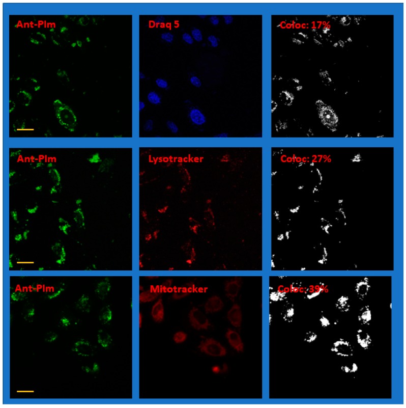
Figure 6.
Representative confocal fluorescence microscope images of CHO-K1 hamster ovary cells after co-staining Draq 5, Lysotracker red, or Mitotracker deep red with Ant-PIm. The left panels are imaging the photo-sensitizer channel and the center panels the co-stained channel. The images in the right panels show the colocalization with the parameter for the whole image given as an inset (%). N.b. The colored images have been modified for clarity by adding brightness. The colocalization data are, by definition, binary and coded black and white. The yellow scale bar is 20 μm. For more details, see the text.

