. 2020 Apr 14;9:e51977. doi: 10.7554/eLife.51977

Figure 7. Loss of Vps29 disrupts lysosomal ultrastructure in the brain.

(A) Transmission electron microscopy (TEM) reveals overall preserved photoreceptor morphology, but aberrant endolysosomal structures in retinae from 30-day-old animals lacking Vps29. Within each ommatidium (yellow outline), the percentage of photoreceptors (PR) with endolysosomal defects was quantified. Asterisks denote photoreceptors with aberrant endolysosomal structures. Quantification based on n = 30 ommatidia from three animals per genotype. See also Figure 7—figure supplement 1A,B. (B) At higher magnification, TEM reveals an increase in lysosomes, multivesicular bodies, autophagic vacuoles, and multilamellar bodies in Vps29 mutant retinae. Lysosomes were frequently observed to be aberrantly enlarged and filled with granular, electron-dense material. (C) Distinct endolysosomal structures and/or compartments were quantified in n = 15 photoreceptors, including five photoreceptors from three animals for each genotype. (D) Increased numbers of multi-lamellar bodies (asterisks) are observed in cortical neurons from brains 30-day-old animals lacking Vps29. Neuronal nuclei (‘N’) are outlined. Quantification based on cell counts from n = 4 animals (50 cells per brain). See also Figure 7—figure supplement 1D. Statistical analysis (A, C, D) based on Student’s t-test. All error bars denote SEM. *, p<0.05; **, p<0.01; ***, p<0.001.

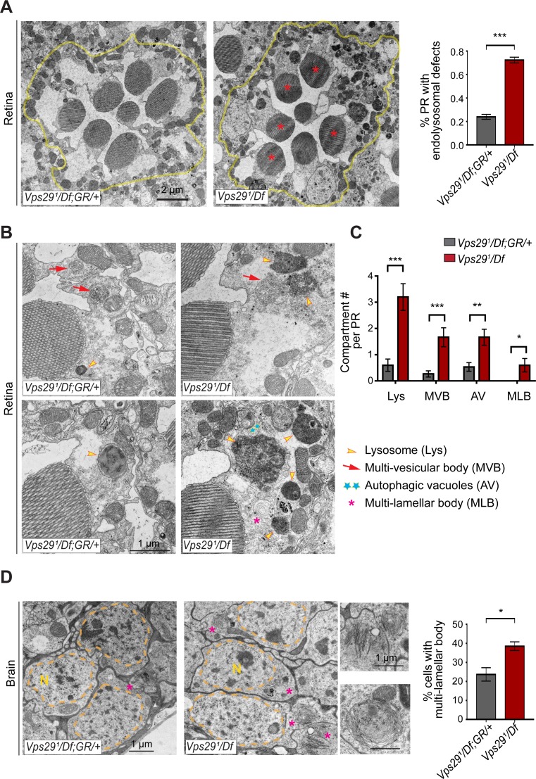
Figure 7.

Figure 7—figure supplement 1. Additional ultrastructural analysis of Vps29 mutants.

Figure 7—figure supplement 1.

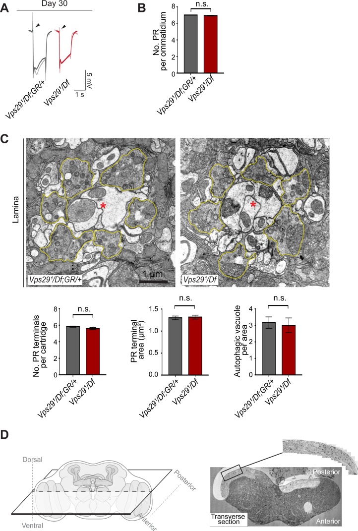
(A) Electroretinogram (ERG) traces of Vps291/Df and Vps291/Df; GR/+ prior to transmission electron microscopy (TEM). Flies were raised under ambient light conditions (~500 lux) for 30 days. Representative traces for n = 2 animals are superimposed for each genotype. (B) Photoreceptor (PR) numbers are preserved in 30-day-old Vps291/Df animals, based on counts from retinal TEM. Quantification based on examination of n = 30 ommatidia (three independent animals) for each genotype. (C) In 30-day-old Vps29 mutants, no overt ultrastructural defects are detected in the lamina, which contains PR presynaptic terminals (yellow outline). Dendritic processes (postsynaptic) are indicated with red asterisks. Quantification based on examination of n = 30 cartridges (three independent animals) for each genotype. No changes were noted in either the number or area of PR terminals or the number of autophagic vacuoles. (D) Left: schematic showing orientation for thin sectioning prior to TEM analysis of fly brain. Right: Representative transverse section of fly brain at low magnification. Boxed region of interest (magnified inset) highlights the dorsal-posterior cortical region with densely packed neuronal cell bodies. This region was imaged at higher magnification in Figure 7D. Statistical analysis (B, C) based on Student’s t-test. All error bars denote SEM. n.s., not significant.