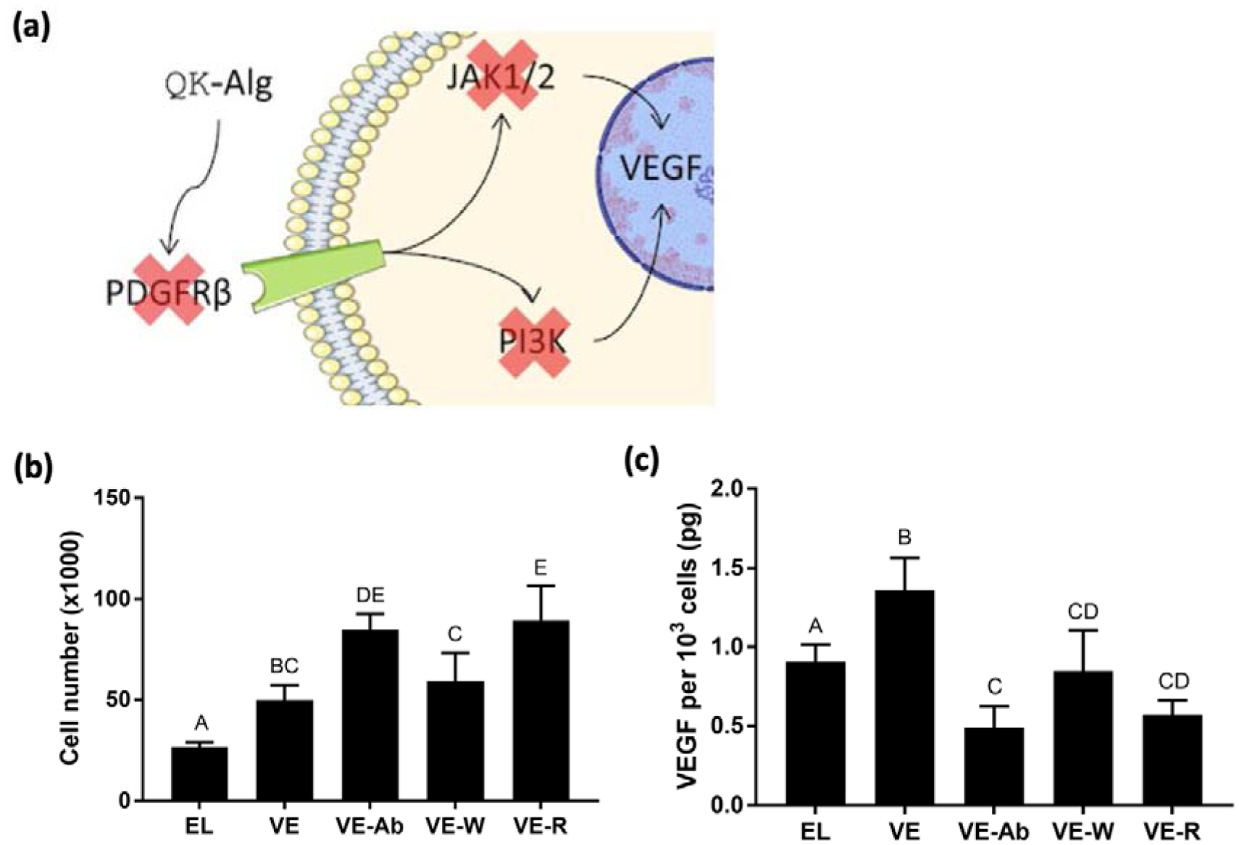
Figure 5: QK exerts its effects through the PDGF receptor.

(a) Schematic of mechanistic studies to interrogate QK signaling pathways. Anti-PDGFRβ (inhibits PDGFRβ binding), wortmannin (inhibits PI3K), and Ruxolitinib (inhibits JAK1/2) were applied to MSCs in 20 kPa ionic gels with 1:1 QK:RGD (VE-Ab, VE-W, and VE-R, respectively) for 7 days. Untreated elastic (EL) and viscoelastic (VE) gels with the same stiffness and peptide content served as controls. (b) Cell number within 1:1 QK:RGD gels (n=4). (c) Quantification of VEGF secretion by entrapped MSCs in 20 kPa viscoelastic gels with 1:1 QK:RGD in presence of inhibitors (n=4). Data points labeled with different letters are significantly different from one another at p<0.05.
