Abstract
Background
Heat shock protein 27 (HSP27) has been proposed as a vital protective factor in atherosclerosis. The objective of the present study was to evaluate the association between circulating HSP27 and carotid intima–media thickness (IMT) in individuals with type 2 diabetes and to determine whether HSP27 represents an independent marker of subclinical atherosclerosis in this patient population.
Methods
We performed a cross-sectional community-based study in 186 Chinese subjects with a median duration of type 2 diabetes of 8.2 years who underwent ultrasound carotid IMT measurement. Serum HSP27 levels were assessed by ELISA.
Results
Serum HSP27 levels were significantly higher in the IMT (+, > 1.0 mm) group than in the IMT (−, ≤1.0 mm) group, with the median values of 8.80 ng/mL (5.62–12.25) and 6.93 ng/mL (4.23–9.60), respectively (P = 0.006). The discriminative value of HSP27 to evaluate IMT was 7.16 ng/mL and the area under the curve was 0.72 (95%CI = 0.64–0.80, P = 0.0065). Spearman’s rank correlation analysis demonstrated that the concentrations of circulating HSP27 were positively associated with carotid IMT (r = 0.198, P = 0.007) and blood urea nitrogen (r = 0.170, P < 0.05). Furthermore, in the logistic model, serum HSP27 levels were found to be independent predictors for carotid IMT in type 2 diabetic patients after adjustment for onset age of diabetes, blood pressure, total cholesterol and C-reactive protein (OR = 1.085, P = 0.022).
Conclusions
Circulating HSP27, positively correlates with carotid IMT, is an independent predictor for early atherosclerotic changes in diabetes, and may represent a novel marker of subclinical atherosclerosis in type 2 diabetes.
Keywords: HSP27, Carotid intima–media thickness, Subclinical atherosclerosis, Type 2 diabetes
Background
Atherosclerotic cardiovascular disease (CVD) is the dominating cause of increasing mortality among patients with type 2 diabetes. Increasing carotid intima–media thickness (IMT), closely associated with CVD, is generally accepted as a surrogate marker of atherosclerosis [1–3].
Heat shock protein 27 (HSP27), also termed HSPB1 [4], is a ubiquitously expressed member of the small heat shock protein family [5]. Originally identified as an intracellular molecular chaperone, HSP27 facilitates the correct folding of proteins [6]. In recent years, the extracellular effects of HSP27 on the cardiovascular system have indicated a protective effect against atherosclerosis [7]. Clinical studies have reported that serum HSP27 levels were dramatically decreased in patients with carotid atherosclerosis compared with healthy controls [8]. Low circulating HSP27 levels were found to be associated with high risk of coronary artery disease [9]. Furthermore, reduced HSP27 levels were observed in unstable plaques versus stable plaques [10].
Although these studies have implicated certain roles of HSP27 in atherosclerosis or coronary heart disease (CHD), little is known about the direct relationship between serum HSP27 concentration and subclinical atherosclerosis in the context of type 2 diabetes. In the present study, we examined the association between circulating HSP27 levels and carotid IMT in patients with type 2 diabetes to determine whether HSP27 may represent a potential predictor for early-stage atherosclerosis in this patient population.
Methods
Participants and study design
This trial was designed as a cross-sectional study. Participants were recruited in Shanghai from February 2004 to July 2004. Twenty residential areas administered by 20 residents’ committees were sampled randomly in the central area of Shanghai. Questionnaires administered by endocrinologists and primary care clinicians were used to identify individuals with type 2 diabetes. Two hundred individuals were randomly selected, and 186 of them with complete information were enrolled in our study [11]. Based on carotid IMT values detected by color ultrasound, subjects were stratified into an IMT (−) group (n = 110) or an IMT (+) group (n = 76). The IMT (−) group included patients with carotid IMT levels ≤1.0 mm while the IMT (+) group contained patients with IMT values > 1.0 mm. All subjects provided written informed consent prior to participation. The research protocol was approved by the Institutional Review Board of Huashan Hospital, Fudan University School of Medicine.
Anthropometric parameters and biochemical indexes
Detailed histories of all the subjects were obtained using questionnaires. Physical examination and anthropometric measurements were performed by trained physicians. Waist circumference (WC) was measured at the midpoint between the lower margin of the least palpable rib and the top of the iliac crest in the late exhalation phase in a standing position. Hip circumference was measured around the widest portion of the buttocks using a tape measure parallel to the floor. Blood pressure measurement was obtained using a standard manual mercury sphygmomanometer at a steady state on the upper arm. Venous blood samples were collected between 7:00 and 8:00 AM from the antecubital vein of each subject after overnight fasting. Blood glucose, serum insulin, total cholesterol (TC), triglycerides (TG), high-density lipoprotein cholesterol (HDL-C), low-density lipoprotein cholesterol (LDL-C), blood urea nitrogen (BUN), and serum creatinine (Scr) were determined using standard methods in a qualified laboratory with a Hitachi 7080 analyzer (Hitachi, Ltd., Tokyo, Japan). C-reactive protein (CRP) was measured in duplicate by ELISA using a Duoset kit (DY1707, R&D Systems, Minneapolis, MN). Glycated hemoglobin A1c (HbA1c) was assessed by high-pressure liquid chromatography (HLC-723G7; Tosoh, Shanghai, China). The homeostasis model assessment of insulin resistance (HOMA-IR) was calculated based on the formula of Matthews et al. [12]. The MDRD Study equation was used for the calculation of estimated glomerular filtration rate (eGFR) [13].
Type 2 diabetes was defined as the presence of ≥1 of the following criteria: fasting plasma glucose ≥ 7.0 mmol/L; plasma glucose ≥11.1 mmol/L 2 h after a 75-g oral glucose load as in a glucose tolerance test (OGTT); and symptoms of high blood sugar and casual plasma glucose ≥11.1 mmol/L. CVD was defined as stroke and CHD, including unstable or stable angina and myocardial infarction, while lipid-lowering drugs referred to statins or fibrates.
Measurement of serum HSP27
Serum HSP27 levels were measured in duplicate using a commercial enzyme-linked immunosorbent assay kit (QIA119, Calbiochem, San Diego, CA) according to the manufacturer’s instructions. The intra- and inter-assay coefficients of variation of the ELISA were 6.2 and 8.3%, respectively.
Measurement of carotid IMT
With reference to the European Mannheim carotid IMT consensus, the intima–media thickness values of the common carotid arteries of the subjects were measured using an Acuson Sequoia 512 system (Siemens Medical Solutions USA, Mountain View, CA). The procedure was performed in subjects in a supine position by an experienced ultra-sonographer who was unaware of the subjects’ demographic and clinical characteristics. Three arterial sites were evaluated: the bilateral distal common carotid arteries, the carotid bulbs, and the proximal internal carotid arteries. Different scanning angles (anterior, lateral, posterior) were used to identify the thickest IMT in each wall. Both left and right carotid IMTs were assessed, and three measurements were performed for each subject. The mean values of the maximum IMT in both left and right sides of the common carotid arteries were defined as carotid artery IMT.
Statistical analysis
Quantitative data were evaluated using the Kolmogorov–Smirnov test to determine whether they followed a normal distribution. Parameters were considered normally distributed if P > 0.05. Normally distributed data were reported as means and standard deviations, while variables with a skewed distribution were expressed as medians (interquartile range). Categorical variables were presented as frequencies and percentages. One-way ANOVA and a Chi-squared test were used for comparisons between the two groups. Spearman’s correlation was used to evaluate the correlation between serum HSP27 concentration and other clinical indexes. Receiver operating characteristics (ROC) curve analysis was performed to calculate the area under the curve (AUC) and the cutoff value of HSP27 for IMT. The determinants of carotid IMT were explored using univariate and multivariate logistic analysis. Linear regression analysis was also used. Variables with statistically significant correlations (P < 0.05) in univariate analysis were examined in the multivariate model. All statistical analyses were conducted using the SPSS version 25 (IBM Corp., Armonk, NY) and Prism 8 software (GraphPad, San Diego, CA). Two-sided values of P < 0.05 were considered statistically significant.
Results
Subject characteristics
The characteristics of the 186 subjects and the two subgroups divided according to carotid IMT (1.0 mm) are shown in Table 1. The low median HbA1c (6.70%) and normal eGFR (121.15 ± 30.98 mL·min− 1 1.73 m− 2) values were indicative of generally well-controlled type 2 diabetes among the study subjects. The mean IMT levels in the IMT (−) and IMT (+) groups were 0.78 mm and 1.41 mm, respectively. Compared with the IMT (−) individuals, the IMT (+) group was 6 years older (P < 0.001) and had a higher onset age of diabetes (P < 0.01). The levels of systolic blood pressure (SBP) (P < 0.05), diastolic blood pressure (DBP) (P < 0.05), TC (P < 0.05), and CRP (P < 0.05) were significantly higher in the IMT (+) group than in the IMT(−) group. There were no statistical differences in sex, smoking status, alcohol consumption, use of lipid-lowering drugs, history of CVD, duration of type 2 diabetes, BMI, WC, waist–hip ratio (WHR), FBG, postprandial blood glucose (PBG), fasting insulin, fasting C peptide, 2 h insulin, 2 h C peptide, HOMA-IR, HbA1c, BUN, Scr, serum uric acid, eGFR, TG, LDL-C, HDL-C between the two groups (all P > 0.05, Table 1).
Table 1.
General characteristics of study subjects
| Characteristic | Total | IMT (−) | IMT (+) | P value |
|---|---|---|---|---|
| Subjects | 186 | 110 | 76 | – |
| Age (years) | 67 (57–73) | 64 (53.75–71) | 70 (64.25–75) | 0.000* |
| Sex (male/female) | 74/112 | 39/71 | 35/41 | 0.147 |
| Smoking (%) | 76.88 | 78.18 | 75.00 | 0.613 |
| Alcohol (%) | 8.60 | 9.09 | 7.89 | 0.078 |
| lipid-lowering drugs (%) | 50.98 | 60.91 | 51.32 | 0.194 |
| CVD (%) | 20.43 | 17.27 | 25.00 | 0.199 |
| Onset age of diabetes (years) | 57.12 ± 11.16 | 54.76 ± 11.29 | 60.47 ± 10.13 | 0.001* |
| Duration of diabetes (years) | 6 (3–10) | 6 (3–10) | 7 (4–12) | 0.370 |
| BMI (kg/m2) | 24.63 ± 3.21 | 24.46 ± 3.19 | 24.88 ± 3.23 | 0.392 |
| WC (cm) | 81.43 ± 8.76 | 81.10 ± 8.75 | 81.90 ± 8.81 | 0.544 |
| WHR | 0.87 ± 0.06 | 0.87 ± 0.07 | 0.88 ± 0.06 | 0.077 |
| SBP (mmHg) | 140 (130–150) | 140 (129.5–150) | 142 (135–160) | 0.001* |
| DBP (mmHg) | 84 (78–92) | 82 (76–90) | 86 (80–94.75) | 0.026* |
| FBG (mmol/L) | 7.80 (6.45–10.00) | 7.55 (6.3–9.65) | 8.60 (6.90–10.10) | 0.236 |
| PBG (mmol/L) | 15.73 ± 6.08 | 15.60 ± 6.16 | 15.93 ± 6.00 | 0.717 |
| Fasting insulin (pmol/L) | 12.20 (7.44–20.06) | 12.22 (7.35–18.81) | 12.20 (7.54–21.77) | 0.387 |
| 2 h insulin (pmol/L) | 46.49 (28.06–69.77) | 47.46 (29.43–70.23) | 43.99 (25.77–69.63) | 0.780 |
| Fasting C peptide (ng/mL) | 2.94 (2.22–3.78) | 2.96 (2.23–3.72) | 2.91 (2.15–3.86) | 0.622 |
| 2 h C peptide (ng/mL) | 8.88 (6.61–11.74) | 8.84 (7.07–10.92) | 8.88 (6.45–12.67) | 0.191 |
| HOMA-IR | 4.56 (2.39–7.99) | 4.11 (2.23–7.42) | 5.27 (2.72–9.22) | 0.359 |
| HbA1c (%) | 6.70 (6.00–7.65) | 6.70 (6.00–7.65) | 6.80 (6.03–7.75) | 0.730 |
| BUN (mmol/L) | 6.00 (5.00–7.15) | 5.85 (5.00–6.70) | 6.20 (5.20–7.70) | 0.224 |
| Scr (μmol/L) | 65 (54–78) | 62 (52–76) | 68 (58–80) | 0.441 |
| Serum uric acid (μmol/L) | 0.27 (0.24–0.33) | 0.28 (0.23–0.33) | 0.27 (0.24–0.33) | 0.887 |
| eGFR (mL·min−1 1.73 m−2) | 121.15 ± 30.98 | 124.15 ± 31.80 | 116.74 ± 29.41 | 0.111 |
| TC (mmol/L) | 5.26 ± 1.18 | 5.07 ± 0.99 | 5.54 ± 1.38 | 0.008* |
| TG (mmol/L) | 1.60 (1.07–2.16) | 1.50 (1.09–2.11) | 1.63 (1.01–2.28) | 0.482 |
| LDL-C (mmol/L) | 3.00 (2.40–3.40) | 2.90 (2.40–3.30) | 3.00 (2.40–3.60) | 0.094 |
| HDL-C (mmol/L) | 1.30 (1.05–1.50) | 1.20 (1.00–1.50) | 1.30 (1.10–1.60) | 0.266 |
| UACR (μg/mg) | 14.42 (6.04–31.06) | 11.29 (4.59–24.94) | 20.17 (7.38–46.28) | 0.651 |
| CRP (mg/L) | 3.05 (1.65–5.29) | 6.92 (4.23–9.60) | 8.80 (5.62–12.25) | 0.019* |
| IMT (mm) | 1.03 ± 0.42 | 0.78 ± 0.19 | 1.41 ± 0.38 | 0.000* |
| HSP27 (ng/mL) | 7.85 (4.78–10.92) | 6.93 (4.23–9.60) | 8.80 (5.62–12.25) | 0.007* |
P < 0.05 (*)
Difference in serum HSP27 between the two groups
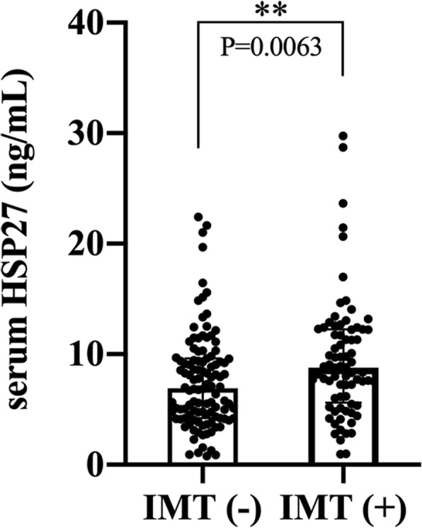
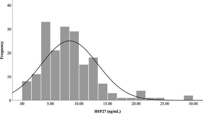
The median value of serum HSP27 in the study population was 7.85 ng/mL (IQR: 4.78–10.92, Fig. 1). As shown in Fig. 2, the median serum HSP27 level in the IMT (+) group was 8.80 ng/mL (IQR: 5.62–12.25, Table 1), significantly higher than that in the IMT (−) group (6.93 ng/mL, IQR: 4.23–9.60, P = 0.006).
Fig. 1.
Distribution of serum HSP27 levels in the participants
Fig. 2.

Serum HSP27 in the IMT (−) and IMT (+) subjects (divided according to the values of IMT 1.0 mm). Data are expressed as median (IQR), P<0.01(**)
Association between serum HSP27 level and carotid IMT
Logistic regression analysis was used to identify independent determinants of carotid IMT. Seven factors were identified as significant predictors using univariate analysis, including age, onset age of diabetes, SBP, DBP, TC, CRP, and HSP27 (all P < 0.05, Table 2). In the multivariate model, these factors were defined as covariates, while carotid IMT (≤1.0 mm or > 1.0 mm) was introduced as a dependent variable. After adjusting for onset age of diabetes, SBP, DBP, TC and CRP, the results illustrated that age and HSP27 were still independently associated with carotid IMT in type 2 diabetes, with adjusted odds ratios of 1.061 (P = 0.028) and 1.085 (P = 0.022), respectively (Table 3). Moreover, a positive relationship between HSP27 and carotid IMT (β = 0.019, P = 0.002) was also observed in the multiple linear regression model (Supplemental Table 1 and Table 2). Age (β = 0.190, P = 0.007) and TC (β = 0.081, P = 0.001) independently correlated with carotid IMT in multiple linear regression analysis.
Table 2.
Univariate logistic analysis: predictors for carotid artery IMT
| B | OR | 95%CI | P value | ||
|---|---|---|---|---|---|
| Lower | Upper | ||||
| Sex | −0.441 | 0.643 | 0.354 | 1.169 | 0.148 |
| Age | 0.067 | 1.069 | 1.035 | 1.105 | 0.000* |
| Onset age | 0.050 | 1.053 | 1.021 | 1.083 | 0.001* |
| Duration of diabetes | 0.019 | 1.019 | 0.978 | 1.062 | 0.370 |
| Smoking | −0.178 | 0.837 | 0.420 | 1.667 | 0.613 |
| Alcohol | 0.154 | 1.167 | 0.405 | 3.358 | 0.775 |
| Lipid-lowering drugs | 0.391 | 1.478 | 0.819 | 2.669 | 0.195 |
| CVD | −0.468 | 0.626 | 0.306 | 1.283 | 0.201 |
| BMI | 0.040 | 1.041 | 0.950 | 1.141 | 0.390 |
| WC | 0.010 | 1.011 | 0.977 | 1.045 | 0.542 |
| WHR | 4.345 | 77.115 | 0.605 | 9837.212 | 0.079 |
| SBP | 0.029 | 1.029 | 1.011 | 1.048 | 0.002* |
| DBP | 0.032 | 1.033 | 1.003 | 1.063 | 0.028* |
| FBG | 0.055 | 1.056 | 0.964 | 1.157 | 0.239 |
| PBG | 0.009 | 1.009 | 0.961 | 1.059 | 0.716 |
| HbA1c | 0.033 | 1.034 | 0.857 | 1.246 | 0.729 |
| Fasting insulin | 0.005 | 1.005 | 0.994 | 1.016 | 0.403 |
| 2 h insulin | 0.001 | 1.001 | 0.994 | 1.007 | 0.779 |
| fasting C peptide | 0.056 | 1.058 | 0.847 | 1.322 | 0.620 |
| 2 h C peptide | 0.026 | 1.027 | 0.986 | 1.069 | 0.207 |
| HOMA-IR | 0.014 | 1.014 | 0.984 | 1.044 | 0.368 |
| BUN | 0.092 | 1.097 | 0.944 | 1.275 | 0.227 |
| Scr | 0.004 | 1.004 | 0.993 | 1.016 | 0.448 |
| Serum uric acid | 0.310 | 1.364 | 0.020 | 94.590 | 0.886 |
| eGFR | −0.008 | 0.992 | 0.982 | 1.002 | 0.113 |
| TC | 0.349 | 1.417 | 1.081 | 1.858 | 0.012* |
| TG | − 0.076 | 0.927 | 0.750 | 1.146 | 0.485 |
| LDL-C | 0.306 | 1.358 | 0.944 | 1.955 | 0.099 |
| HDL-C | 0.409 | 1.505 | 0.733 | 3.089 | 0.265 |
| CRP | 0.099 | 1.105 | 1.011 | 1.207 | 0.028* |
| UACR | 0.000 | 1.000 | 0.998 | 1.001 | 0.655 |
| HSP27 | 0.084 | 1.088 | 1.021 | 1.159 | 0.009* |
P < 0.05 (*)
Table 3.
Multiple logistic regression analysis: independent predictors for carotid IMT in type 2 diabetes
| B | OR | 95%CI | P value | ||
|---|---|---|---|---|---|
| Lower | Upper | ||||
| Age | 0.059 | 1.061 | 1.006 | 1.119 | 0.028* |
| Onset age | 0.005 | 1.005 | 0.958 | 1.053 | 0.851 |
| SBP | 0.004 | 1.004 | 0.977 | 1.031 | 0.795 |
| DBP | 0.036 | 1.037 | 0.994 | 1.081 | 0.091 |
| TC | 0.260 | 1.297 | 0.964 | 1.744 | 0.086 |
| CRP | 0.051 | 1.052 | 0.963 | 1.149 | 0.259 |
| HSP27 | 0.081 | 1.085 | 1.012 | 1.163 | 0.022* |
ROC curve analysis
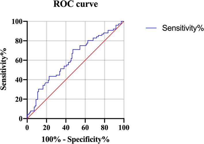
ROC curve analysis was applied to identify an optimal cutoff value for HSP27 to discriminate between > 1 and < 1 IMT (Fig. 3). The AUC of the ROC curve for predicting IMT was 0.72 (95%CI = 0.64–0.80, P = 0.0065) and the optimal cutoff value of HSP27 was 7.16 ng/mL with sensitivity of 75.05% (95% CI =64.04–84.04) and low specificity of 67.73% (95% CI =58.46–76.81).
Fig. 3.
The receiver operating characteristic (ROC) curve of HSP27 in patients with type 2 diabetes with > 1 and < 1 carotid IMT. The area under the curve was 0.72 (95%CI = 0.64–0.80, P = 0.0065)
Correlations between serum HSP27 and other clinical parameters
As presented in Table 4, serum HSP27 levels of the subjects correlated positively with BUN (r = 0.170, P < 0.05) and carotid IMT (r = 0.198, P = 0.007), whereas the relationships between HSP27 and other clinical parameters were not significant.
Table 4.
Correlations between serum HSP27 and other parameters in type 2 diabetes
| variable | HSP27 | |
|---|---|---|
| r | P value | |
| Age | 0.074 | 0.313 |
| Onset age | 0.091 | 0.217 |
| Duration of diabetes | 0.000 | 0.999 |
| BMI | 0.084 | 0.253 |
| WC | −0.005 | 0.949 |
| WHR | 0.023 | 0.753 |
| SBP | 0.041 | 0.578 |
| DBP | 0.018 | 0.811 |
| FBG | 0.08 | 0.281 |
| PBG | 0.005 | 0.945 |
| HbA1c | 0.097 | 0.186 |
| Fasting insulin | −0.142 | 0.054 |
| 2 h insulin | − 0.065 | 0.383 |
| Fasting C peptide | 0.033 | 0.659 |
| 2 h C peptide | 0.072 | 0.332 |
| HOMA-IR | −0.083 | 0.262 |
| BUN | 0.170 | 0.020* |
| Scr | 0.06 | 0.414 |
| Serum uric acid | −0.075 | 0.312 |
| eGFR | −0.018 | 0.812 |
| TC | 0.053 | 0.474 |
| TG | 0.035 | 0.637 |
| LDL-C | 0.092 | 0.215 |
| HDL-C | 0.070 | 0.341 |
| CRP | 0.048 | 0.519 |
| UACR | 0.098 | 0.182 |
| carotid IMT | 0.198 | 0.007* |
r: Spearman’s correlation coefficient. P < 0.05 (*)
Discussion
The major findings of this study were that the median HSP27 level in the IMT (+) group was significantly higher than that in the IMT (−) group, and serum HSP27 level positively correlated with carotid IMT (r = 0.198, P = 0.007). The lack of circulating biomarkers of early-stage atherosclerosis in diabetes requires further clinical investigation. We identified that HSP27 was an independent predictor for subclinical atherosclerosis in patients with type 2 diabetes, even after adjusting for several clinical factors. Therefore, our findings may support the diagnostic value of elevated circulating HSP27. Furthermore, we also found that serum HSP27 concentrations were positively associated with BUN but not with other clinical parameters, which is in alignment with a previous study that showed a correlation between HSP27 and serum creatinine level [14].
HSP27 acts as an antioxidant with the ability to reduce the levels of reactive oxygen species (ROS) through increased intracellular glutathione and decreased intracellular iron [15], which indicates a potential atheroprotective role of HSP27 in atherosclerosis. Another mechanism by which HSP27 exerts its protective function may be attributed to its binding to scavenger receptor-A (SR-A), thus leading to the prevention of acetylated low-density lipoprotein (acLDL) uptake and attenuation of foam cell formation [16]. HSP27 may also reduce the cholesterol content in plaques by more than 30% [17]. In apolipoprotein E null (ApoE−/−) mice, which are prone to atherosclerosis, extracellular HSP27 was reported to activate the NF-κB signaling pathway to induce increased expression of granulocyte-monocyte colony-stimulating factor (GM-CSF), ATP-binding cassette transporter A1 (ABCA1), and ATP-binding cassette transporter G1 (ABCG1), thus facilitating cholesterol efflux [17]. In addition, overexpression of HSP27 in this mouse model contributed to a reduction in lesion formation and plaque stability [9, 16, 18]. Stimulation of the release of anti-inflammatory IL-10 via the p38 signaling pathway may also contribute to this effect [19].
An immediate response to stress, inflammation, and cellular damage is to secrete HSP27 into the blood to protect the body [20]. Given the protective role of HSP27 in protecting vessels from oxidative stress [21] and inhibiting inflammation [22], the higher levels of serum HSP27 observed in the IMT (+) group compared with the IMT (−) group in our study may represent a consequence of a compensatory response to inflammation and oxidative stress in the early stage of atherosclerosis. HSP27 may have been induced to counteract these unfavorable factors in the initiation of atherosclerosis. Similarly, Park et al. reported that circulating HSP27 levels in acute coronary syndrome (ACS) patients were remarkably higher than those in an age- and sex-matched healthy controls [23]. Compared with controls, a significant increase in serum HSP27 in patients with CHD was also observed in a recent study [24].
Contrary to these results, circulating HSP27 levels were reported as decreased > 70% in patients with carotid stenosis [8] and coronary artery diseases (CAD) compared with healthy subjects [25]. In addition to reduced concentrations of HSP27, patients with CAD in these two studies had more severe comorbidities compared with patients having higher HSP27 levels in the studies of Park et al. and Zhang et al. [23, 24], including diabetes and hypertension in both studies and smoking in the former plus hyperlipemia in the latter. Patients in these two studies even had serum HSP27 levels decreased to mean values of 0.19 ng/mL and 1.23 ng/mL, respectively. Therefore, we speculated that the condition of the patients in the two studies was too severe to allow sufficient HSP27 secretion to compensate for inflammation and oxidative stress associated with the reported comorbidities. In addition, the degradation of extracellular HSP27 by proteases such as upregulated plasmin in plaques [26] and matrix metalloproteases (MMPs) [27, 28] may also have accounted for the decline in serum HSP27.
Our study had several limitations. First, this cross-sectional study was confined to a specific time point and was therefore unable to identify cause-effect relationships. Given the lack of follow-up data, whether HSP27 was an independent marker of future cardiovascular events remained to be explored. Second, our study included a small number of subjects. Finally, although we have identified a relationship between circulating HSP27 and carotid IMT in type 2 diabetes, the underlying mechanisms of the direct role of HSP27 remains unclear.
Conclusions
Our study findings demonstrate a positive correlation between circulating HSP27 and carotid IMT, indicating that serum HSP27 may represent a novel biomarker of the progression and diagnosis of subclinical atherosclerosis in type 2 diabetes.
Supplementary information
Additional file 1: Table S1. Univariate linear analysis for variables associated with carotid IMT. Table S2. Multiple stepwise regression analysis showing independent predictors for carotid IMT in type 2 diabetes.
Acknowledgements
We would like to thank all of the study participants of Department of Endocrinology of Huashan Hospital and Xinhua Hospital.
Abbreviations
- IMT
Intima–media thickness
- CVD
Cardiovascular disease
- BMI
Body mass index
- WC
Waist circumference
- WHR
Waist–hip ratio
- SBP
Systolic blood pressure
- DBP
Diastolic blood pressure
- FBG
Fasting blood glucose
- PBG
Postprandial blood glucose
- HOMA-IR
Homeostatic model assessment of insulin resistance
- HbA1c
Glycated hemoglobin A1c
- BUN
Blood urea nitrogen
- Scr
Serum creatinine
- eGFR
Estimated glomerular filtration rate
- TC
Total cholesterol
- TG
Triglycerides
- LDL-C
Low density lipoprotein-cholesterol
- HDL-C
High density lipoprotein-cholesterol
- CRP
C-reactive protein
- UACR
Urine albumin-to-creatinine ratio
- OR
Odds ratio
- CI
Confidence interval
Authors’ contributions
XRW, ZY, XCW proposed the idea and designed the study; BL, RH, JS collected and organized the database; XRW, JS, WZ, JW and YY contributed to the statistical analysis and the interpretation of the data; ZY, XCW, WZ, JW and YY supervised the project administration; XRW wrote the first draft of the paper; ZY, XCW revised the article. The author(s) read and approved the final manuscript.
Funding
The National Natural Science Foundation of China (grant numbers 81370953, 81370936 and 81873645) supported the design of the study and data collection.
The Science and Technology Commission of Shanghai Municipality (grant numbers 16140901200, 18140902100 and 16PJ1401700) supported data analysis and manuscript writing.
Availability of data and materials
Datasets are available from the corresponding author upon reasonable request.
Ethics approval and consent to participate
The research protocol was approved by the Institutional Review Board of Huashan Hospital, Fudan University School of Medicine. All subjects provided written informed consent prior to participation.
Consent for publication
Not applicable.
Competing interests
The authors declare that they have no competing interests.
Footnotes
Publisher’s Note
Springer Nature remains neutral with regard to jurisdictional claims in published maps and institutional affiliations.
Contributor Information
Zhen Yang, Email: yangzhen@xinhuamed.com.cn.
Xuanchun Wang, Email: xuanchunwangfd@126.com.
Supplementary information
Supplementary information accompanies this paper at 10.1186/s12872-020-01456-7.
References
- 1.Cardoso CRL, Salles GC, Leite NC, Salles GF. Prognostic impact of carotid intima-media thickness and carotid plaques on the development of micro- and macrovascular complications in individuals with type 2 diabetes: the Rio de Janeiro type 2 diabetes cohort study. Cardiovasc Diabetol. 2019;18(1):2. doi: 10.1186/s12933-019-0809-1. [DOI] [PMC free article] [PubMed] [Google Scholar]
- 2.Centurion OA. Carotid intima-media thickness as a cardiovascular risk factor and imaging pathway of atherosclerosis. Crit Pathw Cardiol. 2016;15(4):152–160. doi: 10.1097/HPC.0000000000000087. [DOI] [PubMed] [Google Scholar]
- 3.Stein JH, Korcarz CE, Hurst RT, Lonn E, Kendall CB, Mohler ER, Najjar SS, Rembold CM, Post WS. Use of carotid ultrasound to identify subclinical vascular disease and evaluate cardiovascular disease risk: a consensus statement from the American Society of Echocardiography carotid intima-media thickness task force. Endorsed by the Society for Vascular Medicine. J Am Soc Echocardiogr. 2008;21(2):93–111. doi: 10.1016/j.echo.2007.11.011. [DOI] [PubMed] [Google Scholar]
- 4.Van Montfort R, Slingsby C, Vierling E. Structure and function of the small heat shock protein/alpha-crystallin family of molecular chaperones. Adv Protein Chem. 2001;59:105–156. doi: 10.1016/S0065-3233(01)59004-X. [DOI] [PubMed] [Google Scholar]
- 5.Beck FX, Neuhofer W, Muller E. Molecular chaperones in the kidney: distribution, putative roles, and regulation. Am J Physiol Renal Physiol. 2000;279(2):F203–F215. doi: 10.1152/ajprenal.2000.279.2.F203. [DOI] [PubMed] [Google Scholar]
- 6.Ranford JC, Henderson B. Chaperonins in disease: mechanisms, models, and treatments. Mol Pathol. 2002;55(4):209–213. doi: 10.1136/mp.55.4.209. [DOI] [PMC free article] [PubMed] [Google Scholar]
- 7.Batulan Z, Pulakazhi Venu VK, Li Y, Koumbadinga G, Alvarez-Olmedo DG, Shi C, O'Brien ER. Extracellular release and signaling by heat shock protein 27: role in modifying vascular inflammation. Front Immunol. 2016;7:285. doi: 10.3389/fimmu.2016.00285. [DOI] [PMC free article] [PubMed] [Google Scholar]
- 8.Martin-Ventura JL, Duran MC, Blanco-Colio LM, Meilhac O, Leclercq A, Michel JB, Jensen ON, Hernandez-Merida S, Tunon J, Vivanco F, et al. Identification by a differential proteomic approach of heat shock protein 27 as a potential marker of atherosclerosis. Circulation. 2004;110(15):2216–2219. doi: 10.1161/01.CIR.0000136814.87170.B1. [DOI] [PubMed] [Google Scholar]
- 9.Seibert TA, Hibbert B, Chen YX, Rayner K, Simard T, Hu T, Cuerrier CM, Zhao X, de Belleroche J, Chow BJ, et al. Serum heat shock protein 27 levels represent a potential therapeutic target for atherosclerosis: observations from a human cohort and treatment of female mice. J Am Coll Cardiol. 2013;62(16):1446–1454. doi: 10.1016/j.jacc.2013.05.041. [DOI] [PubMed] [Google Scholar]
- 10.Lepedda AJ, Cigliano A, Cherchi GM, Spirito R, Maggioni M, Carta F, Turrini F, Edelstein C, Scanu AM, Formato M. A proteomic approach to differentiate histologically classified stable and unstable plaques from human carotid arteries. Atherosclerosis. 2009;203(1):112–118. doi: 10.1016/j.atherosclerosis.2008.07.001. [DOI] [PMC free article] [PubMed] [Google Scholar]
- 11.Lu B, Yang Y, Yang Z, Feng X, Wang X, Zhang Z, Hu R. Insulin resistance in Chinese patients with type 2 diabetes is associated with C-reactive protein independent of abdominal obesity. Cardiovasc Diabetol. 2010;9:92. doi: 10.1186/1475-2840-9-92. [DOI] [PMC free article] [PubMed] [Google Scholar]
- 12.Matthews DR, Hosker JP, Rudenski AS, Naylor BA, Treacher DF, Turner RC. Homeostasis model assessment: insulin resistance and beta-cell function from fasting plasma glucose and insulin concentrations in man. Diabetologia. 1985;28(7):412–419. doi: 10.1007/BF00280883. [DOI] [PubMed] [Google Scholar]
- 13.National Kidney Foundation. K/DOQI clinical practice guidelines for chronic kidney disease: evaluation, classification, and stratification. Am J Kidney Dis 2002, 39(2 Suppl 1):S1–266. [PubMed]
- 14.Jakhotia S, Sivaprasad M, Shalini T, Reddy PY, Viswanath K, Jakhotia K, Sahay R, Sahay M, Reddy GB. Circulating levels of Hsp27 in microvascular complications of diabetes: prospects as a biomarker of diabetic nephropathy. J Diabetes Complicat. 2018;32(2):221–225. doi: 10.1016/j.jdiacomp.2017.10.004. [DOI] [PubMed] [Google Scholar]
- 15.Arrigo AP, Virot S, Chaufour S, Firdaus W, Kretz-Remy C, Diaz-Latoud C. Hsp27 consolidates intracellular redox homeostasis by upholding glutathione in its reduced form and by decreasing iron intracellular levels. Antioxid Redox Signal. 2005;7(3–4):414–422. doi: 10.1089/ars.2005.7.414. [DOI] [PubMed] [Google Scholar]
- 16.Rayner K, Chen YX, McNulty M, Simard T, Zhao X, Wells DJ, de Belleroche J, O'Brien ER. Extracellular release of the atheroprotective heat shock protein 27 is mediated by estrogen and competitively inhibits acLDL binding to scavenger receptor-a. Circ Res. 2008;103(2):133–141. doi: 10.1161/CIRCRESAHA.108.172155. [DOI] [PubMed] [Google Scholar]
- 17.Pulakazhi Venu VK, Adijiang A, Seibert T, Chen YX, Shi C, Batulan Z, O'Brien ER. Heat shock protein 27-derived atheroprotection involves reverse cholesterol transport that is dependent on GM-CSF to maintain ABCA1 and ABCG1 expression in ApoE(−/−) mice. FASEB J. 2017;31(6):2364–2379. doi: 10.1096/fj.201601188R. [DOI] [PubMed] [Google Scholar]
- 18.Cuerrier CM, Chen YX, Tremblay D, Rayner K, McNulty M, Zhao X, Kennedy CR, de BelleRoche J, Pelling AE, O'Brien ER. Chronic over-expression of heat shock protein 27 attenuates atherogenesis and enhances plaque remodeling: a combined histological and mechanical assessment of aortic lesions. PLoS One. 2013;8(2):e55867. doi: 10.1371/journal.pone.0055867. [DOI] [PMC free article] [PubMed] [Google Scholar]
- 19.De AK, Kodys KM, Yeh BS, Miller-Graziano C. Exaggerated human monocyte IL-10 concomitant to minimal TNF-alpha induction by heat-shock protein 27 (Hsp27) suggests Hsp27 is primarily an antiinflammatory stimulus. J Immunol. 2000;165(7):3951–3958. doi: 10.4049/jimmunol.165.7.3951. [DOI] [PubMed] [Google Scholar]
- 20.Osterloh A, Breloer M. Heat shock proteins: linking danger and pathogen recognition. Med Microbiol Immunol. 2008;197(1):1–8. doi: 10.1007/s00430-007-0055-0. [DOI] [PubMed] [Google Scholar]
- 21.Najemnikova E, Rodgers CD, Locke M. Altered heat stress response following streptozotocin-induced diabetes. Cell Stress Chaperones. 2007;12(4):342–352. doi: 10.1379/CSC-292.1. [DOI] [PMC free article] [PubMed] [Google Scholar]
- 22.Shi C, Ulke-Lemee A, Deng J, Batulan Z, O'Brien ER. Characterization of heat shock protein 27 in extracellular vesicles: a potential anti-inflammatory therapy. FASEB J. 2019;33(2):1617–1630. doi: 10.1096/fj.201800987R. [DOI] [PubMed] [Google Scholar]
- 23.Park HK, Park EC, Bae SW, Park MY, Kim SW, Yoo HS, Tudev M, Ko YH, Choi YH, Kim S, et al. Expression of heat shock protein 27 in human atherosclerotic plaques and increased plasma level of heat shock protein 27 in patients with acute coronary syndrome. Circulation. 2006;114(9):886–893. doi: 10.1161/CIRCULATIONAHA.105.541219. [DOI] [PubMed] [Google Scholar]
- 24.Zhang HL, Jia KY, Sun D, Yang M. Protective effect of HSP27 in atherosclerosis and coronary heart disease by inhibiting reactive oxygen species. J Cell Biochem. 2019;120(3):2859–2868. doi: 10.1002/jcb.26575. [DOI] [PubMed] [Google Scholar]
- 25.Jin C, Phillips VL, Williams MJ, van Rij AM, Jones GT. Plasma heat shock protein 27 is associated with coronary artery disease, abdominal aortic aneurysm and peripheral artery disease. Springerplus. 2014;3:635. doi: 10.1186/2193-1801-3-635. [DOI] [PMC free article] [PubMed] [Google Scholar]
- 26.Martin-Ventura JL, Nicolas V, Houard X, Blanco-Colio LM, Leclercq A, Egido J, Vranckx R, Michel JB, Meilhac O. Biological significance of decreased HSP27 in human atherosclerosis. Arterioscler Thromb Vasc Biol. 2006;26(6):1337–1343. doi: 10.1161/01.ATV.0000220108.97208.67. [DOI] [PubMed] [Google Scholar]
- 27.Lamfers ML, Grimbergen JM, Aalders MC, Havenga MJ, de Vries MR, Huisman LG, van Hinsbergh VW, Quax PH. Gene transfer of the urokinase-type plasminogen activator receptor-targeted matrix metalloproteinase inhibitor TIMP-1.ATF suppresses neointima formation more efficiently than tissue inhibitor of metalloproteinase-1. Circ Res. 2002;91(10):945–952. doi: 10.1161/01.RES.0000041418.51906.57. [DOI] [PubMed] [Google Scholar]
- 28.Choi SH, Lee HJ, Jin YB, Jang J, Kang GY, Lee M, Kim CH, Kim J, Yoon SS, Lee YS, et al. MMP9 processing of HSPB1 regulates tumor progression. PLoS One. 2014;9(1):e85509. doi: 10.1371/journal.pone.0085509. [DOI] [PMC free article] [PubMed] [Google Scholar]
Associated Data
This section collects any data citations, data availability statements, or supplementary materials included in this article.
Supplementary Materials
Additional file 1: Table S1. Univariate linear analysis for variables associated with carotid IMT. Table S2. Multiple stepwise regression analysis showing independent predictors for carotid IMT in type 2 diabetes.
Data Availability Statement
Datasets are available from the corresponding author upon reasonable request.


