Abstract
目的
研究数字化导板应用于前牙美学区种植修复的精确度。
方法
选择50例需接受上前牙种植修复的患者为研究对象,随机分为2组,每组25例,分别实施常规种植修复(对照组,45枚种植体)和数字化导板辅助种植修复(试验组,51枚种植体),术后测量2组植入体术前虚拟设计位置与实际植入位置的颈部距离、根尖部距离、深度和角度偏差。在全瓷冠修复完成后1周(基线)、6个月和1年,观察2组患者术后种植体的临床修复疗效,采用红白美学评分[包括红美学分数(PES)和白美学分数(WES)]评价软组织及牙冠修复的美学效果。
结果
50例患者的96颗种植体术后均取得了良好的骨结合。试验组种植体植入位置的各项偏差均小于对照组(P<0.05);修复完成后1周、6个月和1年,试验组的PES及WES均高于对照组(P<0.05)。
结论
数字化导板辅助种植修复技术可以提高种植体三维位置的准确性和前牙美学区的修复效果。
Keywords: 数字化导板, 前牙, 种植修复, 精确度, 红美学分数, 白美学分数
Abstract
Objective
To study the precision of digital guide plates applied to the implant surgery of anterior teeth.
Methods
Fifty patients scheduled to receive implant restoration treatment in anterior teeth were enrolled in this study and divided into two groups (n=25, each group): those who were given routine implant restoration treatment (control group, 45 implants) and those who received implant restoration treatment using a digital guide plate (test group, 51 implants). After implantation, planned and placed implants were superimposed using digital software, and deviations (corona, apex, depth, degree) were analyzed. Esthetic parameters were assessed at 1 week (baseline), 6 month, and 1 year post final restoration. Pink esthetic (PES) and white esthetic (WES) scores were respectively used to evaluate the soft tissue and restoration esthetic outcome.
Results
The deviation parameters in the test group were significantly lower than those in the control group (P<0.05). PES and WES values recorded for the control group at 1 week, 6 month, and 1 year post final restoration were significantly lower than those in the test group (P<0.05).
Conclusion
The digital guide plate can improve the accuracy of the three-dimensional position of implants in the maxillary esthetic zone. As such, this device may play an important role in obtaining the ideal aesthetic effects of maxillary anterior teeth.
Keywords: digital guide plate, anterior teeth, implant restoration, precision, pink esthetic score, white esthetic score
前牙美学区种植是种植中的难点。一方面,前牙区种植美学风险高[1];另一方面,前牙区种植三维位置要求高[2]。常规种植修复由于单凭医生的经验,可操作性和重复性差,降低了植入的精确度,术后效果难以保证[3]。近年来,随着计算机辅助设计/计算机辅助制造(computer-aided design/computer-assisted manufacture,CAD/CAM)技术的兴起,数字化种植导板开始在口腔种植领域发挥巨大作用。研究[4]提示,数字化种植导板能够协助精确定位种植体的植入位置。种植体植入的位置和角度对美学效果有显著的影响[5]。红白美学评分[包括红美学分数(pink esthetic score,PES)和白美学分数(white esthetic score,WES)]是2005年提出的用于评价短期种植体周围软组织及牙冠修复的美学效果的指标[6]–[7]。为了探讨数字化种植导板应用于前牙美学区种植的精确度及其对最终修复的美学效果的影响,本研究对上前牙种植患者的植入体术前设计位置与术后位置进行对比分析,并应用PES和WES对修复后短期的软组织及修复体的美学效果进行评价。
1. 材料和方法
1.1. 临床资料
选择2015年1月—2018年1月在西南医科大学附属口腔医院种植科就诊的需行上前牙种植修复的50例患者为研究对象。病例的纳入标准:1)要求行上前牙区种植治疗;2)全身情况良好,无种植手术禁忌证;3)对治疗相关情况知情且术前签署手术知情同意书。
采用随机数表法将患者随机分为试验组和对照组,每组25例。试验组中,男13例,女12例,年龄20~52岁,平均年龄(34.0±9.1)岁,共植入51枚种植体;对照组中,男14例,女11例,年龄20~58岁,平均年龄(37.2±11.2)岁,共植入45枚种植体。2组患者的年龄及性别无统计学差异(P>0.05)。
1.2. 术前准备
术前对患者进行全面的口腔检查,包括缺牙数目、缺牙区位置、 龈距、
龈距、 间距、黏膜情况、邻牙及对颌牙情况等,并对患者进行常规洁治术和血常规、凝血功能及传染病筛查等检查。
1.3. 一期种植术
试验组实施数字化导板辅助种植修复。1)数字化导板设计与制作:用锥形束计算机断层扫描(cone-beam computed tomography,CBCT)获得相关数据,结合硅橡胶印模,在计算机上建立一个三维立体模型,导入种植设计软件(3shape公司,丹麦)中确定种植修复方案,根据患者的颌骨、黏膜等具体条件确定种植体合适的三维位置,进行种植体的虚拟放置,以确立导板设计方案。然后再用3D打印技术,打印出数字化导板。2)种植体植入:种植手术开始前先试戴导板,确保导板无误后再行手术。常规消毒铺巾,局部浸润麻醉,沿缺牙区牙槽嵴顶偏腭侧切开黏骨膜,翻瓣,修整牙槽嵴顶后将种植导板戴入,确定导板完全就位后,定点定位,逐级备洞至需要的深度和直径,去除导板,植入Anthogyr Axiom BL REG种植体(Anthogyr公司,法国),根据术前术中评估,同期行引导性骨组织再生(guided bone regeneration,GBR)术,植入骨粉和/或覆盖骨膜,植骨材料选择无机牛骨(Bio-Oss公司,瑞士),屏障骨膜选择胶原生物膜和/或钛膜。减张后对位严密缝合。
对照组实施常规种植修复。术者根据术前CBCT检查及自身经验等确定种植体位置,常规备洞、冲洗等处理后植入Anthogyr种植体,再根据术前术中评估,同期行GBR。
1.4. 种植体植入位置数据采集
种植体植入术后拍摄CBCT,将2组术前及术后数据均导入上述种植设计软件中进行配准(图1)。采用盲法的方式测量并记录植入体的术前虚拟设计位置与实际植入位置的颈部距离、根尖部距离、深度和角度偏差。测量点取种植体横截面的中心点,定点后直接测量两点间的三维距离,深度偏差值取根尖二者垂直距离,角度偏差取2个种植体长轴所形成的角度(图2)。每个数值由同一医生重复测量3次,取平均值。
图 1. 数字化导板辅助下种植体实际植入(左)和虚拟设计(右)位置配准示意图.
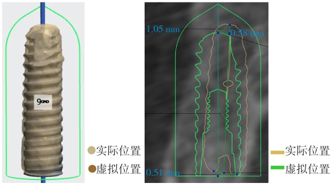
Fig 1 Schematic registration maps of actual (left) and virtual (right) implant positions with the digital guide plate
图 2. 种植体实际和虚拟植入位置偏差测量项目.
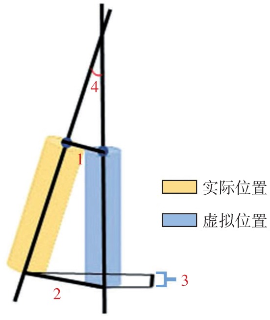
Fig 2 Parameters of deviation between actual and virtual implant positions
1:颈部距离;2:根尖部距离;3:深度;4:角度。
1.5. 二期种植手术及修复
种植体植入4~5个月后进行二期手术,局部麻醉下切开牙龈,翻瓣,暴露种植体颈部,取下封闭螺丝(若有钛膜,也需取下),植入合适穿龈高度的愈合基台。二期术后1个月取硅橡胶印模并制作全瓷冠,修复体制作完成后常规戴牙。
1.6. 术后随访及观察
全瓷冠修复完成后1周(基线)、6个月和1年随访,观察2组患者术后种植体的临床修复疗效和美学效果。美学效果采用PES和WES进行评价。PES包括7个指标:近中龈乳头、远中龈乳头、边缘龈水平、牙槽嵴缺损、软组织形态、软组织颜色和软组织质地[6]。WES包括5个指标:牙冠形态、牙冠外形轮廓、牙冠质地、牙冠颜色和牙冠透明度[8]。每个指标分别打分0、1、2分,其中2分代表龈乳头完整或与邻牙的差异小,0分代表龈乳头缺失或与邻牙的差异大。各个指标分数相加所得的分数,记为PES、WES值,反映修复的美学效果。
1.7. 统计学分析
采用SPSS 17.0软件进行统计学分析。采用独立样本t检验进行2组间的比较,P<0.05表示差异具有统计学意义。
2. 结果
50例患者96颗种植体均顺利完成种植手术。术后随访率为100%。50例患者均表示种植体植入后无明显异常反应,检查可见全瓷冠外观良好,种植体无松动脱落,X线片显示种植体骨结合良好。对照组和试验组各1例患者分别在术后4周和2个月牙龈稍红肿,唇侧牙龈有一瘘管,经胶原膜重新覆盖骨粉、搔刮缝合及口腔卫生教育后愈合。
2.1. 2组种植体植入位置的偏差比较
2组种植体植入位置的偏差比较结果见表1。统计分析结果表明,试验组与对照组植入位置的各项偏差均有统计学差异(P<0.05),试验组的偏差小于对照组。
表 1. 2组种植体植入位置的偏差比较.
Tab 1 Comparison of implant placement deviation between two groups
| 组别 | 颈部距离偏差/mm | 根尖部距离偏差/mm | 深度偏差/mm | 角度偏差/° |
| 对照组 | 1.07±0.25 | 1.38±0.13 | 1.02±0.22 | 6.64±1.72 |
| 试验组 | 0.67±0.13* | 0.86±0.13** | 0.51±0.11** | 3.04±1.65** |
注:试验组与对照组比较,*P<0.05,**P<0.01。
2.2. 2组PES及WES的比较
2组PES及WES的比较结果见表2。统计分析结果表明,试验组与对照组在修复完成后1周、6个月和1年的PES、WES均有统计学差异(P<0.01),试验组的PES及WES高于对照组。
表 2. 2组PES及WES的比较.
Tab 2 PES and WES between two groups
| 项目 | 修复完成后1周 |
修复完成后6个月 |
修复完成后1年 |
|||
| 对照组 | 试验组 | 对照组 | 试验组 | 对照组 | 试验组 | |
| PES | 6.21±1.01 | 7.23±0.92* | 6.92±1.18 | 8.16±1.15** | 7.14±0.90 | 8.20±1.08** |
| WES | 7.23±1.08 | 8.27±0.96* | 7.31±1.05 | 8.42±0.08* | 7.26±1.11 | 8.33±0.72* |
注:同一时间的试验组与对照组比较,*P<0.05,**P<0.01。
2.3. 数字化导板辅助种植修复的典型病例
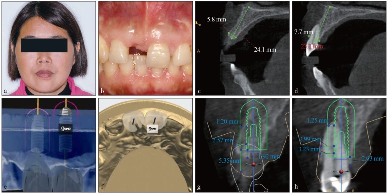
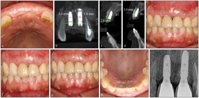
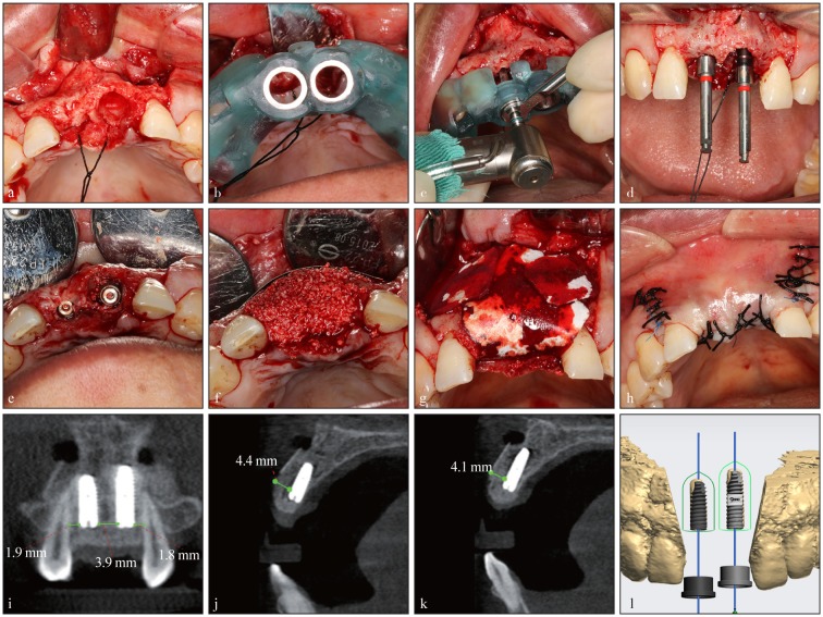
患者徐某,女,48岁,2016年10月11日以“外伤致上前牙缺失1年余”为主诉就诊于西南医科大学附属口腔医院种植科。专科检查:11牙缺失,近远中牙龈乳头不一致;21牙颈部根折,近中唇侧倾斜,Ⅱ度松动。CBCT示:11缺牙区唇侧骨壁薄;21唇侧牙槽突吸收,根尖未见明显低密度暗影。取硅橡胶印模,设计制作牙支持式半程导板后行11种植体植入术+21牙即刻拔除、种植体即刻植入术。术中微创拔除21牙;11和21牙处分别植入Anthogyr种植体3.4 mm×10.0 mm和3.4 mm×12.0 mm各一枚,植入扭矩达35 N·cm。术区唇侧及拔牙窝内骨量少,植入骨粉,唇侧覆盖钛膜,再以胶原生物膜覆盖,对位严密缝合。5个月后行二期手术,取下术区钛膜及封闭螺丝,植入5.0 mm×3.5 mm愈合基台。1个月后行全瓷冠修复。术后随访示11、21牙处牙龈健康,龈缘顶点及龈乳头协调,全瓷冠形态及比列协调,全口卫生条件可,X线片示骨结合良好(图3~5)。
图 3. 数字化导板辅助种植典型病例术前.
Fig 3 Preoperative photos and images of the typical case with the digital guide plate
a:术前正面像;b:术前口内像;c:11牙唇腭侧CBCT切面;d:21牙唇腭侧CBCT切面;e:计算机模拟11、21种植体植入及修复体位置;f:计算机模拟种植体中央螺丝从11、21修复体的舌隆突处穿出;g:计算机模拟设计11唇腭侧植入位置;h:计算机模拟设计21唇腭侧植入位置。
图 5. 数字化导板辅助种植典型病例术后.
Fig 5 Postoperative analysis of the typical case with the digital guide plate
a:一期术后5个月复查口内像;b~d:术后5个月CBCT像,其中b为11、21植入种植体冠状面观,c为11植入种植体的唇腭侧观,d为21植入种植体的唇腭侧观;e:修复体完成1周后口内像,11和21间存在黑三角;f:修复体完成6个月后口内像,黑三角明显减小;g、h:修复体完成1年后口内像,黑三角消失,其中g为前牙咬合面观,h为上前牙 面观;i:修复体完成1年后X线片,种植体骨结合良好。
图 4. 数字化导板辅助种植典型病例一期手术.
Fig 4 Intraoperative photos and images of the typical case with the digital guide plate
a:微创拔除21牙;b:数字化导板在口内就位;c:数字化导板辅助下定点;d:延长杆观测方向;e:植入种植体,旋入覆盖螺丝;f:植入骨粉,唇侧放置钛膜;g:覆盖胶原膜;h:严密对位缝合龈瓣;i~k:术后CBCT影像,其中i为11、21植入种植体冠状面观,j为11植入种植体的唇腭侧观,k为21植入种植体的唇腭侧观;l:术前设计位置与实际植入位置配准。
3. 讨论
近年来随着CAD/CAM技术快速发展,数字化技术在口腔种植领域的应用日益增多,特别是前牙美学区。本研究中使用的数字化导板,即牙种植导向模板,是为了种植手术方案所设计制造的个性化手术辅助工具[8]。美学区是患者微笑时可见的区域,一般指的是上颌前牙区[9]–[10]。此区种植涉及诸多风险,一方面患者对美观要求极高;另一方面前牙美学区的牙齿缺失多伴有牙槽骨的吸收,严重影响种植体植入的位置[11],特别是前牙唇侧骨板的吸收;同时,上前牙区种植手术需避让切牙管等特殊解剖结构,避免造成鼻腭神经的损伤及影响种植体的骨结合。故数字化种植导板对前牙美学区种植手术具有重要意义。
数字化种植导板能够协助精确定位种植体的植入位置。本研究结果显示,试验组与对照组种植体植入位置的偏差有统计学差异(P<0.05),试验组的偏差小于对照组。这证明数字化导板辅助种植较常规种植显著提高了种植体植入的精确性,为获得更好的后期美学修复效果奠定了良好的基础。Park等[12]研究显示,使用数字化导板辅助种植时种植体颈部距离、根尖部距离、深度和角度的偏差分别为(0.60±0.39) mm、(0.89±0.53)mm、(0.39±0.32)mm和(2.97±2.41)°,均小于未使用数字化导板时的偏差[(1.04±0.46)mm、(1.54±0.65)mm、(0.70±0.50)mm和(7.29±5.12)°]。Vercruyssen等[13]研究结果也显示,数字化导板辅助种植时种植体颈部距离、根尖部距离和角度的偏差分别为1.4 mm、1.6 mm和3.0°,均显著小于未使用数字化导板的偏差(2.8 mm、3.1 mm和9.1°)。本研究与Vercruyssen和Park等的研究结论相一致,均说明数字化导板可提高种植体植入的精确度。本研究的偏差结果大于Park等[12]的研究结果,其原因可能与患者剩余牙槽骨条件(包括骨量和骨密度)、植入种植体长度、医师手术水平及经验等有关。Park等[12]的研究中,研究对象包括后牙区等骨量较为充足的牙位,而本研究对象仅为上颌前牙区,此区牙齿缺失后多伴有唇侧骨板的凹陷性吸收,种植窝的预备及种植体植入过程中易向唇侧偏移[14],并且长种植体根方偏差也会较大,本研究对象使用的种植体长度均不小于10 mm。按支持形式不同,数字化导板可以分为牙支持式、骨支持式和黏膜支持式[15];按引导方式不同,可分为全程导板和半程导板[16]。Ozan等[17]研究证实,牙支持式导板辅助种植下的角度和根尖部偏差值小于骨支持式和黏膜支持式导板,牙支持式导板可利用缺失牙邻近的牙齿获得固位,在半程导板的辅助下利用钻头制备种植窝洞的深度、直径和角度,精准度高,适应证广泛,术中如遇特殊情况可及时调整。本研究中采用的是牙支持式半程导板,其稳定性较好,试验组种植体植入的精确度明显高于对照组。
PES和WES用于研究种植修复体的最终美学效果,其是可重复的美学指数,不受观察者的影响[18]。种植体植入的精确度对最终修复的美学效果有显著影响[5]。本研究结果显示,修复体完成后,试验组与对照组在修复完成后1周、6个月和1年的PES、WES均有统计学差异(P<0.01),试验组的PES及WES高于对照组。这说明数字化导板辅助种植相对于常规种植,可以提高软组织及修复体的美学效果。数字化导板能够较为精确地协助医生达到术前所设计的种植位点、深度及角度,获得更好的植入效果和美学效果[19]–[20]。但种植修复最终美学效果的影响因素很多,如牙龈生物型、骨吸收程度及口腔卫生维护等。本研究中2例患者出现唇侧瘘管,分析可能为骨粉暴露及口腔卫生较差引起,经搔刮、重新植入骨粉、覆盖胶原膜缝合及口腔卫生教育后愈合。
数字化导板辅助种植可进行有效的术前评估、精准种植修复设计、保护重要解剖结构、提高手术质量,促进医患及医技良好沟通[21];而且以修复体为指导的数字化导板引导的种植手术并发症(如龈缘萎缩、种植体的颈部骨吸收、基台暴露、疼痛与肿胀等)更少[22]。目前国内外对数字化导板精确性的研究数值不尽相同,很多因素都会影响数字化导板的精确性。影响因素包括有:印模与咬合记录的准确度,软件三维数据重建时的误差,导板加工误差,导板的稳定性,以及套环与导板之间、压板与套环之间、钻与压板之间的间隙等[8],术者水平及经验也可能影响垂直向的偏差和角度偏差[21]–[24]。从术前设计到最终种植体植入的每一步都会有偏差,所以未来努力的方向是将术前设计精准转换,保障种植体的最佳植入位置,进一步全面提高数字化导板的精准度。综上,数字化导板辅助种植修复技术可以提高种植体三维位置的准确性,对获得前牙美学区理想的治疗效果具有重要作用。
Funding Statement
[基金项目] 国家自然科学基金(81870746);中华口腔医学会西部行临床科研基金(CSA-W2017-08);(四川大学-泸州)颅颌面畸形整复中数字化诊疗技术应用和平台建设(2013CDLZ-S19)
Supported by: The National Natural Science Foundation of China (81870746); The Project of the Chinese Stomatological Association West China Clinical Research Fund (CSA-W2017-08); Digital Diagnosis and Treatment Technology Application and Platform Construction in Craniomaxillofacial Deformity (Sichuan University-Luzhou) (2013CDLZ-S19).
Footnotes
利益冲突声明:作者声明本文无利益冲突。
References
- 1.Rousseau P. Flapless and traditional dental implant surgery: an open, retrospective comparative study[J] J Oral Maxillofac Surg. 2010;68(9):2299–2306. doi: 10.1016/j.joms.2010.05.031. [DOI] [PubMed] [Google Scholar]
- 2.Kazor CE, Al-Shammari K, Sarment DP, et al. Implant plastic surgery: a review and rationale[J] J Oral Implantol. 2004;30(4):240–254. doi: 10.1563/0.637.1. [DOI] [PubMed] [Google Scholar]
- 3.袁 治, 陈 一晖. 计算机辅助设计导板修复上颌后种植体头部和角度偏离不影响种植体的植入精度[J] 中国组织工程研究. 2016;20(20):2933–2939. [Google Scholar]; Yuan Z, Chen YH. Implantation accuracy is not affected by head and angle deviation following computer aided design guide for repair of maxillary posterior tooth defects[J] J Clin Rehabil Tissue Eng Res. 2016;20(20):2933–2939. [Google Scholar]
- 4.Block MS, Emery RW. Static or dynamic navigation for implant placement-choosing the method of guidance[J] J Oral Maxillofac Surg. 2016;74(2):269–277. doi: 10.1016/j.joms.2015.09.022. [DOI] [PubMed] [Google Scholar]
- 5.Wittneben JG, Gavric J, Belser UC, et al. Esthetic and clinical performance of implant-supported all-ceramic crowns made with prefabricated or CAD/CAM zirconia abutments: a randomized, multicenter clinical trial[J] J Dent Res. 2017;96(2):163–170. doi: 10.1177/0022034516681767. [DOI] [PubMed] [Google Scholar]
- 6.Fürhauser R, Florescu D, Benesch T, et al. Evaluation of soft tissue around single-tooth implant crowns: the pink esthetic score[J] Clin Oral Implants Res. 2005;16(6):639–644. doi: 10.1111/j.1600-0501.2005.01193.x. [DOI] [PubMed] [Google Scholar]
- 7.Belser UC, Grütter L, Vailati F, et al. Outcome evaluation of early placed maxillary anterior single-tooth implants using objective esthetic criteria: a cross-sectional, retrospective study in 45 patients with a 2- to 4-year follow-up using pink and white esthetic scores[J] J Periodontol. 2009;80(1):140–151. doi: 10.1902/jop.2009.080435. [DOI] [PubMed] [Google Scholar]
- 8.李 晋蒙, 欧 国敏. 计算机辅助设计种植导板精确性及其影响因素[J] 华西口腔医学杂志. 2017;35(1):93–98. [Google Scholar]; Li JM, Ou GM. Accuracy of computer-guided implant placement and influencing factors[J] West China J Stomatol. 2017;35(1):93–98. doi: 10.7518/hxkq.2017.01.015. [DOI] [PMC free article] [PubMed] [Google Scholar]
- 9.Hu XL, Nahles S, Nelson CA, et al. Analysis of soft tissue display during enjoyment smiling. Part 1: caucasians[J] Int J Periodont Restor Dent. 2013;33(1):e9–e15. [PubMed] [Google Scholar]
- 10.Ahn HW, Moon SC, Baek SH. Morphometric evaluation of changes in the alveolar bone and roots of the maxillary anterior teeth before and after en masse retraction using cone-beam computed tomography[J] Angle Orthod. 2013;83(2):212–221. doi: 10.2319/041812-325.1. [DOI] [PMC free article] [PubMed] [Google Scholar]
- 11.Hiroya S. An application of dental implant on alveolar bone defect caused by injury: an esthetic recovery of anterior teeth region without a bone grafting[J] Nihon Hotetsu Shika Gakkai Zasshi. 2005;49(3):510–511. doi: 10.2186/jjps.49.510. [DOI] [PubMed] [Google Scholar]
- 12.Park SJ, Leesungbok R, Cui TX, et al. Reliability of a CAD/CAM surgical guide for implant placement: an in vitro comparison of surgeons' experience levels and implant sites[J] Int J Prosthodont. 2017;30(4):367–369. doi: 10.11607/ijp.5179. [DOI] [PubMed] [Google Scholar]
- 13.Vercruyssen M, Cox C, Coucke W, et al. A randomized clinical trial comparing guided implant surgery (bone- or mucosa-supported) with mental navigation or the use of a pilot-drill template[J] J Clin Periodontol. 2014;41(7):717–723. doi: 10.1111/jcpe.12231. [DOI] [PubMed] [Google Scholar]
- 14.金 杭颖, 吴 英浩, 徐 继文, et al. 国产数字化导板辅助上前牙种植手术的精准度研究[J] 实用口腔医学杂志. 2018;34(5):644–647. [Google Scholar]; Jin HY, Wu YH, Xu JW, et al. Precision evaluation of domestic digital guide assisting in the implantation of maxillary anterior teeth[J] J Pract Stomatol. 2018;34(5):644–647. [Google Scholar]
- 15.Ramasamy M, Giri, Raja R, et al. Implant surgical guides: from the past to the present[J] J Pharm Bioallied Sci. 2013;5(Suppl 1):S98–S102. doi: 10.4103/0975-7406.113306. [DOI] [PMC free article] [PubMed] [Google Scholar]
- 16.Meloni SM, Tallarico M, De Riu G, et al. Guided implant surgery after free-flap reconstruction: four-year results from a prospective clinical trial[J] J Craniomaxillofac Surg. 2015;43(8):1348–1355. doi: 10.1016/j.jcms.2015.06.046. [DOI] [PubMed] [Google Scholar]
- 17.Ozan O, Turkyilmaz I, Ersoy AE, et al. Clinical accuracy of 3 different types of computed tomography-derived stereolithographic surgical guides in implant placement[J] J Oral Maxillofac Surg. 2009;67(2):394–401. doi: 10.1016/j.joms.2008.09.033. [DOI] [PubMed] [Google Scholar]
- 18.Tettamanti S, Millen C, Gavric J, et al. Esthetic evaluation of implant crowns and peri-implant soft tissue in the anterior maxilla: comparison and reproducibility of three different indices[J] Clin Implant Dent Relat Res. 2016;18(3):517–526. doi: 10.1111/cid.12306. [DOI] [PubMed] [Google Scholar]
- 19.Le BT, Borzabadi-Farahani A. Labial bone thickness in area of anterior maxillary implants associated with crestal labial soft tissue thickness[J] Implant Dent. 2012;21(5):406–410. doi: 10.1097/ID.0b013e31826371b5. [DOI] [PubMed] [Google Scholar]
- 20.Jung MS, Lee SP, Kim GT, et al. Three-dimensional analysis of deciduous maxillary anterior teeth using cone-beam computed tomography[J] Clin Anat. 2012;25(2):182–188. doi: 10.1002/ca.21200. [DOI] [PubMed] [Google Scholar]
- 21.Choquet V, Hermans M, Adriaenssens P, et al. Clinical and radiographic evaluation of the papilla level adjacent to single-tooth dental implants. A retrospective study in the maxillary anterior region[J] J Periodontol. 2001;72(10):1364–1371. doi: 10.1902/jop.2001.72.10.1364. [DOI] [PubMed] [Google Scholar]
- 22.Hultin M, Svensson KG, Trulsson M. Clinical advantages of computer-guided implant placement: a systematic review[J] Clin Oral Implants Res. 2012;23(Suppl 6):124–135. doi: 10.1111/j.1600-0501.2012.02545.x. [DOI] [PubMed] [Google Scholar]
- 23.Rungcharassaeng K, Caruso JM, Kan JY, et al. Accuracy of computer-guided surgery: a comparison of operator experience[J] J Prosthet Dent. 2015;114(3):407–413. doi: 10.1016/j.prosdent.2015.04.004. [DOI] [PMC free article] [PubMed] [Google Scholar]
- 24.Cushen SE, Turkyilmaz I. Impact of operator experience on the accuracy of implant placement with stereolithographic surgical templates: an in vitro study[J] J Prosthet Dent. 2013;109(4):248–254. doi: 10.1016/S0022-3913(13)60053-0. [DOI] [PubMed] [Google Scholar]


