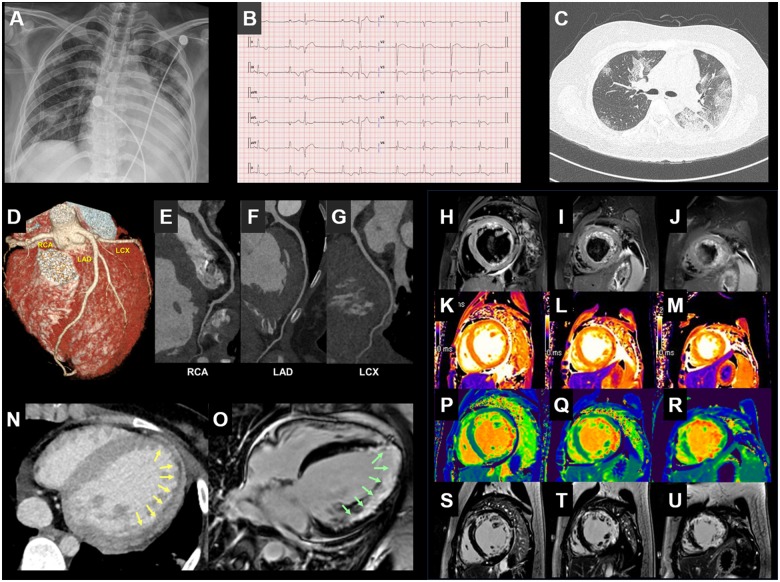
A 21-year-old female patient visited our hospital for febrile sensation, coughing, sputum, diarrhoea, and shortness of breath during the coronavirus disease 2019 (COVID-19) outbreak in Daegu, South Korea. Nasopharyngeal swab was positive for COVID-19. Troponin I level was 1.26 ng/mL (<0.3 ng/mL) and NT-proBNP was 1929 pg/mL (<125 pg/mL). The chest radiograph revealed a multifocal consolidation on both lung fields and cardiomegaly (Panel A). Electrocardiography showed non-specific intraventricular conduction delay and multiple premature ventricular complexes (Panel B). Echocardiography showed severe left ventricular (LV) systolic dysfunction (Supplementary material online, Videos S1–S3). A chest computed tomography (CT) revealed a multifocal consolidation and ground-glass opacification in both lungs in the lower lobe and a peripheral dominant distribution (Panel C). On the cardiac CT, the coronary arteries were normal (Panels D–G), and the myocardium was hypertrophied due to oedema combined with a subendocardial perfusion defect on the lateral left ventricle (Panel N). Cardiac magnetic resonance imaging (MRI) revealed a diffuse high signal intensity (SI) in the LV myocardium on T2 short tau inversion recovery image (Panels H–J; SI ratio of myocardium over skeletal muscle = 2.2), and myocardial wall thickening (LV mass index: 111.3 g/m2), which suggests myocardial wall oedema. On mapping sequence, native T1 (Figure 1K–M; mid-septum, 1431 ms; lateral wall, 1453 ms, reference value ∼1150 ms) and extracellular volume (Panels P–R; mid-septum, 29.7%; lateral wall, 61%; reference value ∼25%) values were diffusely increased (Panel O). Extensive transmural late gadolinium enhancement was noted (Panels S–U). Myocarditis combined with COVID-19 was confirmed by multimodality imaging.
Supplementary material is available at European Heart Journal online.
Supplementary Material
Associated Data
This section collects any data citations, data availability statements, or supplementary materials included in this article.


