Abstract
Schwannoma is a benign neural sheath tumor of the soft tissue, and its intraosseous presentation is very rare. It is estimated that intraosseous schwannomas represent 0.2% of all bone tumors. The tumor may affect any site of the skeleton, including the mandible, the sacrum, vertebral bodies, the ulna, the humerus, the femur, the tibia, the patella, the scapula, the ribs, and small bones of the hand. The involvement of the calcaneus has only been reported four times in the literature. The present study reports the case of a 49-year-old male with right hindfoot pain and a radiological finding of an osteolytic bone lesion in the calcaneus. The diagnosis was confirmed by histopathological study. The treatment of choice was an intralesional resection with adjuvant local control, and bone defect substitution with polymethylmethacrylate and fixation with two cannulated screws. The patient had a satisfactory postoperative evolution; after 1 year, he is asymptomatic, with good functional response and no evidence of disease. The present case report shows the clinical, radiological, and pathological features of a rare benign bone neoplasm. Moreover, intraosseous schwannoma should be included in the differential diagnosis of osteolytic calcaneal lesions.
Keywords: bone neoplasms, neurilemmoma, calcaneus
Introduction
Schwannoma is a benign tumor originating from Schwann cells, which constitute the neural sheath. It is the most common benign tumor in peripheral nerves. It may also affect some cranial nerves, particularly the vestibular branch of the cranial nerve VIII (acoustic).
Schwannoma intraosseous presentation is rare, corresponding to 0.2% of primary bone tumors. 1 2 3 4 5 6 It mainly affects the mandible, the sacrum, vertebral bodies, the ulna, the humerus, the femur, the tibia, the patella, the scapula, the ribs and small bones of the hand. 1
After reviewing the literature, we found only four reported cases of calcaneal intraosseous schwannoma. The present paper aims to review the clinical, radiological and anatomopathological presentation of intraosseous schwannomas, as well as to include it in the differential diagnosis of calcaneus bone tumors.
Case Report
A 49-year-old male patient complained of sudden and severe pain in the right hindfoot, which worsened during gait. One month after the onset of the condition, his right hindfoot was swollen, his pain got worse, and the patient was unable to walk.
The patient had a history of congenital clubfoot (PTC) ipsilateral to the lesion, conservatively treated during childhood.
Physical examination revealed a sequela deformity related to PTC, with subtalar joint block and plantar fixed, 20° flexion. The patient presented pain during palpation of the lateral aspect and especially the medial aspect of the hindfoot.
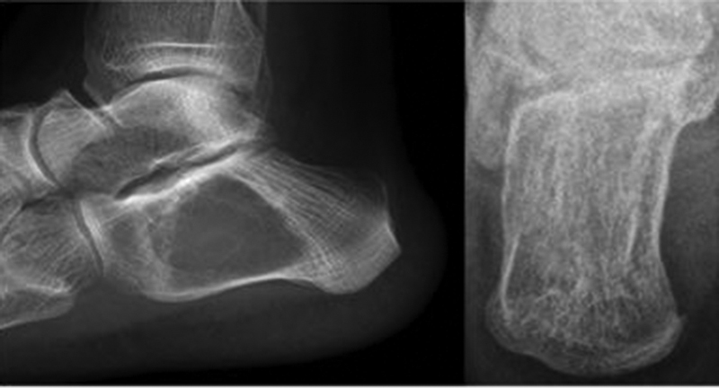
Radiographic images showed a well-delimited lytic bone lesion in the body and part of the anterior calcaneus process, with areas of cortical rupture, and no signs of intralesional calcification ( Fig. 1 ).
Fig. 1.
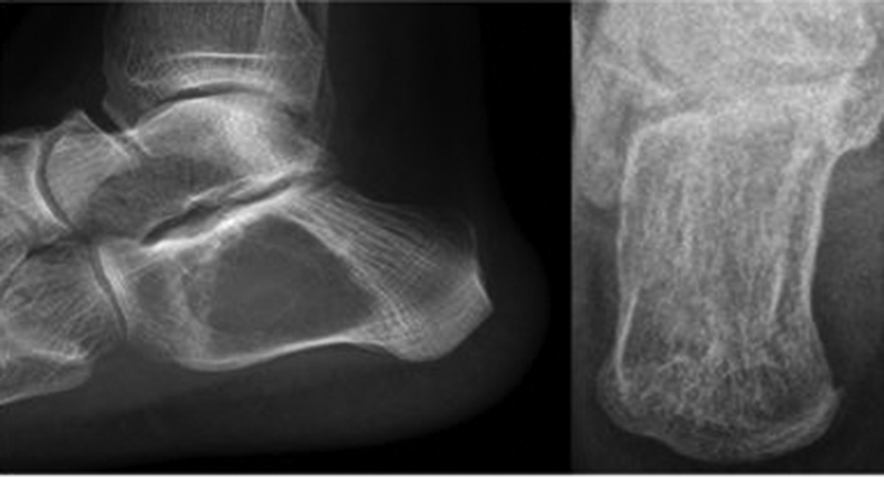
Lateral and axial calcaneal radiographic images.
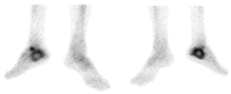
Bone scintigraphy showed an anomalous and unique uptake area in the right calcaneus ( Fig. 2 ).
Fig. 2.
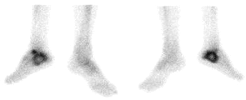
Images from the late phase of bone scintigraphy.
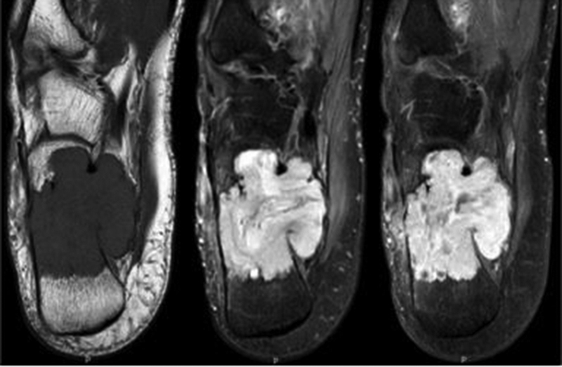
A nuclear magnetic resonance (NMR) showed a lesion with hypointense signal in T1-weighted sequences, which was heterogeneous in T2-weighted sequences, in addition to intense contrast enhancement, with a mean diameter of 50 mm, lateral and medial cortices rupture, extensive medial extraosseous component, plantaris muscle infiltration, partial involvement of the flexor hallucis longus tendon and close contact with the posterior tibial neurovascular bundle ( Fig. 3 ).
Fig. 3.
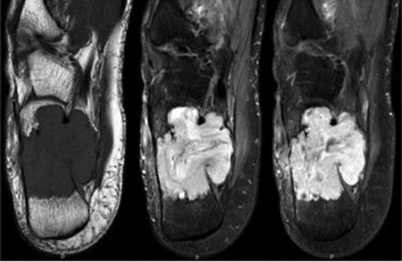
Axial nuclear magnetic resonance, T1-weighted (A), T2-weighted (B) and T1-weighted with fat saturation and enhanced with gadolinium (C) images.
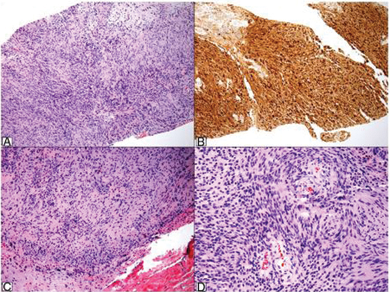
A computed tomography (CT)-guided percutaneous biopsy revealed the presence of spindle cells with eosinophilic and fibrillar cytoplasm, arranged in short bundles, with no atypia, and elongated and normochromic nuclei. Areas of lower cellularity were occasionally observed, in addition to regions with parallel-arranged nuclei. An immunohistochemical study revealed strong, diffuse positivity for S-100 protein, leading to an intraosseous schwannoma diagnosis ( Fig. 4 ).
Fig. 4.
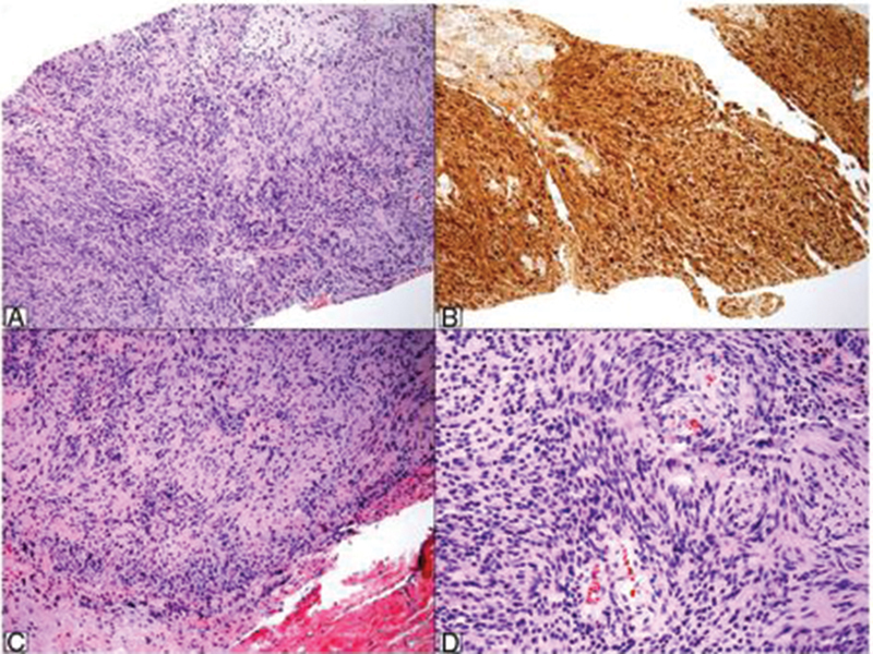
Histopathological study of biopsy specimen showing a spindle cells neoplasm with no atypia and alternating higher and lower cellularity areas (A, hematoxylin & eosin [H&E], 100×). An immunohistochemical study reveals the strong, diffuse positivity for S-100 protein (B, S-100, 100×). The surgical piece exhibited the same aspects from the biopsy, that is, a spindle cell neoplasm with short bundles, hypocellular areas and nuclear palisade foci (C and D, H&E, 100× and 200×).
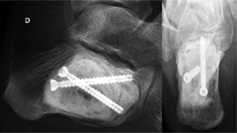
Thus, we opted for an intralesional resection for double access to the calcaneus (medial and lateral parts) with local, adjuvant treatment by electrocauterization of the tumor bed. The resulting bone defect was filled with polymethyl methacrylate (PMMA) and fixed with two cannulated screws for greater stability ( Fig. 5 ). The anatomopathological study of the surgical specimen showed the same biopsy findings, confirming the diagnosis.
Fig. 5.
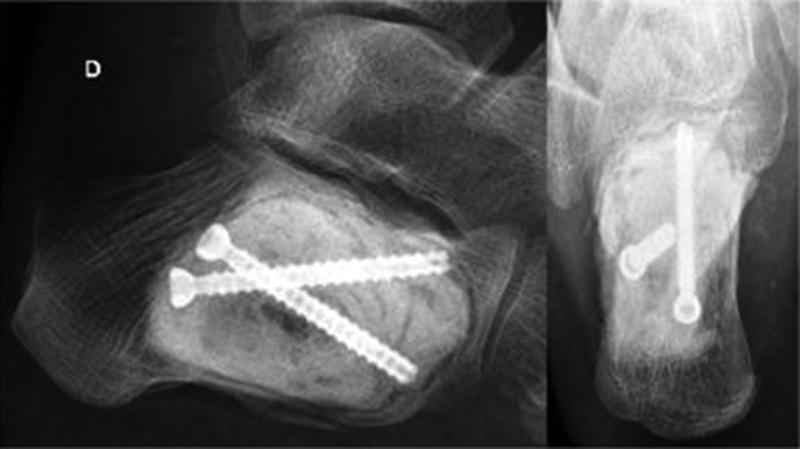
Postsurgical appearance, lateral and axial calcaneal radiographic images.
The patient presented good postoperative evolution with improvement of the previous symptoms. One year after the procedure, he is asymptomatic, and radiography and CT showed no signs of local recurrence.
Discussion
Intraosseous schwannoma is a rare condition. The most widely accepted theory to justify such rarity is that these tumors appear almost exclusively in sensory fibers, which are scarce in bones, prevailing in the periosteum. 1 In bones, these fibers are mostly located near vessels accounting for bone nutrition. 3
The most affected bone is the mandible. Although some authors believe that the long intraosseous course of the mandibular nerve result in a greater incidence of the tumor in this region, it has been reported that the same happens in other bones, with no increased incidence in these other locations. The sacrum is the second most affected site, which can be justified by the numerous sensory roots passing through its foramina.
These tumors can affect any age group and are more frequent between the 4 th and 6 th decades of life. It has no predilection for gender or ethnicity, 7 although some series show a higher incidence in females 1 3
Schwannomas can manifest in bone tissue through three mechanisms: extraosseous lesion resulting in secondary bone erosion; a lesion arising within the nutrition canal that grows and takes on a dumbbell shape; and a lesion arising within the bone.
Regarding the clinical presentation, these tumors may cause pain associated or not with edema. Some cases may present with pathological fracture at diagnosis. Tumor growth is slow and insidious, with long periods of evolution. 1 2 3
Radiographically, these tumors manifest as well-circumscribed lytic lesions, possibly with a sclerotic halo, which sometimes tapers or destroys the cortical layer. Adjacent soft tissues are rarely invaded. There is no calcification or bone tissue formation within the lesion. 1 2 3
Nuclear magnetic resonance is a better imaging method to assess cortical and soft tissue involvement when compared with CT. Schwannoma lesions present hypointense signal on T1-weighted images, heterogeneous signal on T2-weighted images and diffuse paramagnetic contrast enhancement. 3
Histopathological findings do not usually differ from other soft tissue neoplasms. Schwannomas usually exhibit spindle cells with elongated nuclei and alternation of hypercellular and hypocellular areas, known as Antoni A and Antoni B pattern, respectively. Some more typical cases present nuclear palisades alternating with fibrillar areas, which constitute the so-called Verocay bodies.
Cases consisting only of Antoni A areas with no nuclear palisades may simulate malignant neoplasms, such as synovial sarcoma or leiomyosarcoma. They are diagnosed as cellular Schwannomas, a variant with no higher biological aggressiveness. Diagnosis is aided by the strong and diffuse immunohistochemical positivity for S-100 protein. Other markers may be used, such as SOX-10, and pericellular positivity to anti-collagen IV antibody can be observed.
Treatment is based on intralesional resection and filling with bone graft or PMMA, associated or not with synthesis material for support and stability. Relapse is rare and the prognosis is good. Schwannoma malignant transformation was not described. 4 5 8 9 10
This case resembles the other intraosseous Schwannomas described regarding clinical presentation, evolution, imaging, pathological findings and instituted treatment. Intralesional resection associated with local adjuvant therapy and bone filling seems to be an effective method for surgical treatment.
Conflitos de Interesses Os autores declaram não haver conflitos de interesses.
Originalmente Publicado por Elsevier Editora Ltda.
Originally Published by Elsevier Editora Ltda.
Referências
- 1.de la Monte S M, Dorfman H D, Chandra R, Malawer M. Intraosseous schwannoma: histologic features, ultrastructure, and review of the literature. Hum Pathol. 1984;15(06):551–558. doi: 10.1016/s0046-8177(84)80009-x. [DOI] [PubMed] [Google Scholar]
- 2.Kashima T G, Gibbons M R, Whitwell D et al. Intraosseous schwannoma in schwannomatosis. Skeletal Radiol. 2013;42(12):1665–1671. doi: 10.1007/s00256-013-1712-6. [DOI] [PubMed] [Google Scholar]
- 3.Ida C M, Scheithauer B W, Yapicier O et al. Primary schwannoma of the bone: a clinicopathologic and radiologic study of 17 cases. Am J Surg Pathol. 2011;35(07):989–997. doi: 10.1097/PAS.0b013e31821fcc0c. [DOI] [PubMed] [Google Scholar]
- 4.Sochart D H. Intraosseous schwannoma of calcaneum. Foot. 1995;5:38–40. [Google Scholar]
- 5.Wirth W A, Bray C B., Jr Intra-osseous neurilemoma. Case report and review of thirty-one cases from the literature. J Bone Joint Surg Am. 1977;59(02):252–255. [PubMed] [Google Scholar]
- 6.Knight D M, Birch R, Pringle J. Benign solitary schwannomas: a review of 234 cases. J Bone Joint Surg Br. 2007;89(03):382–387. doi: 10.1302/0301-620X.89B3.18123. [DOI] [PubMed] [Google Scholar]
- 7.Antonescu C R, Perry A A, Woodruff J M. Lyon: IARC Press; 2013. Schwannoma (including variants) pp. 170–172. [Google Scholar]
- 8.Pyati P S, Sanzone A G. Intraosseous neurilemoma of the calcaneus. Orthopedics. 1996;19(04):353–355. doi: 10.3928/0147-7447-19960401-16. [DOI] [PubMed] [Google Scholar]
- 9.Li K, Yang Q, Gao B et al. Giant intraosseous Schwannoma of the calcaneus: case report. Int J Clin Exp Med. 2016;9(03):6897–6901. [Google Scholar]
- 10.Salunkhe R, Limaye S, Biswas S K, Mehta R P. A rare case of calcaneal intraosseous schwannoma. Med J DY Patil Univ. 2012;5:76–78. [Google Scholar]


