This case series describes intratumoral spatial heterogeneity of tumor cell populations and investigates its association with survival among patients with nonmetastatic gastroesophageal adenocarcinoma.
Key Points
Question
Is there an association between tumor cell heterogeneity at time of diagnosis in nonmetastatic gastroesophageal adenocarcinoma and prognosis?
Findings
In this case series of 41 patients with gastroesophageal adenocarcinoma, a high degree of intratumoral heterogeneity was identified. The presence of clonal populations coexisting at submillimeter distances was associated with worse survival.
Meaning
These findings suggest that understanding intratumoral heterogeneity is highly relevant for future precision medicine neoadjuvant strategies in gastroesophageal adenocarcinoma, and single-cell analytic approaches are recommended.
Abstract
Importance
Intratumoral heterogeneity has been recognized as a significant barrier in successfully developing targetable biomarkers for gastroesophageal adenocarcinoma (GEA) and may affect neoadjuvant precision medicine approaches.
Objective
To describe intratumoral spatial heterogeneity of tumor cell populations in nonmetastatic GEA and its association with survival.
Design, Setting, and Participants
This case series retrospectively identified 41 patients with GEA who underwent up-front surgical resection at a tertiary referral cancer center from January 1, 1989, through December 31, 2013. Survival was calculated from date of surgery to date of death through June 1, 2017. Data were analyzed from June 2, 2017, to March 1, 2019.
Main Outcomes and Measures
Overall survival, intratumoral clonal composition determined by genomic single-nucleotide variation array and bioinformatic analysis, and intercellular tumoral distances determined by multiprobe fluorescence in situ hybridization.
Results
Among the 41 patients included in the analysis (22 men [54%]; mean [SD] age, 63 [12] years), a high proportion (19 [46%]) presented with tumors possessing high intratumoral heterogeneity. Kaplan-Meier analysis demonstrated that cases with an intratumoral clonal composition count of at least 2 exhibited worse survival compared with cases with a clonal composition count of 0 to 1 (univariate hazard ratio, 3.92; 95% CI, 1.27-12.08; P = .02). This finding remained significant on multivariate analysis controlling for stage, Lauren histologic subtype, receipt of adjuvant therapy, and age (multivariate hazard ratio, 4.55; 95% CI, 1.09-19.04; P = .04). Multiprobe fluorescence in situ hybridization demonstrated intratumoral clonal populations coexisting at submillimeter distances with differing relevant oncogenic copy number alterations, such as EGFR, JAK2, FGFR2, MET, CCND1, KRAS, MYC, PIK3CA, CD274, and PDCD1LG2.
Conclusions and Relevance
This study found that spatial intratumoral heterogeneity of oncogenic copy number alterations exists before metastatic dissemination, and increased heterogeneity was associated with worse outcomes in resected GEA. Baseline heterogeneity illustrates the challenges in GEA targeted therapy. Further study may offer insight into strategies on combinatorial and/or sequential targeted and immunotherapeutic approaches.
Introduction
Precision medicine efforts in gastroesophageal adenocarcinoma (GEA) have been hampered by the recognition that intratumoral heterogeneity confounds results of genomic testing yielded from limited sampling of the tumor.1 Such intratumoral heterogeneity of actionable receptor tyrosine kinases, including ERBB2, MET, and FGFR2, affects targeted therapeutic strategies.1,2,3,4 Knowledge of intratumoral heterogeneity in gastric cancer at de novo nonmetastatic disease presentation remains limited, with recent studies providing emerging evidence.1 The Cancer Genome Atlas (TCGA) has laid the framework of copy number alterations (CNAs) predominating the mutational landscape of GEA, particularly in the chromosomal instability subtype of tumors.5,6 Pancancer TCGA studies have also deciphered intratumoral heterogeneity from whole-exome sequencing data, with worse survival observed in patients with higher heterogeneity.7 With this understanding, we undertook an in-depth study of GEA intratumoral heterogeneity and its association with survival using a genome-wide single-nucleotide variation (SNV; formerly termed single-nucleotide polymorphism) array panel. We focused on cases treated without neoadjuvant therapy before surgery to best capture de novo disease presentation and the molecular landscape that may affect neoadjuvant strategies incorporating targeted agents. We pursued multiprobe fluorescence in situ hybridization (FISH) analyses to discern at a single-cell level the presence and patterns of spatial genomic heterogeneity.
Methods
Study Population
For this case series, 41 patients with nonmetastatic GEA who underwent up-front surgical resection with curative intent were retrospectively identified from the City of Hope Biospecimen Repository. Waiver of consent was granted under a protocol approved by the institutional review board of City of Hope, as the proposed research presented no more than minimal risk of harm, and the waiver did not adversely affect the rights and welfare of the participants. Patients who underwent surgical resection from January 1, 1989, to December 31, 2013, without receipt of neoadjuvant chemotherapy, radiotherapy, and/or targeted therapy were selected to best capture innate heterogeneity at disease presentation. The explicit selection of patients to test the hypothesis that intratumoral heterogeneity at disease presentation is associated with survival follows published guidelines for case series studies.8
Available demographic, clinical, and pathologic characteristics were extracted from retrospective review of patients’ medical records and pathology reports. The staging system of the American Joint Committee on Cancer, 7th edition, was applied from pathology reports and used to define pathologic stage. Overall survival was calculated as time from the date of surgery to the date of death due to any cause through a cutoff date of June 1, 2017.
SNV Array Panel Copy Number Analysis
For genomic tumor DNA isolation, hematoxylin-eosin–stained slides were evaluated by the pathologist (P.C.), and tumor areas were circled. Tumor tissue was microdissected from two 10-μm formalin-fixed, paraffin-embedded (FFPE) sections. The DNA was isolated using the FFPE plus low elution volume DNA isolation kit (Maxwell 16; Promega) and quantified with the manufacturer’s fluorometer (Quantus; Promega). The FFPE assay (OncoScan; ThermoFisher Scientific) was run according to the manufacturer’s directions using 80 ng of DNA. This molecular inversion probe technology enabled reliable analysis of 30-year-old FFPE specimens.9 The OSCHP files were analyzed with Nexus Express software, version 4.0 (Biodiscovery, Inc) and Chromosome Analysis Suite, version 3.1 (ThermoFisher Scientific). We assayed the CNAs of 891 cancer-related genes at 50- to 100-kilobase resolution using the Affymetrix OncoScan platform per the manufacturer’s instructions. Copy number gains were defined as CNAs of greater than 2 and 5 or less; amplifications, as CNAs of 6 or greater; shallow deletions, as CNAs of less than 2 and 1 or greater; and deep deletions, as CNAs of 0.
Clonal Composition Analysis
Clonal composition was determined using a combination of 4 software tools: Chromosome Analysis, version 3.1 (ChAS; ThermoFisher Scientific), Nexus Express software for OncoScan (Biodiscovery, Inc), Oncoclone Composition,10 and Tuscanator, version 6 (provided by Sam Dougaparsad, PhD). Tuscanator script is derived from the published ASCAT (allele-specific copy number analysis of tumors) algorithm from Van Loo et al,11 whereas for a given aberrant segment, Tuscanator uses the mean smooth signal values (gaussian smoothed calibrated copy number estimate from the log2 ratio data) and the mean B-allele frequency (BAF) SNV values, with a range of copy numbers from 1 to 6. Briefly, Nexus Express was used to visualize a whole genome view and determine BAF patterns across each sample. OncoClone Composition was used to estimate the number of clones within a patient’s primary gastric tumor. OncoClone Composition is less predictive for high-ploidy samples; therefore, BAF and smooth signal average per segment were entered into Tuscanator software to generate a representative percentage of abnormal cells for each segment and subsequently determine clonal composition.
Simultaneous and Sequential FISH Analyses
Copy number alterations were confirmed by FISH in select samples to confirm spatial heterogeneity signal patterns. The method used to pursue multiprobe FISH on FFPE has been previously described.12 Briefly, hematoxylin-eosin–stained slides were scanned on an image analyzer system (Duet; BioView). Fluorescent images were matched with hematoxylin-eosin staining and captured. For sequential hybridizations, the probe was removed by incubating in 70% formamide/2 × sodium saline citrate for 5 minutes at 75 °C. Images were matched with previous targets and captured. Two-dimensional spatial coordinates were noted for FISH images at micron resolution afforded by image-capturing software. Distances (d) between 2 coordinates (x1, y1) and (x2, y2) were calculated using the pythagorean theorem:
| d = √(x2 – x1)2 + (y2 – y1)2 |
Statistical Analysis
Data were analyzed from June 2, 2017, to March 1, 2019. All data were analyzed in R statistical software, version 3.4.3 (R Project for Statistical Computing). For continuous variables, Mann-Whitney tests were used. Continuous data are presented as mean (SD) unless otherwise stated, and 2-tailed P ≤ .05 was considered statistically significant. Overall survival was analyzed by Kaplan-Meier estimates, and hazard ratios were calculated from Cox proportional hazards regression models.
Results
Interpatient Tumoral Heterogeneity
Gastroesophageal adenocarcinoma samples from 41 patients (22 men [54%] and 19 women [46%]; mean [SD] age, 63 [12] years) were analyzed using the OncoScan platform. Thirty-seven samples were within quality control metric standards. Four samples failed the SNV quality control of normal diploid markers (ndSNVQC) metric. The allelic data for these samples was otherwise clear, and failure of ndSNVQC was likely owing to a lower number of normal diploid markers, high ploidy, or multiple clones in the sample. Four cases had normal copy number results. The percentage of abnormal cells in the remaining 37 cases ranged from 16% to 60%.
The Table lists the clinicopathologic characteristics of the study population and corresponding percentage of genomic changes, percentage loss of heterozygosity, and clonal composition count. Further analysis demonstrated a higher percentage of genomic changes associated strongly with Lauren intestinal subtype histology compared with diffuse subtype histology (median, 39.9% vs 4.2%; P = .001 by Mann-Whitney test) (eFigure 1A in the Supplement), and in tumors arising from the gastroesophageal junction, cardia, or proximal stomach vs tumors arising from the gastric body or antrum (median, 39.3% vs 14.4%; P = .01 by Mann-Whitney test) (eFigure 1B in the Supplement), consistent with TCGA trends. We had good representation of cases with TCGA chromosomal instability subtype with detection of multiple CNAs across the genome (eFigure 2 in the Supplement, top panel). In this case, multiple chromosomes also exhibited alternating disruption of BAFs, indicating both retention and loss of heterozygosity throughout the genome, pathognomonic for a chromothripsis event having occurred in this tumor’s evolution (eFigure 2 in the Supplement, bottom panel).
Table. Clinicopathologic Characteristics and Genomic Analyses Yielded From the Oncoscan Platform of 41 Gastric Cancer Cases.
| Patient No./sex/age at diagnosis, y | Year of surgery | Lauren classification | Location | Staging AJCC7 | Received adjuvant therapy | LOH, % | Genome changed, % | Clonal composition count |
|---|---|---|---|---|---|---|---|---|
| 1/M/60s | 1989 | Intestinal | GEJ | T3N3 | No | 20.30 | 48.30 | 2 |
| 2/M/60s | 1989 | Indeterminate | GEJ | T3N1 | No | 0.54 | 0.52 | 0 |
| 3/M/60s | 1990 | Mixed | GEJ/fundus | T2N1 | NR | 17.80 | 39.00 | 3 |
| 4/F/50s | 1994 | Intestinal | Stomach/distal body lesser curve | T3N0 | No | 2.29 | 49.60 | 2 |
| 5/M/70s | 1994 | Intestinal | GEJ | T3N0 | No | 12.00 | 73.00 | 3 |
| 6/F/30s | 1994 | Intestinal | Stomach/proximal | T4N2 | Yes | 1.01 | 18.10 | 2 |
| 7/M/60s | 1995 | Diffuse | GEJ/stomach/cardia | T3N2 | No | 1.49 | 20.30 | 1 |
| 8/M/70s | 1995 | Indeterminate | Stomach/cardia | T4aN3 | No | 5.10 | 34.40 | 2 |
| 9/F/70s | 1996 | Indeterminate | Stomach/antrum | T3N2 | No | 0.43 | 4.70 | 0 |
| 10/F/80s | 1996 | Indeterminate | GEJ | T3N3 | No | 2.05 | 13.30 | 1 |
| 11/F/70s | 1996 | Mixed | Stomach/antrum | T4aN3 | No | 1.99 | 12.60 | 2 |
| 12/F/50s | 1997 | Intestinal | GEJ | T3N2 | Yes | 0.72 | 27.70 | 2 |
| 13/F/50s | 1997 | Diffuse | Stomach/body (linitis) | T4aN3 | Yes | 0.86 | 0.69 | 1 |
| 14/M/70s | 1999 | Diffuse | Stomach/antrum | T4aN3 | No | 0.38 | 1.39 | 1 |
| 15/M/60s | 2000 | Mixed | Stomach/cardia | T2N1 | Yes | 7.43 | 2.71 | 1 |
| 16/M/70s | 2001 | Indeterminate | GEJ | T3N2 | Yes | 26.90 | 60.90 | 3 |
| 17/M/50s | 2001 | Intestinal | GEJ | T3N2 | Yes | 16.10 | 46.50 | 2 |
| 18/F/50s | 2001 | Diffuse | Stomach/body | T2N2 | Yes | 0.74 | 4.66 | 1 |
| 19/M/60s | 2003 | Intestinal | Stomach/distal lesser curve | T3N3 | Yes | 1.84 | 14.40 | 1 |
| 20/M/60s | 2004 | Indeterminate | GEJ | T3N3 | No | 13.40 | 66.30 | 3 |
| 21/M/70s | 2004 | Intestinal | Stomach/antrum | T3N0 | NR | 19.30 | 62.10 | 2 |
| 22/M/50s | 2005 | Diffuse | Stomach/lesser curvature | T3N1 | Yes | 0.11 | 0.24 | 0 |
| 23/F/30s | 2005 | Diffuse | Stomach/middle body greater curve | T3N1 | Yes | 2.80 | 17.60 | 1 |
| 24/M/80s | 2006 | Diffuse | Stomach/antrum | T3N0 | No | 14.90 | 44.30 | 1 |
| 25/M/70s | 2008 | Intestinal | Stomach/distal body | T3N1 | Yes | 30.10 | 63.10 | 3 |
| 26/F/30s | 2008 | Diffuse | Stomach/distal lesser curve | T3N1 | Yes | 14.40 | 34.50 | 1 |
| 27/M/60s | 2008 | Indeterminate | Stomach/distal lesser curve | T2N2 | Yes | 13.50 | 22.60 | 1 |
| 28/F/60s | 2009 | Indeterminate | Stomach/proximal greater curve | T3N1 | No | 24.70 | 68.80 | 2 |
| 29/F/50s | 2009 | Diffuse | GEJ | T3N0 | Yes | 14.30 | 51.20 | 3 |
| 30/M/50s | 2010 | Intestinal | GEJ | T2N1 | Yes | 4.99 | 41.30 | 3 |
| 31/F/40s | 2010 | Intestinal | Stomach/distal body | T3N2 | Yes | 41.00 | 48.80 | 4 |
| 32/M/50s | 2011 | Diffuse | Stomach/antrum | T2N0 | Yes | 0.44 | 0.15 | 0 |
| 33/F/70s | 2012 | Diffuse | Stomach/body | T1N3 | Yes | 0.72 | 31.00 | 1 |
| 34/F/70s | 2012 | Diffuse | Stomach/antrum | T3N1 | Yes | 0.83 | 3.18 | 0 |
| 35/M/60s | 2012 | Mixed | Stomach/antrum | T3N3 | Yes | 0.23 | 0.18 | 0 |
| 36/F/40s | 2013 | Diffuse | Stomach/body | T3N0 | Yes | 0.68 | 0.00 | 0 |
| 37/M/50s | 2013 | Intestinal | Stomach/cardia | T2N3 | Yes | 9.43 | 58.40 | 3 |
| 38/M/60s | 2013 | Diffuse | Stomach/middle distal body | T3N3 | Yes | 0.41 | 3.58 | 0 |
| 39/F/60s | 2014 | Intestinal | Stomach/distal body | T2N1 | Yes | 3.78 | 24.10 | 1 |
| 40/F/60s | 2014 | Diffuse | Stomach/antrum | T4aN2 | Yes | 2.92 | 46.00 | 2 |
| 41/F/60s | 2014 | Diffuse | Stomach/lesser curve (linitis) | T3N0 | Yes | 3.07 | 2.52 | 0 |
Abbreviations: AJCC7, American Joint Committee on Cancer Cancer Staging Manual, Seventh Edition, GEJ, gastroesophageal junction; LOH, loss of heterozygosity; NR, not recorded.
We observed interpatient tumoral heterogeneity across nearly all cases, with a variety of genomic alterations occurring in selected genes of interest as represented in the Oncoprint diagram (eFigure 3 in the Supplement).13,14 Multiple oncogenic CNAs were observed across patients, including high copy gains in EGFR, (OMIM 131550), ERBB2 (OMIM 164870), JAK2 (OMIM 147796), FGFR2 (OMIM 176943), MET (OMIM 164860), VEGFA (OMIM 192240), KRAS (OMIM 190070), NRAS (OMIM 164790), PIK3CA (OMIM 171834), CCNE1 (OMIM 123837), CCND1 (OMIM 168461), CDK4 (OMIM 123829), CDK6 (OMIM 603368), AURKA (OMIM 603072), MDM2 (OMIM 164785), CD274 (OMIM 605402), and PDCD1LG2 (OMIM 605723).
Intratumoral Heterogeneity Determined by SNV Array Analyses
To further analyze the presence of intratumoral heterogeneity across our cohort, we calculated clonal composition using a combination of Chromosome Analysis, version 3.1, Nexus Express software for OncoScan, OncoClone Composition,10 and Tuscanator, version 6. We identified a clonal composition count of 4 in 1 case (2%), 3 in 8 cases (20%), and 2 in 10 cases (24%) (Table). Nine cases were classified as being composed of a single homogeneous cell population (clonal composition count of 0). Four of these 9 cases did not have any detectable copy number changes. Of the 5 remaining cases, 3 were notable for small genomic areas of focal DNA amplification, with the rest of the genome exhibiting normal copy number data. The remaining 2 cases had 16% abnormal cells, which is below the manufacturer’s limit of sensitivity of 20%; however, the percentage of abnormal cells was resolved using BAF and smooth signal averages. Thirteen samples had 1 clone identified ranging from 18% to 55% of the cells within the respective tumor sample. In total, intratumoral heterogeneity as represented by a clonal composition count greater than 1 was confirmed in 19 of 41 samples (46%).
Association of Increased Heterogeneity With Shorter Overall Survival
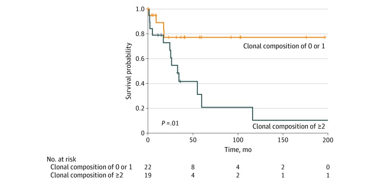
To examine the association of clinical outcome with increased heterogeneity, we stratified patients by clonal composition and performed survival analyses. Kaplan-Meier analysis demonstrated that cases with a clonal composition count of 2 or greater exhibited much worse survival compared with cases with a clonal composition count of 0 to 1 (univariate hazard ratio, 3.92; 95% CI, 1.27-12.08; P = .02) (Figure 1). This observation remained significant on multivariate analysis controlling for stage (T4 vs other), Lauren histologic subtype (intestinal vs other), receipt of adjuvant therapy (yes vs no), and age (continuous) (multivariate hazard ratio, 4.55; 95% CI, 1.09-19.04; P = .04). This finding aligns well with pancancer analyses of exome-sequencing data in which bioinformatic approaches have associated worse survival with higher intratumoral clone numbers in other tumor types.7
Figure 1. Kaplan-Meier Analysis of Survival in the Study Patient Population.
Data are stratified based on clonal composition count.
Confirmation of Intratumoral Heterogeneity and Ascertainment of Spatial Relationships by Multiprobe FISH
With evidence of significant intratumoral heterogeneity determined by the OncoScan SNV array calculation of clonal composition, we pursued multiprobe FISH analyses to ascertain whether detected multiple CNAs were restrained to a single cell or distributed among differing clonal populations. We initially focused on patient 29, who had a pT3N0 diffuse subtype adenocarcinoma arising from the gastroesophageal junction with amplification of MET, FGFR2, and CCND1 (eFigure 4A in the Supplement). Copy number gains in EGFR and copy number loss in CD274 and PDCD1LG2, which encode programmed death ligands 1 and 2 (PD-L1 and PD-L2), respectively, were also detected. Analysis of clonal composition had determined the existence of 3 clones, although this may be an underestimate owing to increased ploidy of the sample. In-depth multiprobe FISH analysis revealed intratumoral heterogeneity for MET gene amplification and heterogeneity in coamplification of other oncogenes among differing spatial regions. In the primary tumor region (designated target 1), tumor cells exhibited only 3 copies of MET, 4 to 7 copies of EGFR, 3 to 4 copies of FGFR2, single copies of the genes encoding PD-L1/PD-L2, and significant amplification of CCND1 (Figure 2 and eFigure 5 in the Supplement). Target 2, which resided 1.06 mm from target 1, exhibited tumor cells containing both strongly amplified MET and CCND1, 3 to 4 copies of EGFR, 3 to 4 copies of FGFR2, and loss of both copies of the genes encoding PD-L1/PD-L2. To localize the strong FGFR2 amplification reported by OncoScan but not exhibited in targets 1 or 2, we scanned the tumor area finding target 3, which resided 0.67 mm from target 1 and exhibited strong FISH amplification of FGFR2 along with CCND1. To further confirm whether other intratumoral areas may be heterogeneous for CCND1 amplification, we identified target 4, which resided 4.86 mm away from target 1, in which tumor cells harbored only 2 copies of CCND1 and 2 copies of FGFR2. Thus, in total for this case we observed 4 differing FISH oncogene coamplification patterns exemplifying significant spatial intratumoral heterogeneity (Figure 2).
Figure 2. Patient 29 .
The patient had a pT3N0 Lauren diffuse subtype adenocarcinoma arising from the gastroesophageal junction with copy number alterations in MET, FGFR2, CCND1, EGFR, CD274, and PDCD1LG2. A, Target 1 images reside at coordinates x = 14 124 μm and y = 23 706 μm and exhibited 3 copies of MET, amplified CCND1, and 3 to 4 copies of FGFR2. Target 2 image resides at coordinates x = 15 145 μm and y = 23 424 μm and exhibited amplified MET. Based on OncoScan data reporting FGFR2 amplification, additional targets were captured after initial analysis. An area of highly amplified FGFR2 was subsequently identified in target 3 (x = 13 505 μm and y = 23 449 μm). Target 4 resides the greatest distance from target 1 at coordinates x = 11 002 μm and y = 27 428 μm and exhibited a normal copy number for CCND1 and FGFR2. B, The tumor area of interest is circled on the hematoxylin-eosin–stained (H&E) slide section with the x, y reference coordinates at the 4 corners of the image displayed in micrometer distances. All fluorescent images were obtained at magnification x60; whole H&E tumor slide section image, magnification x5.
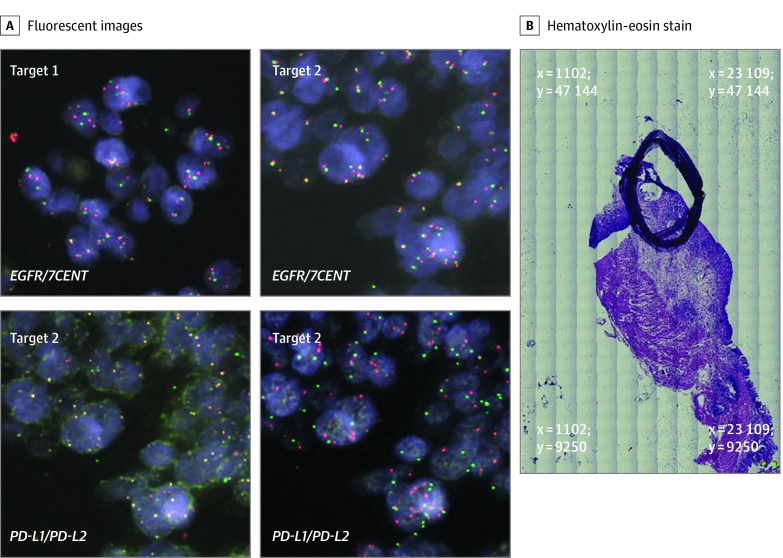
The complementary role of multiprobe FISH was confirmed in patient 37, with a pT2N3a intestinal subtype adenocarcinoma arising from the cardia. OncoScan analysis revealed gain of the genes encoding PD-L1 and PD-L2 and EGFR, MET, and PIK3CA (eFigure 4B in the Supplement). Testing for Epstein-Barr virus was also pursued, with no evidence of infection by viral RNA in situ hybridization. Clonal composition analysis had determined the existence of 3 clones. In depth multiprobe FISH analysis observed 2 major spatial regions spaced 2.52 mm apart in the tumor that exhibited variability in low and high copy gains of multiple oncogenes (Figure 3 and eFigure 6 in the Supplement). Target 1 demonstrated amplification of the genes encoding PD-L1 and PD-L2 but only modest copy gains in EGFR, MET, and PIK3CA. However, target 2 exhibited amplification of all 5 genes of interest. As such, in this sample we observed 2 FISH coamplification patterns in which the genetic events for PD-L1 and PD-L2 appear to be truncal alterations common to both target regions. However, amplification of EGFR, MET, and PIK3CA appear to be subclonal, in which all 3 oncogenes are coamplified within the same tumor cells as opposed to being each individually amplified across mutually exclusive clonal populations.
Figure 3. Patient 37.
This patient had a pT2N3a Lauren intestinal subtype adenocarcinoma arising from the gastric cardia. OncoScan analysis revealed major copy number alterations of CD274, PDCD1LG2, EGFR, MET, and PIK3CA. A, Target 1 image at coordinates x = 13 654 μm and y = 33 594 μm exhibited modest copy number gains of EGFR. Target 2 images at coordinates x = 11 292 μm and y = 34 466 μm exhibited amplification in all 5 oncogenes. B, The tumor area of interest is circled on the hematoxylin-eosin–stained (H&E) slide section with the x, y reference coordinates at the 4 corners of the image displayed in micrometer distances. All fluorescent images were obtained at magnification x60, and whole H&E tumor slide section image, magnification x5.
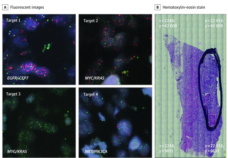
Finally, we focused on patient 21, with a pT3N0 intestinal histologic subtype tumor arising from the gastric antrum characterized by gains of EGFR, MYC (OMIM 190080) KRAS, MET, and PIK3CA by OncoScan (eFigure 4C in the Supplement). Clonal composition analysis had determined the existence of 2 clones. Target area 1 exhibited amplification in EGFR only with normal copy numbers of MET, MYC, and PIK3CA and modest copy number gain of KRAS (Figure 4 and eFigure 7 in the Supplement). Target 2 demonstrated amplification of MYC but normal copy numbers of KRAS as well as EGFR, MET, and PIK3CA. Target 3 exhibited amplification of KRAS only, but a normal copy number of MYC. Target 4 demonstrated amplification of MET, but a normal copy number of PIK3CA. In terms of intratumoral distances from target 1, target 2 resided at 0.86 mm, target 3 resided at 0.99 mm, and target 4 resided at 0.57 mm. As such, although OncoScan analysis of the entire tumor section reported coamplification of multiple oncogenes, in-depth multiprobe FISH analysis discerned that differing subclonal tumor cell populations each contain amplification of a mutually exclusive single oncogene.
Figure 4. Patient 21.
This patient had a pT3N0 Lauren intestinal subtype adenocarcinoma arising from the gastric antrum. OncoScan analysis reported amplification of EGFR, MYC, KRAS, MET, and PIK3CA. A, Target 1 image resides at coordinates x = 16 854 μm and y = 24 633 μm and exhibited amplified EGFR. Target 2 image resides at coordinates x = 17 387 μm and y = 25 310 μm and demonstrated amplified MYC with normal KRAS copy number. Target 3 image resides at coordinates x = 17 682 μm and y = 25 188 μm and exhibited the converse of target 2 with amplified KRAS but normal MYC copy number. Target 4 image resides at coordinates x = 17 407 μm and y = 24 485 μm and demonstrated amplified MET with normal copy number for PIK3CA. B, The tumor area of interest is circled on the hematoxylin-eosin–stained (H&E) slide section with the x, y reference coordinates at the 4 corners of the image displayed in micrometer distances. All fluorescent images were obtained at 60 × magnification; whole H&E tumor slide section image, 5 × magnification.
Discussion
The inherent spatial intratumoral heterogeneity of oncogenic CNAs present in de novo disease illustrates the challenges in developing targeted GEA therapies. In our data set, we observed nearly one-half (46%) of the cases exhibiting significant subclones detected via SNV array analysis and computational tools to estimate the number of clonal populations. Our spatial FISH analyses also exemplify intercellular genomic heterogeneity of actionable oncogenes at distances of less than 1 mm and greater than 4 mm. Detection of heterogeneity across large intratumoral distances can be abrogated with multiregion sampling of a tumor. However, determination of heterogeneity at submillimeter distances will likely require single-cell resolution as demonstrated herein to offer insight into strategies on combinatorial and/or sequential targeted and immunotherapeutic approaches. Variation in CNAs within differing clonal populations adds to the literature of caution needed in informing clinical treatment decisions based on next-generation sequencing analyses of small biopsy samples that pool tumor DNA, invariably representing metagenomes of multiple clonal populations.7 Our present data set supports in-depth analysis of the spatial intratumoral landscape at de novo GEA presentation to provide a road map for initial clonal composition. The development of intratumoral heterogeneity has been attributed to the gradual accumulation of mutations over time selecting for favorably growing subclones.15,16,17 Other reports also support a single catastrophic genome-wide mutational and chromosomal rearrangement event (chromothripsis), which subsequently drives the outgrowth of a selectively favorable clone.18,19,20 In our data set via OncoScan analysis, we observed an example of a chromothripsis event having occurred during tumor evolution. An improved understanding of mechanisms driving heterogeneity will be key to developing therapeutic strategies in all stages of GEA because appreciation of factors influencing innate and acquired drug resistance is necessary to optimize outcomes.
Kwak et al3 reported patient cases of MET and ERBB2 coamplification occurring in the same tumor cells, including a single case of MET, ERBB2, and EGFR coamplification all occurring within a single tumor cell population. We also observed these findings, but in addition demonstrated cases at initial diagnosis in whom the primary tumor contains heterogeneity in which only a single oncogene is amplified in some tumor cells, and other tumor cells contain amplification of a differing oncogene. This finding hypothetically can affect targeted therapy strategies in which combination therapy is likely needed, in cases of coamplification within the same tumor cell, compared with sequential targeted therapy potentially used to eliminate successive clones in cases of coamplification occurring among heterogeneous tumor cell populations. Circulating tumor DNA may also have limitations in distinguishing these 2 mechanisms of coamplification, and as such repeated tumor biopsies may still need to be considered to provide spatial heterogeneity and enhance the temporal heterogeneity demonstrated by liquid biopsy. One may be able to infer through single-cell analyses that if only single oncogene amplification is captured, then identification of coamplification in circulating tumor DNA is likely accounted for by another clonal population residing in a nonsampled site. Thus, biopsy of at least a single metastatic lesion complemented by circulating tumor DNA analysis may provide a sufficient composite of tumoral heterogeneity at a given point in a patient’s therapy and avoid the invasiveness of sampling every metastatic site. Although limited by sample size, the immediate clinical implications of heterogeneity assessment are highlighted by our survival analyses, which suggest that more heterogeneous tumors carry a worse prognosis.
Limitations
Weaknesses of our study include the retrospective nature of the analysis without pairing of our genomic findings to clinical response data from targeted therapeutics. However, the restriction of our sample selection to cases treated with up-front surgical resection avoids confounding the evolution of the tumor’s mutational profile that may arise under the pressure of antineoplastic therapies. Furthermore, no molecularly targeted agents as yet have a proven effect in the nonmetastatic setting for GEA that would have provided meaningful clinical annotation of response to such targeted strategies. Despite this limitation, our data further support the existence of significant intratumoral heterogeneity within untreated primary GEAs, and genomic analysis to the depth of single-cell approaches can be useful for cases in which surgical resection of the primary tumor has been performed. Understanding the spatial mapping of intratumoral heterogeneity will support ongoing efforts incorporating targeted therapeutics in the neoadjuvant setting for resectable disease. If such efforts fail, our demonstration of significant intratumoral heterogeneity of multiple oncogene coamplification events existing at nonmetastatic disease presentation may account for primary resistance to such approaches. Pectasides et al1 similarly observed heterogeneity in oncogene amplification within geographically distinct regions of the primary tumor and lymph node metastases, although such observations were limited to large intratumoral distances.11 As such, our finding of heterogeneity occurring at submillimeter distances in early-stage GEA is novel and should guide single-cell analytic efforts in refining genomic heterogeneity events.
Conclusions
Our report represents, to our knowledge, one of the first efforts in GEA to demonstrate significant spatial intratumoral heterogeneity of relevant CNAs with clonal tumor cell populations coexisting at submillimeter distances. More heterogeneous tumors (clonal composition count of ≥2) exhibit worse clinical outcomes. Recognition of heterogeneous spatial coamplification patterns of intratumoral clonal populations existing at submillimeter distances lends support for single-cell analyses to guide precision medicine efforts.
eFigure 1. Distribution of Percentage of Genomic Changes
eFigure 2. A Case With Significant Alterations in Whole-Genome Copy Number and B-Allele Frequency Plot
eFigure 3. Oncoprint of CNAs and Mutations Detected With the OncoScan Platform Demonstrating Interpatient Tumoral Heterogeneity of Genomic Alterations of Selected Genes of Interest
eFigure 4. OncoScan Genomic Data
eFigure 5. Patient 29 Representing a pT3N0 Lauren Diffuse Subtype Adenocarcinoma Arising From the GEJ With CNAs in MET, FGFR2, CCND1, EGFR, CD274, and PDCD1LG2
eFigure 6. Patient 37 Representing a pT2N3a Lauren Intestinal Subtype Adenocarcinoma Arising From the Gastric Cardia
eFigure 7. Patient 21 Representing a pT3N0 Lauren Intestinal Subtype Adenocarcinoma Arising From the Gastric Antrum
References
- 1.Pectasides E, Stachler MD, Derks S, et al. Genomic heterogeneity as a barrier to precision medicine in gastroesophageal adenocarcinoma. Cancer Discov. 2018;8(1):-. doi: 10.1158/2159-8290.CD-17-0395 [DOI] [PMC free article] [PubMed] [Google Scholar]
- 2.Bartley AN, Washington MK, Colasacco C, et al. HER2 testing and clinical decision making in gastroesophageal adenocarcinoma: guideline from the College of American Pathologists, American Society for Clinical Pathology, and the American Society of Clinical Oncology. J Clin Oncol. 2017;35(4):446-464. doi: 10.1200/JCO.2016.69.4836 [DOI] [PubMed] [Google Scholar]
- 3.Kwak EL, Ahronian LG, Siravegna G, et al. Molecular heterogeneity and receptor coamplification drive resistance to targeted therapy in MET-amplified esophagogastric cancer. Cancer Discov. 2015;5(12):1271-1281. doi: 10.1158/2159-8290.CD-15-0748 [DOI] [PMC free article] [PubMed] [Google Scholar]
- 4.Van Cutsem E, Bang YJ, Mansoor W, et al. A randomized, open-label study of the efficacy and safety of AZD4547 monotherapy versus paclitaxel for the treatment of advanced gastric adenocarcinoma with FGFR2 polysomy or gene amplification. Ann Oncol. 2017;28(6):1316-1324. doi: 10.1093/annonc/mdx107 [DOI] [PubMed] [Google Scholar]
- 5.Cancer Genome Atlas Research Network Comprehensive molecular characterization of gastric adenocarcinoma. Nature. 2014;513(7517):202-209. doi: 10.1038/nature13480 [DOI] [PMC free article] [PubMed] [Google Scholar]
- 6.Cancer Genome Atlas Research Network Integrated genomic characterization of oesophageal carcinoma. Nature. 2017;541(7636):169-175. doi: 10.1038/nature20805 [DOI] [PMC free article] [PubMed] [Google Scholar]
- 7.Andor N, Graham TA, Jansen M, et al. Pan-cancer analysis of the extent and consequences of intratumor heterogeneity. Nat Med. 2015;22(1):105-113. doi: 10.1038/nm.3984 [DOI] [PMC free article] [PubMed] [Google Scholar]
- 8.Kempen JH. Appropriate use and reporting of uncontrolled case series in the medical literature. Am J Ophthalmol. 2011;151(1):7-10.e1. doi: 10.1016/j.ajo.2010.08.047 [DOI] [PMC free article] [PubMed] [Google Scholar]
- 9.Wang Y, Carlton VE, Karlin-Neumann G, et al. High quality copy number and genotype data from FFPE samples using Molecular Inversion Probe (MIP) microarrays. BMC Med Genomics. 2009;2:8. doi: 10.1186/1755-8794-2-8 [DOI] [PMC free article] [PubMed] [Google Scholar]
- 10.Sakai K, Ukita M, Schmidt J, et al. Clonal composition of human ovarian cancer based on copy number analysis reveals a reciprocal relation with oncogenic mutation status. Cancer Lett. 2017;405:22-28. doi: 10.1016/j.canlet.2017.07.013 [DOI] [PubMed] [Google Scholar]
- 11.Van Loo P, Nordgard SH, Lingjærde OC, et al. Allele-specific copy number analysis of tumors. Proc Natl Acad Sci U S A. 2010;107(39):16910-16915. doi: 10.1073/pnas.1009843107 [DOI] [PMC free article] [PubMed] [Google Scholar]
- 12.Bedell V, Forman SJ, Gaal K, Pullarkat V, Weiss LM, Slovak ML. Successful application of a direct detection slide-based sequential phenotype/genotype assay using archived bone marrow smears and paraffin embedded tissue sections. J Mol Diagn. 2007;9(5):589-597. doi: 10.2353/jmoldx.2007.070050 [DOI] [PMC free article] [PubMed] [Google Scholar]
- 13.Cerami E, Gao J, Dogrusoz U, et al. The cBio cancer genomics portal: an open platform for exploring multidimensional cancer genomics data. Cancer Discov. 2012;2(5):401-404. doi: 10.1158/2159-8290.CD-12-0095 [DOI] [PMC free article] [PubMed] [Google Scholar]
- 14.Gao J, Aksoy BA, Dogrusoz U, et al. Integrative analysis of complex cancer genomics and clinical profiles using the cBioPortal. Sci Signal. 2013;6(269):pl1-pl1. doi: 10.1126/scisignal.2004088 [DOI] [PMC free article] [PubMed] [Google Scholar]
- 15.Bozic I, Antal T, Ohtsuki H, et al. Accumulation of driver and passenger mutations during tumor progression. Proc Natl Acad Sci U S A. 2010;107(43):18545-18550. doi: 10.1073/pnas.1010978107 [DOI] [PMC free article] [PubMed] [Google Scholar]
- 16.Greaves M, Maley CC. Clonal evolution in cancer. Nature. 2012;481(7381):306-313. doi: 10.1038/nature10762 [DOI] [PMC free article] [PubMed] [Google Scholar]
- 17.Hunter KW, Amin R, Deasy S, Ha N-H, Wakefield L. Genetic insights into the morass of metastatic heterogeneity. Nat Rev Cancer. 2018;18(4):211-223. doi: 10.1038/nrc.2017.126 [DOI] [PMC free article] [PubMed] [Google Scholar]
- 18.Stephens PJ, Greenman CD, Fu B, et al. Massive genomic rearrangement acquired in a single catastrophic event during cancer development. Cell. 2011;144(1):27-40. doi: 10.1016/j.cell.2010.11.055 [DOI] [PMC free article] [PubMed] [Google Scholar]
- 19.Rausch T, Jones DT, Zapatka M, et al. Genome sequencing of pediatric medulloblastoma links catastrophic DNA rearrangements with TP53 mutations. Cell. 2012;148(1-2):59-71. doi: 10.1016/j.cell.2011.12.013 [DOI] [PMC free article] [PubMed] [Google Scholar]
- 20.Molenaar JJ, Koster J, Zwijnenburg DA, et al. Sequencing of neuroblastoma identifies chromothripsis and defects in neuritogenesis genes. Nature. 2012;483(7391):589-593. doi: 10.1038/nature10910 [DOI] [PubMed] [Google Scholar]
Associated Data
This section collects any data citations, data availability statements, or supplementary materials included in this article.
Supplementary Materials
eFigure 1. Distribution of Percentage of Genomic Changes
eFigure 2. A Case With Significant Alterations in Whole-Genome Copy Number and B-Allele Frequency Plot
eFigure 3. Oncoprint of CNAs and Mutations Detected With the OncoScan Platform Demonstrating Interpatient Tumoral Heterogeneity of Genomic Alterations of Selected Genes of Interest
eFigure 4. OncoScan Genomic Data
eFigure 5. Patient 29 Representing a pT3N0 Lauren Diffuse Subtype Adenocarcinoma Arising From the GEJ With CNAs in MET, FGFR2, CCND1, EGFR, CD274, and PDCD1LG2
eFigure 6. Patient 37 Representing a pT2N3a Lauren Intestinal Subtype Adenocarcinoma Arising From the Gastric Cardia
eFigure 7. Patient 21 Representing a pT3N0 Lauren Intestinal Subtype Adenocarcinoma Arising From the Gastric Antrum


