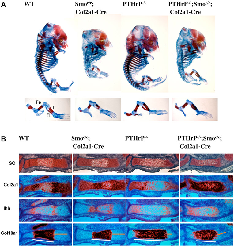
Fig. 4. Removal of Hh signaling delays chondrocyte hypertrophy in the absence of PTHrP.
(A) Skeletal preparation of E15.5 embryos. Hindlimbs are shown at higher magnifications in the lower panel. (B) Serial sections of tibia were stained with Safranin O and hybridized with 35S labeled Ihh and Col10a1 riboprobes. Smoc/c;Col2a1-Cre mutant tibia showed a slight delay in chondrocyte hypertrophy compared with that of wild-type embryos. PTHrP−/−;Smoc/c;Col2a1-Cre mutant tibia also showed a delay of chondrocyte hypertrophy, as compared with that of the PTHrP−/− mutant. The proliferating chondrocyte region is indicated by the yellow line; the hypertrophic region is indicated by the white line. Fe, femur; T, tibia; Fi, fibula.

