Abstract
Elastofibroma dorsi is a benign lesion commonly presents as a palpable enlarging mass at the inferior pole of the scapula. Clinical presentation and radiological characteristics are often enough to suggest an accurate diagnosis. Increased awareness of the characteristic appearance and location of these benign lesions will increase radiologic diagnosis and decrease the need for biopsy.
Ten patients were admitted with a complaint of asymptomatic or painful subcutaneous masses localized at subscapulary region. Thorax computed tomography, magnetic resonance imaging (MRI) and a new feasible technique in differential diagnosis with malignancy and probable diagnosis of elastofibroma dorsi and diffusion-weighted MRI were used for diagnosis.
Surgery was applied to all patients, frozen-section biopsies of the lesions at the preoperative period, and final pathologies were all benign. Totally resection of whole lesions as en-bloc excision without any rest was performed at all patients. Postoperative and follow-up periods were uneventful.
Diffusion MRI can play an important role in the future and save the patients, especially medically poor ones, from the potential risks of surgery. Necessary further examinations for probable bilaterally lesions will save the patient from the risk of a second operation.
Keywords: Chest wall, imaging, tumour
Elastofibroma dorsi (ED) is a rare, slow-growing, non-capsulated, benign soft tissue tumour of the chest wall that arises from connective tissue forming collagen bundles. ED is most commonly located in the periscapular region, beneath the rhomboid major and latissimus dorsi muscles, especially in older women. ED can be bilaterally, but not always in synchronous localizations. ED may be densely adherent to surrounding muscle and bone. Thus, the suspicion for malignancy is often raised. In addition to careful clinical investigation, radiology is the method of choice leading to a presumptive diagnosis.[1] In addition to a few sporadic cases, the only literature reporting a large series in the literature is with 170 cases.[2] Ultrasound, computed tomography (CT) and magnetic resonance imaging (MRI) have all been used to characterize elastofibroma. Surgical extirpation is usually curative.[2,3] We report 10 cases of ED treated with surgery and want to point out a new promising diagnostic technique to differentiate benign lesions without surgery.
Methods
Eight female and two male patients, ages between 44-72 years-old (median: 54), were admitted to the clinic with the complaint of asymptomatic or painful subcutaneous masses localized subscapular. Lesions were palpated at the right side in three patients, left in two and bilaterally in five patients. Thorax CT and/or MR (Fig. 1) performed to all. Five patients were detected with MR (Fig. 2a, b) and one patient with also diffusion (Fig. 2c). All revealed heterogeneous soft tissue masses under the latissimus dorsi muscle. Diffusion MRI, which can be a new diagnostic technique for the ED, has been used to differentiate malign lesions in one case. The diffusion-weighted sequence showed a 6x2.5cm mass with an intense restriction at right between the scapula and the chest wall. The mass is characterized by a moderately heterogeneous hyperintense signal in T2. The fusion of the diffusion-weighted sequence in T2 demonstrates more clearly the intense restriction in the mass.
Figure 1.
Thorax CT (a) and MRI (b, c) sections of left-sided soft tissue mass under latissimus dorsi.
Figure 2.
Thorax MRI (a, b) of bilaterally heterogeneous soft tissue masses and diffusion MR (c) of another case with right-sided mass under latissimus dorsi.
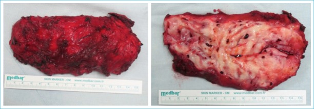
Surgical resection was performed by a posterolaterally incision directly over the mass at the prone position with arms at abduction. Transverse incision and dissection parallel to latissimus dorsi muscle made the tumour visible then totally resection was performed. Although varying in size, all of the lesions had the same anatomical placement and were lying under the scapula, adherent to the ribs. They were all mobile and like tempered rubber with palpation (Fig. 3). Resections of bilaterally lesions in five patients were performed during the same session. Pathologies were benign ED (Fig. 4). The postoperative period was uneventful; patients were pain-free with a completely normal shoulder range of motion. Median follow up was 32 (12-80) months, and there was no recurrence.
Figure 3.
Resected and incised specimen just after surgery.
Figure 4.
(a) Dark-colored elastic fibers between hyalinised collagen tissue; seen heterogeneous at horizontal sections and circular at transverse sections with Haematoxylin-Eosining (x100). (b) Darker elastic fibers detected between fat tissues inside heterogeneous dense collagen connective tissue with Mason trichrome (x100).
Discussion
Elastofibroma dorsi is a subcutaneous, benign nodular lesion, firstly, reported by Jarvi in 1961.[4] Although more than 90% of the cases are localized to the lower subscapular region, deep in the rhomboid and latissimus dorsi muscles, unusual locations, such as deltoid muscle, ischial tuberosity, olecranon, thoracic wall, axilla, foot, stomach, rectum, spinal canal, mediastinum, and cornea, were noted in the literature.[5]
Our findings support previous reports suggesting that a preoperative diagnosis is not necessary in most cases since the lesion can be confidently diagnosed by CT or MRI when interpreted in the light of appropriate clinical findings. Surgical excision in symptomatic patients is preferred.
Elastofibroma is not a true neoplastic process. Although it was suggested that repetitive microtrauma - might be because of the working status - by friction between the lower part of the scapula and the thoracic wall or hereditary enzyme defects might cause the reactive hyperproliferation of fibroelastic tissue, the pathogenesis of the lesion still remains unclear and thought to be multifactorial.[3,6] Microtrauma theory was not suitable for our patients. They were housewife, teachers, security, and retired people.
Elastofibromas are often seen in older women and mostly asymptomatic besides the appearance of a subcutaneous bump. In other cases, there may be moderate pain, and limited functions in hard efforts, or even in daily performance.[5,7] Compatible with the literature, our patients were mostly female and older than 45 years of age. All our lesions were anatomically located at the back of the shoulder adjacent to the scapula.
When it is detected at one side, necessary further investigations should be performed by considering lesions bilaterally. Thus, patients should be protected from the risk of a second operation, by operating on both sides in the same session. Our five patients had bilaterally lesions and this was compatible with the reports of possibility in 10-66% of all cases.[8]
Radiology takes an important role in the ED. The scapula may appear raised on a chest x-ray and the scapulo-thoracic space may appear enlarged. An opacity between the two scapulas may be identified, without bone lesions or associative calcifications. Thorax CT shows the typical characteristics of an unencapsulated lentiform shaped tumour with hypodense strands, which appears isodense to muscular structures.[2] All our CT or MRI scans were similar to those typical characteristics with indicating isodense, soft tissue masses under muscularis latissimus dorsi. Diffusion-weighted magnetic resonance imaging (DWI) is one such technique, which explores the translational mobility of water molecules, thereby shedding light on the microstructural features of the tissue of interest, whether they facilitate or restrict such freedom of proton mobility. DWI has been primarily used in neuroradiology, but applications in other body areas have also been increasing. Fast imaging techniques, such as echo-planar imaging (EPI), facilitate the use of DWI in thoracic imaging by decreasing the deleterious effects of motion.[9,10] This may be a new diagnostic tool for also elastofibroma with more specific findings by providing functional information about the diffusivity of water molecules and can highlight high cellularity lesions throughout the body.[11] Like in one of our patients, diffusion MRI revealed benign characteristics of the soft tissue mass. Elastofibromas display typical diagnostic histologic, cytologic, and electron microscopic features. Heterogenous, thick eosinophilic elastic bands of fat, muscle and collagen tissue can be detected.[12] As in our surgical specimens, H-E and mason trichrome staining can be strongly positive. In more recent articles, some characteristic imaging results may help in differentiating malignant from benign masses noninvasively.[13,14]
Conclusion
Patients with ED are often asymptomatic; and enlarging palpable mass is the common physical sign. When a mass lesion observed in the subscapular regions of the old patients, ED, a rare soft tissue tumour, should be considered. Surgery is essential in symptomatic patients, and although tumours of ED are benign, the histological study is advisable to establish a differential diagnosis with malignant neoplastic processes. Complete surgical excision is the treatment of choice. Thus, it is important to say with certainty that it is benign. Diffusion MRI can be a new tool to define the diagnosis as benign and prevent surgery in small, asymptomatic lesions or in patients with poor conditions for the operation without suspicion of malignancy.
Also, when it is detected unilaterally, bilaterally cases must be kept in mind and further examinations should be performed by considering that it may be bilaterally. In these cases, if surgery is needed, patients should be avoided from the risk of a second operation by operating on both sides in the same session.
Disclosures
Informed Consent: Written informed consent was obtained from the patient for the publication of the case report and the accompanying images.
Peer-review: Externally peer-reviewed.
Conflict of Interest: None declared.
Authorship Contributions: Concept – A.G.A.; Design – A.G.A.; Supervision – S.T.; Materials – A.G.A., U.T.; Data collection &/or processing – U.T.; Analysis and/or interpretation – A.G.A.; Literature search – A.G.A., U.T.; Writing – A.G.A., U.T.; Critical review – A.G.A.
References
- 1.Mortman KD, Hochheiser GM, Giblin EM, Manon-Matos Y, Frankel KM. Elastofibroma dorsi:clinicopathologic review of 6 cases. Ann Thorac Surg. 2007;83:1894–7. doi: 10.1016/j.athoracsur.2006.11.050. [DOI] [PubMed] [Google Scholar]
- 2.Nagamine N, Nohara Y, Ito E. Elastofibroma in Okinawa A clinicopathologic study of 170 cases. Cancer. 1982;50:1794–805. doi: 10.1002/1097-0142(19821101)50:9<1794::aid-cncr2820500925>3.0.co;2-l. [DOI] [PubMed] [Google Scholar]
- 3.Muratori F, Esposito M, Rosa F, et al. Elastofibroma dorsi:8 case reports and a literature review. J Orthop Traumatol. 2008;9:33–7. doi: 10.1007/s10195-008-0102-7. [DOI] [PMC free article] [PubMed] [Google Scholar]
- 4.Jarvi OH, Saxen AE. Elastofibroma dorse. Acta Pathol Microbiol Scand Suppl. 1961;51:83–4. [PubMed] [Google Scholar]
- 5.Briccoli A, Casadei R, Di Renzo M, Favale L, Bacchini P, Bertoni F. Elastofibroma dorsi. Surg Today. 2000;30:147–52. doi: 10.1007/PL00010063. [DOI] [PubMed] [Google Scholar]
- 6.Muramatsu K, Ihara K, Hashimoto T, Seto S, Taguchi T. Elastofibroma dorsi:diagnosis and treatment. J Shoulder Elbow Surg. 2007;16:591–5. doi: 10.1016/j.jse.2006.12.010. [DOI] [PubMed] [Google Scholar]
- 7.Soler R, Requejo I, Pombo F, Sáez A. Elastofibroma dorsi:MR and CT findings. Eur J Radiol. 1998;27:264–7. doi: 10.1016/s0720-048x(97)00074-0. [DOI] [PubMed] [Google Scholar]
- 8.Hayes AJ, Alexander N, Clark MA, Thomas JM. Elastofibroma:a rare soft tissue tumour with a pathognomonic anatomical location and clinical symptom. Eur J Surg Oncol. 2004;30:450–3. doi: 10.1016/j.ejso.2004.01.006. [DOI] [PubMed] [Google Scholar]
- 9.Tanaka R, Horikoshi H, Nakazato Y, Seki E, Minato K, Iijima M, et al. Magnetic resonance imaging in peripheral lung adenocarcinoma:correlation with histopathologic features. J Thorac Imaging. 2009;24:4–9. doi: 10.1097/RTI.0b013e31818703b7. [DOI] [PubMed] [Google Scholar]
- 10.Ohno Y, Sugimura K, Hatabu H. MR imaging of lung cancer. Eur J Radiol. 2002;44:172–81. doi: 10.1016/s0720-048x(02)00267-x. [DOI] [PubMed] [Google Scholar]
- 11.Hochhegger B, Marchiori E, Soares Souza L. MR Diffusion in Elastofibroma Dorsi. Arch Bronconeumol. 2011;47:535–6. doi: 10.1016/j.arbres.2011.06.006. [DOI] [PubMed] [Google Scholar]
- 12.Hoffman JK, Klein MH, McInerney VK. Bilateral elastofibroma:a case report and review of the literature. Clin Orthop Relat Res. 1996:245–50. [PubMed] [Google Scholar]
- 13.Gümüştaş S, Inan N, Sarisoy HT, Anik Y, Arslan A, Ciftçi E, et al. Malignant versus benign mediastinal lesions:quantitative assessment with diffusion weighted MR imaging. Eur Radiol. 2011;21:2255–60. doi: 10.1007/s00330-011-2180-9. [DOI] [PubMed] [Google Scholar]
- 14.Gümüştaş S, Inan N, Akansel G, Ciftçi E, Demirci A, Ozkara SK. Differentiation of malignant and benign lung lesions with diffusion-weighted MR imaging. Radiol Oncol. 2012;46:106–13. doi: 10.2478/v10019-012-0021-3. [DOI] [PMC free article] [PubMed] [Google Scholar]


