Abstract
Inadvertent dural puncture (IDP) is one of the complications of lumbar epidural steroid injections (ESIs). We report a case in which pneumocephalus and chemical meningitis developed at the same time after an IDP during a lumbar interlaminar ESI. A 60-year-old woman presented to the emergency room with thunderclap headache and febrile sensation 3 hours after receiving a lumbar interlaminar ESI. Brain computed tomography (CT) scan showed multiple small foci of air within the subarachnoid space and ventricle. After the admission, the patient was afebrile and reported mild improvement of headache with analgesics. However, 2 days after the admission, headache worsened and fever recurred. Follow-up brain CT scan revealed resolution of the pneumocephalus. A diagnostic lumbar puncture for cerebrospinal fluid (CSF) examination revealed the findings suggestive of aseptic (chemical) meningitis rather than bacterial meningitis. With symptomatic treatment, headache improved and there was no fever after 48 hours. No bacteria, Mycobacterium, or fungi grew in the CSF for 7 days. This case shows an IDP during a lumbar ESI can cause pneumocephalus and chemical meningitis at the same time and efforts should be made to reduce the risk of IDP during lumbar ESIs.
Keywords: Pneumocephalus, Meningitis, Dura mater, Punctures, Injections
INTRODUCTION
Epidural steroid injections (ESIs) have been used for the symptomatic relief of back pain with a radicular component in patients with herniated disk, foraminal stenosis, and central canal stenosis.2,13) ESIs are relatively safe, and serious complications are uncommon.2,10,13) Inadvertent dural puncture (IDP) is one of the complications of ESIs and can cause postdural puncture headache (PDPH) or pneumocephalus.1,2,13,17) Although extremely rare, meningitis is also one of the complications of ESIs.5,16) Although there were clinical studies and case reports that reported these two complications separately, there were only a few case reports in which both complications occurred at the same time after ESIs.5,16) We report a case in which pneumocephalus and chemical meningitis developed at the same time after an IDP during a lumbar interlaminar ESI.
CASE REPORT
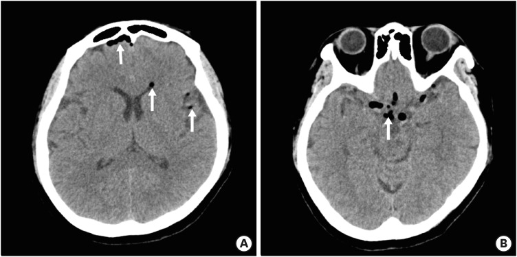
A 60-year-old woman presented to the emergency room (ER) with thunderclap headache and febrile sensation 3 hours after receiving a lumbar interlaminar ESI with mepivacaine and dexamethasone at an outpatient pain clinic. The patient reported there was no abnormal symptoms except mild discomfort in the lower back during or immediately after the procedure. When leaving the clinic 30 minutes after the procedure, a mild headache began. Over time, the headache got worse, and a thunderclap headache with febrile sensation began. The headache was global, constant, and throbbing. Changes in the posture and position were not related to the intensity of the headache. On arrival at the ER, the patient's body temperature was 40.1°C, and blood pressure, pulse rate and respiration rate were normal. On examination, no neck stiffness and focal neurologic deficits were found. In the laboratory tests, white blood cell (WBC) count was 9400/uL (normal range: 4,000–10,000/uL) with 83.9% of neutrophils and C-reactive protein (CRP) was 0.11 (normal range: 0–0.5 mg/dL). Although postdural puncture headache (PDPH) was suspected, the patient underwent a non-contrast brain computed tomography (CT) scan to rule out other causes of thunderclap headache. Brain CT scan showed multiple small foci of air within the subarachnoid space and lateral ventricle (FIGURE 1). The patient had a fever on arrival at the ER. Therefore, we suspected a possibility of meningitis and recommended the patient a diagnostic lumbar puncture (LP) for cerebrospinal fluid (CSF) study at the ER. However, the patient refused the LP stubbornly because the patient still had the discomfort in the lower back due to the ESI and appealed fear of the LP. Therefore, while monitoring the body temperature, we decided to do the diagnostic LP when the fever recurred.
FIGURE 1. Brain CT at the emergency room. (A) CT shows multifocal air density in the lateral ventricle and subarachnoid space (arrows). (B) CT shows multifocal air density in the basal cisterns (arrow).
CT: computed tomography.
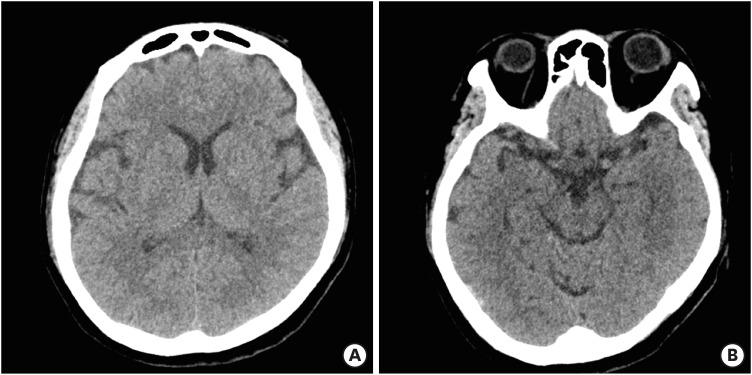
The patient admitted for the symptomatic treatment, monitoring vital signs, and serial neurologic examinations. The following day, the patient was afebrile and reported mild improvement of the headache with analgesics. However, the next day, the headache got worse and there was a fever accompanied by vomiting. In the blood tests, an increase in CRP from 0.11 to 3.72 mg/dL was found. Follow-up brain CT scan revealed resolution of the pneumocephalus (FIGURE 2). A diagnostic LP for CSF study was performed to rule out meningitis. Opening pressure was 26 cmH2O and CSF examination showed WBC 300/mm3 with 59.5% neutrophils, 18% lymphocyte, 22% monocyte, protein 62.1 mg/dL, and glucose 49.1 mg/dL with serum glucose 119 mg/dL. Gram's staining of the CSF showed no organisms. Under the diagnosis of chemical meningitis rather than bacterial meningitis, antibiotic treatment was withheld until the CSF culture results were confirmed. With symptomatic treatment with hydration and analgesics, the headache improved and there was no fever after 48 hours. The patient remained afebrile for the next 5 days and was discharged without headache and fever. No bacteria, Mycobacterium, or fungi grew in the CSF for 7 days.
FIGURE 2. Brain CT (2 days later) shows resolution of pneumocephalus.
CT: computed tomography.
DISCUSSION
IDP is one of the complications of epidural anesthesia and ESIs.1,2,3,13,17) Choi et al.3) reported that the incidence of IPD during obstetrical epidural anesthesia was 1.5% in their meta-analysis. Aida et al.1) reported that the incidence of IDP during interlaminar ESI was 2.7% of 3730 patients in the retrospective study. Manchikanti et al.10) reported that the incidence of IDP was 0.8% in a prospective evaluation of 1,450 fluoroscopically directed lumbar interlaminar ESIs.
IDP can cause PDPH.3,14) PDPH is typically secondary to a CSF leak after an IDP, but can be associated with pneumocephalus after an IDP in rare cases.14) Two main mechanisms have been postulated for the development of pneumocephalus in relation to lumbar anesthesias.7,14) The ball-valve theory is air movement in one direction from outside into the cranial cavity, which then creates an air trap.7,14) In the other mechanism, the inverted soda bottle theory, excessive CSF loss leads to a negative intracranial pressure gradient that may be relieved by the influx of air.7,14) In the present case, the air might have been injected into the subarachnoid space when an unrecognized IDP occurred during the interlaminar ESI.
Headaches associated with pneumocephalus usually have a rapid onset, are severe, and may not be relieved with recumbency or supine position.14) On the other hand, headache associated with persistent CSF leakage usually has a later onset, and is relieved when the patient lies down, but is provoked or worsens with sitting or standing.14) In the present case, the patient's headache on arrival at the ER might be due to the pneumocephalus, because the headache was a rapid onset, severe, and not relieved when the patient was lying down. It was possible that headache got worse to thunderclap headache as the air in the spinal subarachnoid space moved to cephalad during standing or sitting while returning to home after the procedure.6)
Loss of resistance to air (LORA) technique and loss of resistance to saline (LORS) technique are used for the identification of the epidural space during ESIs or epidural anesthesias.1,4,15) Some authors reported a higher frequency of IDP when using the LORA technique to identify the epidural space, while others report no difference in the frequency of IDP between the 2 techniques.1,4,15) However, even if there is no difference in the frequency of IDP between the two techniques, when an IDP occurs during EISs or epidural anesthesia, the incidence of pneumocephalus and resultant headache is higher when using the LORA technique.1,15) In the present case, the LORA technique was used for the interlaminar lumbar ESI.
When an IDP occurs during ESIs or epidural anesthesias using LORA technique, air can enter the subarachnoid space or subdural space, resulting in pneumocephauls.1,17) The intracranial air bubbles can behave like a space-occupying lesion, causing meningeal irritation and resultant headache.12) In 2014, Verdun et al.17) reviewed the literature on pneumocephalus after ESIs published from 1919 to 2013. In their review, onset of pneumocephalus-related headache could begin from immediately following the procedure and symptom resolution was variable from 1 to 3 hours to 7 days.17) Time to radiological resolution of pneumocephalus was also variable from 1 day to 2 weeks.17) In the present case, pneumocephalus disappeared within 2 days based on the brain CT findings.
When an unrecognized IDP during ESIs or epidural anesthesia, not only air, but drugs can also enter the subdural or subarachnoid space.5,16) It is a commonly held belief that the instillation of any substance into the CSF may result in chemical irritation and meningitis.11) Steroid or local anesthetic agents such as bupivacaine, lidocaine, or mepivacaine can cause chemical meningitis when entering the thecal sac.5,8,16) Although there were many reports of chemical meningitis due to local anesthetics after spinal anesthesia or due to steroid after intrathecal steroid injections, reported cases of chemical meningitis after lumbar ESIs are extremely rare.5,16) In addition, reported cases in which chemical meningitis and pneumocepahus occurred at the same time after lumbar ESIs were even rarer.5,16) Gutknecht5) reported a case of chemical meningitis in which symptoms of meningitis developed 4 hours after the epidural injection of methylprednisolone for the treatment of lumbar radiculopathy. In this case, brain CT scan showed air droplets in the subarachnoid space and probable dural puncture was noted during the injection in the author's opinion.5) Shah et al.16) reported a case of pneumocephalus and chemical meningitis in which symptoms of meningitis developed 1.5 hours after the epidural injection of lidocaine, methylprednisolone and betamethasone. In the present case, mixed solution with dexamethasone and mepivacaine was used for the interlaminar ESI. It is unclear, however, which component of the injected solution induced the chemical meningitis.
In the present case, the patient had a fever. It is not known whether or not there was chemical meningitis on arrival at the ER because the diagnostic LP for CSF study was not performed at the ER. In addition, intraventricular air itself has been reported to cause fever.9) It was suggested that the hypothalamic temperature regulating center is stimulated by intraventricular air.9) It is also possible that irritation of either the ependyma or the meninges may be responsible for the fever.9) Therefore, it is unclear whether the cause of fever on arrival at the ER was due to chemical meningitis, pneumocephalus, or both. The worsening headache and recurred fever 2 days after the admission might be due to chemical meningitis because the headache and fever developed even after air was lost on the brain CT scan.
In the present case, pneumocephalus and chemical meningitis were most likely caused by an IDP during an interlaminar ESI. The risk of IDP can be reduced by the use of a blunt needle, the use of the LORS technique rather than the LORA technique, and accurate placement of the needle on live fluoroscopy.13,17) Some authors recommend, in case of significant central canal stenosis from a disk herniation, bilateral transforaminal ESIs rather than interlaminar ESIs or positioning the needle 1 interspace cephalad to reduce the risk of IDP in the narrowed epidural space.17)
CONCLUSION
Pneumocephalus and chemical meningitis can occur at the same time after an IDP during ESIs. Best efforts should be made to reduce the risk of IDP during ESIs. This can prevent pneumocephalus or meningitis as well as more serious complications such as paresis, cardiopulmonary arrest, or respiratory depression.
Footnotes
Conflict of Interest: The authors have no financial conflicts of interest.
References
- 1.Aida S, Taga K, Yamakura T, Endoh H, Shimoji K. Headache after attempted epidural block: the role of intrathecal air. Anesthesiology. 1998;88:76–81. doi: 10.1097/00000542-199801000-00014. [DOI] [PubMed] [Google Scholar]
- 2.Benyamin RM, Manchikanti L, Parr AT, Diwan S, Singh V, Falco FJ, et al. The effectiveness of lumbar interlaminar epidural injections in managing chronic low back and lower extremity pain. Pain Physician. 2012;15:E363–E404. [PubMed] [Google Scholar]
- 3.Choi PT, Galinski SE, Takeuchi L, Lucas S, Tamayo C, Jadad AR. PDPH is a common complication of neuraxial blockade in parturients: a meta-analysis of obstetrical studies. Can J Anaesth. 2003;50:460–469. doi: 10.1007/BF03021057. [DOI] [PubMed] [Google Scholar]
- 4.Evron S, Sessler D, Sadan O, Boaz M, Glezerman M, Ezri T. Identification of the epidural space: loss of resistance with air, lidocaine, or the combination of air and lidocaine. Anesth Analg. 2004;99:245–250. doi: 10.1213/01.ANE.0000120084.56136.15. [DOI] [PubMed] [Google Scholar]
- 5.Gutknecht DR. Chemical meningitis following epidural injections of corticosteroids. Am J Med. 1987;82:570. doi: 10.1016/0002-9343(87)90468-2. [DOI] [PubMed] [Google Scholar]
- 6.Kozikowski GP, Cohen SP. Lumbar puncture associated with pneumocephalus: report of a case. Anesth Analg. 2004;98:524–526. doi: 10.1213/01.ANE.0000095153.75625.1F. [DOI] [PubMed] [Google Scholar]
- 7.Kwon J, Rha HK, Park HK, Chough CK, Joo WI, Cho SH, et al. Proper management of posttraumatic tension pneumocephalus. Korean J Neurotrauma. 2017;13:158–161. doi: 10.13004/kjnt.2017.13.2.158. [DOI] [PMC free article] [PubMed] [Google Scholar]
- 8.Lee SH, Lee MJ, Kim WJ. Chemical meningitis after spinal or epidural anesthesia. J Neurocrit Care. 2014;7:40–43. [Google Scholar]
- 9.Lipton MJ, Crowther D. Fever after air encephalography. Br J Radiol. 1968;41:672–673. doi: 10.1259/0007-1285-41-489-672. [DOI] [PubMed] [Google Scholar]
- 10.Manchikanti L, Malla Y, Wargo BW, Cash KA, Pampati V, Fellows B. A prospective evaluation of complications of 10,000 fluoroscopically directed epidural injections. Pain Physician. 2012;15:131–140. [PubMed] [Google Scholar]
- 11.Marinac JS. Drug- and chemical-induced aseptic meningitis: a review of the literature. Ann Pharmacother. 1992;26:813–822. doi: 10.1177/106002809202600613. [DOI] [PubMed] [Google Scholar]
- 12.Nafiu OO, Urquhart JC. Pneumocephalus with headache complicating labour epidural analgesia: should we still be using air? Int J Obstet Anesth. 2006;15:237–239. doi: 10.1016/j.ijoa.2005.10.011. [DOI] [PubMed] [Google Scholar]
- 13.Pountos I, Panteli M, Walters G, Bush D, Giannoudis PV. Safety of epidural corticosteroid injections. Drugs R D. 2016;16:19–34. doi: 10.1007/s40268-015-0119-3. [DOI] [PMC free article] [PubMed] [Google Scholar]
- 14.Reddi S, Honchar V, Robbins MS. Pneumocephalus associated with epidural and spinal anesthesia for labor. Neurol Clin Pract. 2015;5:376–382. doi: 10.1212/CPJ.0000000000000178. [DOI] [PMC free article] [PubMed] [Google Scholar]
- 15.Schier R, Guerra D, Aguilar J, Pratt GF, Hernandez M, Boddu K, et al. Epidural space identification: a meta-analysis of complications after air versus liquid as the medium for loss of resistance. Anesth Analg. 2009;109:2012–2021. doi: 10.1213/ANE.0b013e3181bc113a. [DOI] [PubMed] [Google Scholar]
- 16.Shah AK, Bilko A, Takayesu JK. Epidural steroid injection complicated by intrathecal entry, pneumocephalus, and chemical meningitis. J Emerg Med. 2016;51:265–268. doi: 10.1016/j.jemermed.2016.05.040. [DOI] [PubMed] [Google Scholar]
- 17.Verdun AV, Cohen SP, Williams BS, Hurley RW. Pneumocephalus after lumbar epidural steroid injection: a case report and review of the literature. A A Case Rep. 2014;3:9–13. doi: 10.1213/XAA.0000000000000055. [DOI] [PubMed] [Google Scholar]


