Development of bone marrow (BM) fibrosis and transformation to accelerated/blast phase are the main forms of disease progression in myeloproliferative neoplasms (MPN). Chronic myeloid leukemia, BCR-ABL1-positive has several well-defined criteria that qualify for a diagnosis of accelerated phase (AP) disease according to the updated World Health Organisation (WHO) Classification.1 However, the only WHO criterion for diagnosing AP BCR-ABL1 negative MPN is the presence of 10-19% blasts in the BM and/or peripheral blood (PB).1 Clinical characteristics and outcomes of patients with increased blasts falling below the 10% cut-off are not well-known. Prognostic schemes such as the International Prognostic Scoring System (IPSS) and the dynamic prognostic model (DIPSS), which are used to estimate survival and risk of leukemic transformation in MPN patients, include presence of ≥1% circulating blasts to predict disease progression.2,3 Other schemes have suggested circulating blast cut-offs of 2% or 3%.2–7 However, the current WHO classification that forms the basis for the pathology practice does not take into account increased blasts when they fall short of 10%.
Other less common types of disease progression have also been recently reported, but their characteristics in relation to AP have not yet been characterized. They include the development of persistent absolute monocytosis in patients with established primary myelofibrosis (PMF), an occurrence that has been associated with rapid progression and short overall survival.8–10 Absolute monocytosis was also found to be a high-risk prognostic factor in patients with polycythemia vera (PV).11 In addition, persistent neutrophilic leukocytosis (≥13×109/L) occurring around the time of progression to post-polycythemic myelofibrosis (post-PV MF) has also been associated with an aggressive course and shorter survival.12 An elevated leukocyte count (≥15×109/L) at disease onset or early during the polycythemic phase of PV has also been identified as an independent unfavorable prognostic factor.13,14
In this study we searched the files of four large academic medical centers in the United States and Italy for cases of BCR-ABL1 negative MPN showing progression-associated features. One hundred fourteen patients showed one of the following findings: circulating PB (2-19%) and/or BM (≥5-19%) blasts; persistent (≥3 months) neutrophilic leukocytosis (white blood cell count [WBC] ≥15×109/L); or persistent (≥3 months) absolute monocytosis (≥1×10⁹/L with monocytes accounting for ≥10% of the leukocytes). In the latter two settings, careful review of the patients’ clinical data was performed to exclude clinical conditions or treatments known to cause reactive neutrophilia or monocytosis. Patients presenting in blast phase (>20% PB or BM blasts) were excluded. Cases were also excluded if any of the above progression-associated features were already present at the disease onset. The original diagnosis in all cases was established in accordance the WHO criteria.
The cases were divided into four groups: increased blasts-1 (IB-1) with 2-4% PB blasts and <5% BM blasts; increased blasts-2 (IB-2) with 5-9% blasts in BM and/or PB; accelerated phase (AP) with 10-19% blasts in BM and/or PB; and other types of progression (OP) without increased blasts. A control group of 93 patients with BCR-ABL1 negative MPN lacking increased blasts, monocytosis, or leukocytosis was identified from the archives of Weill Cornell Medicine over the same time period. The controls were matched with respect to age and MPN type, including PV, essential thrombocytemia (ET), PMF, post-PV MF and post-essential thrombocythemia myelofibrosis (post-ET MF).
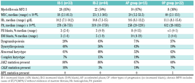
The patients’ median age at disease progression was 69 (range 38-88 years). There were 72 men and 42 women (1.7:1). The initial diagnoses included PV (n=35, 31%), ET (n=9, 8%), MPN unclassifiable (n=4, 4%), post-PV MF (n=37, 32%), prefibrotic PMF (n=5, 4%), fibrotic PMF (n=19, 17%) and post-ET MF (n=5, 4%) (see Table 1). The diagnosis of disease progression was rendered after a median of 11 (range: <1-40) years of follow-up after patient’s initial diagnosis. Ninety-two (79%) patients had increased blasts and were classified as IB-1, IB-2 or AP group (see Table 1). The OP group included 22 patients with persistent absolute monocytosis or neutrophilic leukocytosis. In addition to defined signs of progression above, the most common additional clinical signs at the time of disease progression were increasing symptomatic splenomegaly (76%), anemia (35%), and decreasing platelet count (15%).
Table 1.
The summary of clinicopathologic and molecular characteristics of the patients (n=114).
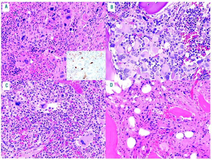
Groups IB-1 and IB-2 comprised 16 cases of PV, 4 ET, 2 MPN-U and 22 PMF (see Figure 1A). Myelofibrosis (MF 2-3) was significantly more prevalent in the IB-1 group (85%) compared to the IB-2 group (50%) (P=0.0017), AP group (67%, P=0.005) and OP group (36%, P<0.0001). Morphologic dysplasia was frequently seen (Table 1), and was overall similar in all groups. The AP group comprised two cases of PV, 1 ET, 2 MPN-U and 10 PMF/Post-ET MF/Post-PV MF (see Figure 1B). The OP group comprised 12 cases of PV, two ET and eight PMF/Post-ET MF/Post-PV MF. Twelve patients had neutrophilia (eight PV, two post PV MF, one ET, one post ET MF) (see Figure 1C) with the median WBC of 58 (range: 18.5 to 151×109/L) and a median neutrophil count of 81% (range: 62-94%). None of these patients had monocytosis. The remaining OP patients (four PMF and one ET) had persistent absolute monocytosis (see Figure 1D) with the median WBC of 52×10⁹/L (range: 16.8-120×109/L); a median absolute monocyte count of 10.5×10⁹/L (range: 2.0-38.4) and a median monocyte percentage of 30% (range: 11-81%).
Figure 1.
Morphologic characteristics of the bone marrow biopsy findings. (A) Patient with post-polycythemia myelofibrosis who developed 7% bone marrow blasts (group IB-2). CD34 highlights an increase in blasts (insert). (B) Patient with post-polycythemia myelofibrosis who developed accelerated phase (group AP). (C) Patient with polycythemia vera with neutrophilic progression (group OP). (D) Patient with primary myelofibrosis with monocytic progression (group OP).
At the time of disease progression, the most common cytogenetic abnormalities included a complex karyotype, deletion 20q, abnormalities of chromosome 7, trisomy 8 and trisomy 9. An abnormal karyotype was more frequent in groups with increased blasts, but this association was not statistically significant (P=0.15). A complex karyotype was more frequent in the AP group compared to IB-1 (P=0.02). Molecular analysis demonstrated that 80% of the patients had JAK2 mutations, while 13% had CALR and <1% had MPL mutations (see Table 1). The mean JAK2 variant allele frequency (VAF) at the time of progression was 75% (range: 3-99%). The JAK2 VAF was generally stable in each patient, regardless of disease phase or the presence of progression. Thirthy patients underwent next generation sequencing. Additional mutations were present in all groups (75% IB1, 40% IB2, 75% OP and 100% AP patients) and included TET2 (40% of all cases), followed by ASXL1 and DNMT3A in 17% each, EZH2, KRAS and CBL in 10%, as well as many other mutations in a smaller number of patients. There was no relationship between the type of mutation and the patient group or clinical outcome, with the caveat that the study size was small. Further studies are needed to clarify these findings.
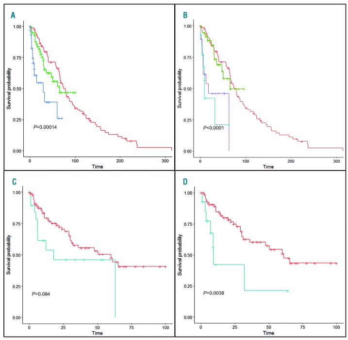
We compared the cases to a matched control group of MPN with a similar follow-up duration (median 105.8 months versus 88.5 months, respectively; P=0.07). Although the control cases were matched to the disease subtypes of the progression cases, there was a trend for a younger age, female sex, and a shorter observation period in the controls compared to the cases (Online Supplementary Table S1); this may be related to the longer clinical course of the MPN patients who progressed. In the study group, 47 of 113 (42%) patients died of disease (median 11 months, range of 1-63 months following disease progression), compared to 19 of 93 (20%) control patients. Patients in group IB-2, had a significantly worse overall survival (OS) than control patients (P=0.023, Figure 1A), while patients in group IB-1 had similar OS to controls (Figure 1A). Patients in the OP group had a significantly shorter OS compared to controls (P<0.0001) and the IB-1 group (P=0.023), but similar OS to the AP group (P=0.85, Figure 1B-C). As expected, AP patients had significantly shorter OS than controls (P<0.0001), and combined IB-1/IB-2 patients (P=0.0038, Figure 1D). The presence of myelofibrosis or specific MPN type did not affect patient survival in this cohort (data not shown
Figure 2.
Survival analysis in patients with disease progression. (A) Comparison between groups IB-1 (green), IB-2 (blue) and the control patients (red) shows a significantly worse survival for patients in group IB-2 (5-9% blasts) compared to both group IB-1 and controls. Conversely, there is no significant difference in survival between controls and IB-1 patients (P=0.24). (B) The OP group patients (purple) have significantly shorter survival than controls (red) (P<0.0001) and the IB-1 group (green) (P=0.023), but similar survival to AP group (blue) (P=0.85). (C) Comparison between IB-1 and IB-2 groups (combined, in red) and OP group (in blue) shows that patients with alternative forms of progression have outcomes similar to those with excess blasts with a trend toward even poorer survival. (D) Comparison between AP group (blue) and the combined IB patients (red) shows a significant difference in survival.
In conclusion, our results validate the current WHO cut-off of 10% blasts that defines AP in MPN.15 We found that the OS of MPN patients with 2-4% PB blasts was similar to matched control MPN patients without increased blasts. However, patients with 5-9% BM or PB blasts had intermediate survival between MPN AP cases and the MPN controls. Thus, we propose that MPN patients developing 5-9% PB or BM blasts, persistent neutrophilic leukocytosis, or absolute monocytosis experience worse outcomes than patients in chronic phase MPN and warrant closer clinical follow-up and possibly earlier therapeutic interventions. These new parameters should be considered for future inclusion among pathologic criteria for diagnosing disease progression in MPN and in future modifications to dynamic MPN scoring systems.
Footnotes
Information on authorship, contributions, and financial & other disclosures was provided by the authors and is available with the online version of this article at www.haematologica.org.
References
- 1.Swerdlow SH CE, Harris NL, Jaffe ES, Pileri SA, Stein H, Thiele J. (Eds). WHO classification of tumours of haematopoietic and lymphoid tissues (revised 4th edition). Lyon: IARC; 2017. [Google Scholar]
- 2.Cervantes F, Dupriez B, Pereira A, et al. New prognostic scoring system for primary myelofibrosis based on a study of the International Working Group for Myelofibrosis Research and Treatment. Blood. 2009;113(13):2895–2901. [DOI] [PubMed] [Google Scholar]
- 3.Gangat N, Caramazza D, Vaidya R, et al. DIPSS plus: a refined Dynamic International Prognostic Scoring System for primary myelofibrosis that incorporates prognostic information from karyotype, platelet count, and transfusion status. J Clin Oncol. 2011; 29(4):392–397. [DOI] [PubMed] [Google Scholar]
- 4.Vallapureddy RR, Mudireddy M, Penna D, et al. Leukemic transformation among 1306 patients with primary myelofibrosis: risk factors and development of a predictive model. Blood Cancer J. 2019;9(2):12. [DOI] [PMC free article] [PubMed] [Google Scholar]
- 5.Mudireddy M, Gangat N, Hanson CA, Ketterling RP, Pardanani A, Tefferi A. Validation of the WHO-defined 20% circulating blasts threshold for diagnosis of leukemic transformation in primary myelofibrosis. Blood Cancer J. 2018;8(6):57. [DOI] [PMC free article] [PubMed] [Google Scholar]
- 6.Tefferi A, Mudireddy M, Mannelli F, et al. Blast phase myeloproliferative neoplasm: Mayo-AGIMM study of 410 patients from two separate cohorts. Leukemia. 2018;32(5):1200–1210. [DOI] [PMC free article] [PubMed] [Google Scholar]
- 7.Guglielmelli P, Lasho TL, Rotunno G, et al. MIPSS70: mutation-enhanced International Prognostic Score System for transplantation-age patients with primary myelofibrosis. J Clin Oncol. 2018; 36(4): 310–318. [DOI] [PubMed] [Google Scholar]
- 8.Boiocchi L, Espinal-Witter R, Geyer JT, et al. Development of monocytosis in patients with primary myelofibrosis indicates an accelerated phase of the disease. Mod Pathol. 2013;26(2):204–212. [DOI] [PubMed] [Google Scholar]
- 9.Tefferi A, Shah S, Mudireddy M, et al. Monocytosis is a powerful and independent predictor of inferior survival in primary myelofibrosis. Br J Haematol. 2018;183(5):835–838. [DOI] [PubMed] [Google Scholar]
- 10.Elliott MA, Verstovsek S, Dingli D, et al. Monocytosis is an adverse prognostic factor for survival in younger patients with primary myelofibrosis. Leuk Res. 2007;31(11):1503–1509. [DOI] [PubMed] [Google Scholar]
- 11.Barraco D, Cerquozzi S, Gangat N, et al. Monocytosis in polycythemia vera: Clinical and molecular correlates. Am J Hematol. 2017;92(7):640–645. [DOI] [PubMed] [Google Scholar]
- 12.Boiocchi L, Gianelli U, Iurlo A, et al. Neutrophilic leukocytosis in advanced stage polycythemia vera: hematopathologic features and prognostic implications. Mod Pathol. 2015;28(11):1448–1457. [DOI] [PubMed] [Google Scholar]
- 13.Bonicelli G, Abdulkarim K, Mounier M, et al. Leucocytosis and thrombosis at diagnosis are associated with poor survival in polycythaemia vera: a population-based study of 327 patients. Br J Haematol. 2013;160(2):251–254. [DOI] [PubMed] [Google Scholar]
- 14.Tefferi A, Rumi E, Finazzi G, et al. Survival and prognosis among 1545 patients with contemporary polycythemia vera: an international study. Leukemia. 2013;27(9):1874–1881. [DOI] [PMC free article] [PubMed] [Google Scholar]
- 15.Iurlo A, Cattaneo D, Gianelli U. Blast transformation in myeloproliferative neoplasms: risk factors, biological findings, and targeted therapeutic options. Int J Mol Sci. 2019;20(8):1839–1852. [DOI] [PMC free article] [PubMed] [Google Scholar]


