

Abstract
The ability to perform reverse genetics in the zebrafish model organism has been greatly advanced with the advent of the CRISPR (clustered regularly interspaced short palindromic repeats)/Cas9 (CRISPR-associated) system. The high level of efficiency in generating mutations when using the CRISPR/Cas9 system combined with the rapid generation time of zebrafish model organism has made the possibility of performing F0 screens in this organism a reality. This Unit describes a detailed protocol for performing an F0 screen using the CRISPR/Cas9 system in zebrafish starting with the design and production of custom CRISPR/Cas9 reagents for injection. Next, two approaches for determining the efficiency of mutation induction by the custom CRISRP/Cas9 reagents that is easily performed using standard molecular biology protocols are detailed. Finally, screening for F0 induced phenotypes using the zebrafish flh gene as an example is discussed.
Keywords: CRISPR/Cas9, zebrafish, FO screen, loss-of-function, recessive mutant, gene editing
Introduction
Clustered regularly interspaced small palindromic repeats (CRISPR)/Cas9 based genome editing tools are extensively used for generating mutations in target genomic regions in model systems ranging from cell lines to model organisms. The zebrafish (Danio rerio) is uniquely suited to study gene function, due to its rapid development rate, the ability to obtain large number of embryos, the transparent nature of embryos in the first 48 hours and its diploid status. Combining the power of CRISPR/Cas9 technology with the zebrafish as a well-defined model organism has transformed the field of developmental biology, the study of human genetic diseases and the application of high-throughput genomic screens. Prior to the advent of CRISPR/Cas9 technology, the methods of choice for reverse genetics analysis of specific gene function in zebrafish were less efficient genome editing technologies or a morpholino-based gene knockdown (Nasevicius & Ekker, 2000). The introduction of of morpholino-based gene knock down approaches revolutionized the use of zebrafish as a model system to perform large-scale in-vivo, reverse genetic screens. However, morpholino-based approaches have drawbacks such as lack of germ line transmission, and the effects of morpholino knockdown are both transient and restricted to early developmental stages. The advent of CRISPR/Cas9 based genome editing in 2012 was a huge step forward in addressing the shortcomings of the morpholino approach (Jinek et al., 2012). This Unit describes the fundamentals of CRISPR/Cas9 technology and its application to high-throughput genetic screens in zebrafish in the context of a detailed methodological approach for the practical application of CRISPR/Cas9 techniques.
Overview of CRISPR/Cas9 biology
The CRISPR/Cas9 system takes advantage of the ability of Cas9 endonuclease to cleave DNA by directing cleavage to specific targeted regions of the genome. As outlined in Figure 1, a typical CRISPR construct consists of a single guide RNA that includes a 20-nucleotide sequence specific to the DNA target and an 80-nucleotide Cas9 interaction loop (1) that binds to a recombinant form of Cas9 protein. Cas9 induces DNA breaks 3bp upstream from a proto-spacer adjacent motif (PAM) sequence adjacent to the target sequence, consisting of 5′-NGG-3′ motif (2). The requirement for a PAM sequence limits the number of possible target sequences, although from a practical standpoint, there are usually sufficient numbers of possible targets within a gene to design CRISPRs for appropriate gene knockout. The combination of CRISPR and Cas9 results in a double-stranded break (DSB) at the target DNA site (3). In the absence of donor DNA, the DSB is repaired by the non-homologous end joining (NHEJ) DNA repair pathway, which is an error-prone process. NHEJ typically results in insertions/deletions (indels), which may lead to disruption of target gene function (4).
Figure 1.

CRISPR/Cas9-mediated gene disruption - principle and components. CRISPR/Cas9 is a two component system consisting of a single guide RNA (sgRNA) that includes a 20-nucleotide target sequence specific to the DNA target and a 80-nucleotide Cas9 interaction loop that binds to a recombinant form of Cas9 protein with DNA endonuclease activity. The resulting complex induces a target-specific double-stranded break (DSB). In the absence of donor DNA, the DSB is repaired by the non-homologous end joining (NHEJ) DNA repair pathway, which is an error-prone process. This typically results in insertions/deletions (INDELs), which may lead to disruption of target gene function.
The CRISPR/Cas9 system is very effective at inducing site-specific indels in zebrafish (Chang et al., 2013; Hwang et al., 2013). A mix of site-specific CRISPR sgRNA and Cas9 protein is injected into 1-cell stage zebrafish embryos to create indels at the target site, resulting in a pool of F0 embryos that are genetically mosaic. Although CRISPR sgRNA and Cas9 mRNA may be co-injected, this protocol uses the injection of the Cas9 protein instead of Cas9 mRNA in order to initiate cleavage as early as possible, with the goal of achieving as close to a null mutant phenotype as possible in the F0. If mutagenesis is highly efficient, then biallelic gene disruptions are sufficiently frequent to generate mutant phenotypes in this population of genetically mosaic F0 embryos (Jao, Wente, & Chen, 2013). Depending upon the phenotype, the mosaic F0 embryos can provide initial clues regarding the function of candidate genes. In addition, the CRISPR/Cas9 system can effectively generate stable mutant lines for genes of interest by screening these mosaic F0 adults to identify carriers of germline mutations, followed by outcrossing to generate heterozygous carriers of the mutations of interest.
In this protocol, the CRISPR/Cas9 system has been optimized with the goal of performing moderate-high throughput F0 screens in the zebrafish model organism. Basic Protocol 1 describes the identification of optimal zebrafish CRISPR/Cas9 target sites. Basic Protocol 2 describes the synthesis of the gene specific sgRNA RNA and the preparation of recombinant Cas9 protein. Basic protocol 3 describes zebrafish microinjections and scoring F0 phenotypes at 48hrs. Basic protocol 4 describes identification of mutations in CRISPR/Cas9 injected embryos by High Resolution Melt Analysis (HRMA). Basic protocol 5 describes identification of mutations in CRISPR/Cas9 injected embryos by mobility shift assays.
BASIC PROTOCOL 1
Identification of zebrafish CRISPR/Cas9 target site
Prior to choosing a CRISPR target site, it is helpful to first gain as much background information as possible on the gene of interest. Start by performing a literature search for existing data on relevant phenotypes, expression patterns and protein domain information of the target gene. Ideally, the goal is to target one of the initial exons such that introduced indels lead to a premature stop codon resulting in a truncated protein that fails to function normally, and often results in nonsense mediated mRNA decay. Avoid targeting the first exon of multi-exonic genes given the possibility of multiple initiation sites and/or alternative transcripts. If for technical reasons initial exons cannot be targeted, it is recommend to target highly conserved, functional domains, which when disrupted are most likely to have functional consequences.
-
1
Check ZFIN (www.zfin.org) for information on RNA in situ expression, available mutants, and publications on gene function and interactions.
-
2
Look for information on duplicated genes, which are referred to as paralogs. Often gene paralogs function in a redundant fashion. If the target gene of interest has a paralog or multiple paralogs, consider targeting all genes at the same time instead of individually.
-
3
Check the ENSEMBL zebrafish page (www.uswest.ensembl.org/Danio_rerio) to gain knowledge on alternate transcripts, exon-intron architecture, 5′ and 3′ untranslated regions, and the amino acid sequence of the target protein. If the target gene has multiple alternative transcripts, try to choose an exon sequence to target that is conserved across all transcripts.
Design appropriate sgRNA targeting sequence
There are multiple free design tools available for researchers to use to predict if a specific sgRNA target site will be functional or if it will also bind at off-target sites causing unwanted mutations. Each design tool is unique in the information it provides and the predictive algorithm it uses. Most predictive design tools are based on experimentally assembled databases, meaning the design tool is only as good as that database. It is strongly recommended to read the primary literature associated with any design tool to fully understand the experimental data it is based on and to gauge its utility. It is preferred to use a combinatory approach that draws predictive information from several different sources to help in identifying the most optimal CRISPR/Cas9 target site. Below is an example of one design approach that utilizes four freely available design tools to streamline optimal target site identification.
-
4
Identify the exonic sequence to be targeted along with 25bp +/− intronic sequence. sgRNA target sites can start in intronic sequence, but should cut in exonic sequence.
-
5Input the target sequence into the Broad Institutes sgRNA Designer tool to identify all the possible CRISPR target sites in the sequence, http://portals.broadinstitute.org/gpp/public/analysis-tools/sgrna-design (Doench et al., 2016). This is the second incarnation of this design tool and gives a predictive Score between 0 and 1. The probability of a CRISPR target site being highly mutagenic increases with higher Scores. Download the text file this tool generates and then open it using Microsoft Excel. Delete all columns except: Position of Base After Cut (1-based), Strand, sgRNA Target Sequence, On-Target Efficacy Score, and On-Target Rank. Determine the GC percentage and the number of G and A nucleotides in the last four positions of the target sequence, 3′ end.
-
–Delete sites with GC content below or above 35-80%.
-
–Delete sites with no G/A nucleotides in last four positions.
-
–Delete sites with tracks of T nucleotides of 4 or more (possible in vitro transcription (IVT) termination signal).
-
–Delete sites cutting in intronic sequence.
-
–
-
6Input the target sequence into the MIT Optimized CRISPR Design tool, http://CRISPR.mit.edu, and choose the zebrafish (GRCz10) option (Hsu et al., 2013). This design tool analyzes 250bp of sequence at a time, identifies all CRISPR target sites in the sequence, and then ranks (0-100) these sites based on the potential for binding and cutting at off-target sites. A higher Score indicates less potential for off target cutting.
-
–Delete sites with a score less then 55
-
–Give preference to sites with higher Scores.
-
–
-
7Choose the View UCSC tracks option on the CRISPRScan design tool site: http://www.CRISPRscan.org (Moreno-Mateos et al., 2015). Choose the Zebrafish (GRCz10) option and enter the target gene’s Ensembl gene ID to visualize all the CRISPRScan scored sgRNA sites for the target gene. Navigate to the Tools option above and choose Table Browser. This allows retrieval of a list of all the CRISPRScan scored sgRNA sites by selecting “get output”. Determine the CRISPRScan score for each sgRNA remaining.
-
–Delete sites with CRISPRScan scores less than 40.
-
–If an sgRNA site is not found using the CRISPRScan tool do not delete it. CRISPRScan only scores sites that have G nucleotides in the first two 5′ end positions of the sgRNA sequence.
-
–
-
8Several sites may remain and a list of each site’s predicted off-target profile can be obtained using the CRISPR/Cas9 system (Cas9/sgRNA) Off-Targeter (CasOT), http://eendb.zfgenetics.org/casot/ (Xiao et al., 2014).
-
–Download the CasOT Perl script form website.
-
–Download the most current version of the zebrafish genome (Danio_rerio.GRCz10.dna.toplevel.fa) using the FTP download option on Ensembl: http://www.ensembl.org/info/data/ftp/index.html.
-
–Use the following parameters when using CasOT:
- Allow 2 mismatches in seed region
- Allow 4 mismatches in non-seed region
- Search for NGG PAM associated sites only
-
–Analyze the resulting list:
- Delete sites with three or less mismatches total.
- Allowances are made if two mis-matches are in the 3′ end of the CRISPR target site (in the seed region).
-
–
-
9Preference is given to CRISPR target sites that:
-
–Have high predictive scores for all three design tools.
-
–Have low chances of off target complication based on the CasOT off target profile.
-
–Start with GG and end with GG.
-
–Target early in the gene, before conserved enzymatic function, or specifically target nucleotides responsible for enzymatic function.
-
–
-
10
If no target sites start with GG, which is required for IVT from the T7 promoter, either two Gs can be added at the beginning or the first two nucleotides of the CRISPR target site can be replaced by Gs. There are pros and cons to both approaches. This protocol will proceed with the option of adding Gs to the end instead of replacing nucleotides.
The CRISPRScan tool has been updated several times, first to incorporate the new Zebrafish genome releases and more recently to incorporate aspects of the MIT Optimized CRISPR Design tool and three separate ways to identify optimal CRISPR/Cas9 target sites. The CRISPRScan tool is specific to the Zebrafish model organism with its rules and design algorithm trained on a large database of empirically determined sgRNA mutagenesis rates in zebrafish (Moreno-Mateos et al., 2015). To speed up the design process by using just one design tool the CRISPRScan tool is recommended.
BASIC PROTOCOL 2
Preparation of the sgRNA DR274 plasmid
The CRISPR/Cas9 system consists of two components: sgRNA and Cas9 protein. DR274 is a customizable empty backbone construct used to produce custom sgRNA RNA via IVT from a T7 promoter. Details of plasmid preparation and cloning strategy are detailed in Figure 2.
Figure 2.
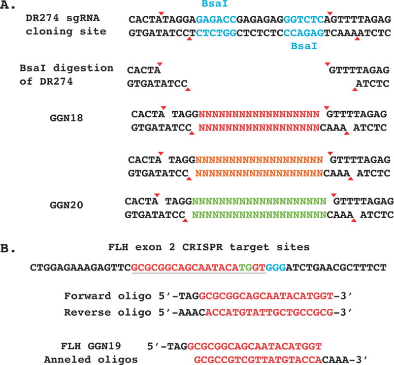
DR274 sgRNA digestion and cloning scheme. A. The DR274 sgRNA plasmid has two BsaI restriction enzyme recognition sites that cut outside the binding site to leave 4bp overhangs. Complementary oligos are ordered, annealed, and then ligated into the overhang sites. B. Optimal CRISPR target site (Red) in exon 2 of flh gene showing PAM sequence (blue) and cut site (green). Design of oligos for cloning flh target site into the BsaI digested DR274 backbone.
Reagents and materials
DR274 is obtained from Addgene, Plasmid #42250
Bunsen Burner
LB agar plate containing 50 μg/ml kanamycin
LB liquid medium containing 50 μg/ml Kanamycin
Inoculation Loop
Inoculation Stick
37°C incubator-shaker
17x100mm bacterial culture tube, polystyrene, sterile
15ml conical tube
Omega Bio-tek E.Z.N.A. Plasmid Mini Kit I, V Spin. Cat# D6943-02
Bacterial cryogenic vials
Sterile 60% Glycerol
BsaI with Cutsmart buffer from New England Biolabs (NEB). Cat# R0535S
Sterile dH2O
0.2 ml sterile PCR tube
1.5 ml eppendorf tubes
PCR machine or 37°C and 50°C water baths
10 mg/ml Proteinase K
10% Sodium dodecyl sulfate (SDS)
Omega Bio-tek E.Z.N.A. Cycle Pure Kit. Cat# D6492-02
Agarose
Gel electrophoresis equipment
Electrophoresis tracking dye
Ethidium bromide
NanoDrop Spectrophotometer (http://www.nanodrop.com)
M13 Forward (−20) universal sequencing primer: 5′- GTAAAACGACGGCCAGT-3′
Access to Sanger sequencing facility
Ape Sequence analysis software: http://biologylabs.utah.edu/jorgensen/wayned/ape/
SOC liquid media
5M NaCl
STE solution (1mM EDTA, 100mM NaCl, 10mM Tris pH 8.0)
Takara DNA Ligation Kit, Version 2.1 from Clontech. Cat# 6022
DH5alpha chemically competent E. coli cells
DraI with CutSmart buffer from NEB. Cat# R0129S
HiScribe T7 Quick High Yield RNA Synthesis Kit form NEB. Cat# E2050S
RNase free dH2O
Qiagen RNeasy RNA purification kit. Cat# 74104
100% RNase Free Ethanol
Obtain DR274 plasmid
-
1
Order constructs from Addgene
-
2
Use an inoculation loop to streak from the Addgene bacterial stab onto LB plates with 50 μg/ml kanamycin. Incubate streaked plates at 37°C overnight.
-
3
Use an inoculation stick to pick one isolated colony into 6 ml of LB liquid medium containing 50 μg/ml Kanamycin in a 17x100mm bacterial culture tube. Grow the culture overnight at 37°C with shaking at 250rpm.
-
4
Transfer 5 ml of the bacterial culture to a 15 ml centrifuge tube and spin at 2800 × g for 9 min. Decant supernatant and isolate plasmid DNA from the bacterial pellet using a commercial Miniprep Kit.
-
5
Use a NanoDrop Spectrophotometer to measure the DNA concentration and use these constructs in the following protocol steps
-
6
Optional: sequence the constructs using the universal M13F primer
-
7
Use the remaining 1 ml of bacterial culture to make a glycerol stock by combining 250 μl 60% sterile glycerol with 750 μl culture, mixing, and storing at −80°C.
BsaI Digestion of DR274 sgRNA Plasmid
-
8
In a 0.2 ml PCR tube pipet 5 μl NEB CutSmart Buffer, 10 μg of DR274 plasmid, 2 μl of BsaI, and dH2O to 50 μl. Vortex the reaction and spin down briefly.
-
9
Incubate the digestion at 37°C overnight in a PCR machine or water bath.
-
10
Add 0.5 μl Proteinase K and 2.5 μl SDS to digest the restriction enzyme.
-
11
Incubate at 50°C for 20 min in a PCR machine or water bath.
-
12
Purify the digested plasmid using the Omega Bio-tek ENZA Cycle Pure column purification kit (or other commercially available kit), eluting in 30μl of 1:10 diluted Elution Buffer.
-
13
Use a Nanodrop microspectrophometer to measure the DNA concentration.
-
14
Run the purified digest on a 1% agarose gel to check for complete digestion. BsaI linearization results in a 2426bp fragment.
-
15
Dilute digested DR274 to 20 ng/μl
-
16
Optional: the DR274 construct can be dephosphorylated prior to column purification to minimize vector re-ligation. Dephosphorylation requires ordering 5′-phosphorylated oligos in the ligation step below.
Ligation of CRISPR target site cloning oligos into BsaI digested DR274
The BsaI digested DR274 construct can be repeatedly customized to express optimal CRISPR target site sgRNA RNA by ligation of annealed short oligos into the BsaI site. Refer to Figure 2 showing the design of both Forward and Reverse oligos for cloning into the BsaI digested DR274 plasmid.
Anneal forward and reverse oligos
-
17
Order crude oligos and resuspend to 100 uM.
-
18
Combine 15 μl each forward and reverse oligo together with 1 μl 5M NaCl in a sterile 0.2 ml PCR tube.
-
19In a PCR machine anneal the oligos using the following program:
- Incubate at 70°C for 5min
- Cool to 25°C as slow as possible (0.1°C ramp rate)
- Incubate at 25°C for 5 sec
- Cool to 10°C at fastest ramp rate possible and hold
-
20
Dilute annealed oligos 1:50 in STE (1 μl annealed oligos into 49 μl STE).
-
21Dilute again 1:100 in STE (1 μl 1:50 dilution into 99 μl STE).
- Final dilution is 1:5000 or 0.02 μM
Ligate annealed oligos into BsaI digested DR274
-
22
In a sterile 0.2 ml PCR tube combine 1.5 μl Takara ligation Sol II, 1.0 μl 1:100 diluted anneal oligos, and 0.5 μl BsaI digested DR274 backbone (20 ng/μl). Vortex and spin down briefly.
-
23
Incubate at 16°C for 30 min in a PCR machine.
-
24
Add 0.3 μl Sol III transformation enhancer. Vortex and spin down briefly.
Transform DH5alpha chemically competent E. coli cells
-
25
Add entire ligation reaction to 40 μl of DH5alpha cells in a 1.5 ml eppendorf tube.
-
26
Flick tube gently, but completely to mix.
-
27
Incubate on ice for 30 min.
-
28
Heat shock at 42°C for 30 sec.
-
29
Incubate on ice for 2 min.
-
30
Add 160 μl of SOC.
-
31
Incubate at 37°C for 1 hour with shaking at 250 rpm.
-
32
Plate 50 μl on LB agar plate containing 50 μg/ml kanamycin and incubate at 37°C overnight.
Miniprep customized DR274
-
33
Use an inoculation stick to inoculate two colonies into 6 ml LB liquid medium containing 50 μg/ml Kanamycin in 17x100 mm bacterial culture tubes.
-
34
Grow the cultures overnight at 37°C with shaking at 250 rpm.
-
35
Transfer 5 ml of the bacterial culture to a 15 ml centrifuge tube and spin down at 2800 × g for 9 min, decant supernatant. Store remaining 1 ml of bacterial culture at 4°C.
-
36
Isolate plasmid DNA using a commercial Miniprep Kit.
-
37
Use a NanoDrop Spectrophotometer to measure the DNA concentration.
-
38
Sequence plasmid prep using universal M13F primer.
-
39
Make a glycerol bacterial stock of correct clones by combining 250 μl 60% sterile glycerol with 750 μl left over bacterial culture, mixing, and storing at −80°C.
Production of sgRNA RNA from customized DR274
Customized DR274 constructs that now contain an optimal CRISPR target site sequence can be digested to produced a small T7-promoter containing fragment for the production of sgRNA RNA by IVT using RNA polymerase.
DraI Digestion of custom DR274 sgRNA construct
-
40
In a 0.2 ml PCR tube pipet 5 μl NEB Cutsmart Buffer, up to 43 μl custom DR274 sgRNA construct, 2 μl of DraI, and dH2O to 50ul. Vortex the reaction and spin down briefly.
-
41
Incubate the digestion at 37°C overnight in a PCR machine or water bath.
-
42
Add 0.5 μl Proteinase K and 2.5 μl SDS to digest the restriction enzymes.
-
43
Incubate at 50°C for 20 min in a PCR machine or water bath.
-
44
Purify the digested plasmid using the Omega Bio-tek ENZA Cycle Pure column purification kit (or other commercially available kit), eluting in 30 μl of 1:10 diluted Elution Buffer.
-
45
Run the purified digest on a 1% agarose gel. DraI digestion will release a T7-sgRNA fragment of 285 bp and a backbone fragment of 1862 bp. Gel purification of the 285 bp fragment is optional, but not required. Yield of the following RNA IVT will not be significantly increased with gel purification, Figure 3.
Figure 3.

Example of DraI restriction enzyme digestion of custom sgRNA constructs showing uncut plasmid and DraI digested plasmid with 1862 bp backbone and 285 bp T7-sgRNA fragment.
Production of sgRNA RNA by T7 IVT
-
46
In a 0.2 ml PCR tube, setup the following IVT reaction using the NEB HiScribe T7 Quick High Yield RNA Synthesis Kit. Add reagents in the following order: 10 μl RNase free dH2O, 10 μl NTP Buffer Mix, 8 μl DraI digested custom sgRNA construct. Vortex and briefly spin down.
-
47
Add 2 μl T7 RNA Polymerase Mix. Vortex and briefly spin down.
-
48
Incubate at 37°C overnight in a PCR machine or water bath.
-
49
The following morning add 20 μl RNase Free dH2O and 2 μl of DNase I (provided with the HiScribe kit).
-
50
Incubate at 37°C for 15 min in a PCR machine or water bath.
Purification of sgRNA RNA using Qiagen RNeasy Kit
-
51
Add the 52 μl of RNA IVT reaction to 48 μl RNase free dH2O (provided with RNeasy kit) in a new RNase free 1.5 ml eppendorf tube (not provide with kit).
-
53
Add 350 μl RLT buffer and pipet gently to mix.
-
54
Add 550 μl of 100% RNase Free Ethanol and pipet gently to mix.
-
55
Add 500 μl to column and spin for 22 sec at full speed (≥10,000 × g) in a tabletop centrifuge.
-
56
Aspirate flow through liquid.
-
57
Add remaining sample to column and spin for 22 sec at full speed (≥10, 000 × g).
-
58
Aspirate flow through liquid.
-
59
Add 500 μl of RPE buffer and spin for 22 sec at full speed (≥10, 000 × g).
-
60
Aspirate flow through liquid.
-
61
Add 500 μl of RPE buffer and spin for 2min at full speed (≥10,000 × g)
-
62
Carefully transfer column without transferring any of the flow through to a clean RNase free 1.5 ml tube (provided with kit).
-
63
Add 30 μl RNase free dH2O (provide with kit). Spin 1min at full speed (≥10, 000 × g).
-
64
Use a NanoDrop Spectrophotometer to measure the RNA concentration. Expected yield should be between 1–4 μg/μl.
-
65
Run 150-300ng of RNA on a 1% agarose gel at 120V to check RNA integrity. There may be two bands due to secondary structure of the RNA. Run DraI custom sgRNA undigested and digested plasmid as controls, Figure 4.
Figure 4.

Example of sgRNA RNA production as compared DraI digested and uncut plasmid customized DR274 plasmid. sgRNA is approximately 103bp but will not run according to size due to its single stranded nature.
Obtain Cas9 protein
There are several sources for commercially produced recombinant Cas9 protein. Each vendor provides differing purification levels and concentrations. Recombinant Cas9 protein from IDT (Cat# 1074181) is suggested.
BASIC PROTOCOL 3
Zebrafish microinjections and scoring phenotype
To assess the targeting and mutation efficiency of the gene specific sgRNA and Cas9 enzyme, they are mixed together and injected into 1-cell zebrafish embryos. The most important factor leading to high mutation rates is the injection technique. The embryos are oriented at roughly a 45° downward angle and injected at the junction of the first cell and the dividing yolk cells. The injection solution contains phenol red, which allows determination of correct injection location.
Reagents and materials
Gene specific sgRNA RNA and recombinant Cas9 protein from Basic Protocol 2
Phenol Red
RNase Free dH2O
Zebrafish mating cages with dividers
9 cm petridishes
Borosilicate micro capillaries to pull into injection needles using a micropipette puller
E3 embryo buffer containing 5mM NaCl, 0.17 mM KCl, 0.33 mM CaCl and 0.33mM MgSO4
Bright field stereomicroscope with diascopic illumination
Setting up zebrafish mating
Wild type AB or TU zebrafish (Danio rerio) are reared at 28.5°C in accordance with an appropriate IACUC protocol and kept under a 14h light and 10h dark cycle.
Injecting 1-cell zebrafish embryos
Set up matings the evening before by placing adult two male fish and two female fish into a breeding tank separated by dividers.
The next morning change out the water in each of the breeding tanks, remove the dividers and wait for the fish to mate and lay eggs. This should take approximately 15–30 min.
Rinse the newly laid eggs with E3 egg water and collect them in a petri dish. These eggs start dividing into 1-cell stage embryos within 45 min of fertlization
Inject into 1-cell zebrafish embryos using a micro capillary needle with an injection solution containing sgRNA, Cas9 protein, RNAse free water and phenol red dye.
To ensure high mutation rates and low levels of injection toxicity, titrating the concentration of sgRNA and Cas9 protein is very important.
- A good starting point is to prepare multiple tubes of injection solution containing
- sgRNA in a concentration range: 200–400 ng.
- Recombinant Cas9 protein in the concentration range: 600-1000ng.
- 1 μl Phenol Red Dye.
- Make up injection solution volume to 10 μl with RNAse free H2O.
Inject 1 nl of injection solution per embryo for a final injection amount of 200–400 pg sgRNA and 600–1000 pg recombinant Cas9 protein.
After injections, collect embryos into petri dish containing E3 egg water and transfer them into a 28.5°C incubator and let them grow for 24–48 hours.
After 24–48 hours, observe the embryos under a light microscope and score for visible embryo phenotypes. An example is shown in Figure 5 where the wild type embryo in panel A clearly differs from the injected mutant embryo in panel B, in a number of observable and scorable embryonic features. This includes the length of the embryo, presence of anatomical structures such as different brain regions, shape and size of organs such as the eyes, heart and rate of blood flow.
After scoring phenotype, collect these embryos for genomic DNA extraction and HRMA analysis which is described in the next section
Figure 5.

Example of zebrafish embryonic phenotype at 48 hours post fertilization. (A) wild type uninfected embryo. (B) Embryo injected with a mix of sgRNA and Cas9 protein and showing phenotypic differences when compared to wt embryo. This embryo has a shorter body axis and lacks a visible notochord (C) Diagram showing precise injection technique for higher mutation rates. The injection solution shown in red is injected at the junction between the dividing first cell and the yolk.
BASIC PROTOCOL 4
Identification of mutations in CRISPR/Cas9 injected embryos by High Resolution Melt Analysis (HRMA)
HRMA is a mutation detection assay that can detect the presence of mutant alleles in a mosaic population of wild type and mutant genomes (Dahlem et al., 2012; Parant, George, Pryor, Wittwer, & Yost, 2009). The first step is to perform a PCR amplification of the region around the CRISPR target site in the presence of a second-generation fluorescent dye that fluoresces when bound to dsDNA, i.e. PCR amplicon. The second step is to perform a melt analysis of the PCR amplicon that tracks loss of fluorescence as the temperature is increased. Figure 6 illustrates the fundamental principle of HRMA that in a mixed population of PCR amplicons heteroduplex species consisting of one WT strand and one mutant strand will form. Their lack of full complementary sequence will cause mismatches that destabilize the heteroduplex and reduce its Tm, i.e. they melt at lower temperatures compared to wild type homoduplexes and mutant homoduplexes that are fully complementary. This shift in Tm is easily identified if loss of fluorescence is tracked in high resolution. HRMA is a high-throughput and rapid assay that allows efficient identification of embryos with CRISPR induced mutations and semi-quantitative determination of mutation induction rates. Below, the use of BioFire Biodefense reagents and equipment is described, however most modern q-PCR machines are capable of performing HRMA and several commercial companies provide the necessary reagents.
Figure 6.

High Resolution Melt Analysis (HRMA) scientific basis and mutation detection. A. Heterozygous locus with a SNP change/mutation will produce two different PCR duplex amplicon populations. When denatured and re-annealed the parental duplexes will be formed, as will heteroduplexes consisting of one strand of “type” (WT) DNA bound to one strand of “Mutant” DNA. Mismatch at the site of the SNP difference will cause a bulge/bubble in the DNA duplex that lowers the Tm. B. Example HRMA data showing the identification of CRISPR/Cas9 induced mutations as samples that melt sooner than wild type counterparts.
Reagents and materials
Embryos
Proteinase K lysis buffer: (final concentrations) 20 mM Tris-HCL (pH 8.0), 50 mM KCL, 0.3% Tween-20, 0.3% NP-40, 0.5 mg/ml Proteinase K. Proteinase K is added the day of use. Other components can be premixed and stored at room temperature for one month.
30 mg/ml Pronase
20 μl pipette tips
P20 Pipette
96-well plates
96-well plate centrifuge
Sealing films
E3 embryo buffer
BioRad 96-well black shell white well PCR plate. Cat# HSP9665
BioRad Sealing Tape for Optical Assays. Cat# 2239444
Sigma Mineral Oil. Cat# M5904
BioFire LightScanner MasterMix. Cat# HRLS-ASY-0003
8-well PCR strips
1.5 ml eppendorf tubes
Multichannel pipette
PCR machine
Invitrogen TOPO-TA cloning kit. Cat# 450641
0.5 ml PCR tube
10 μl pipette tips
LB agar plate containing 100 μg/ml ampicillin
LB liquid medium containing 100 μg/ml ampicillin
Bunsen Burner
37°C incubator-shaker
Inoculation Stick
17×100 mm bacterial culture tube, polystyrene, sterile
15ml conical tube
Omega Bio-tek E.Z.N.A. Plasmid Mini Kit I, V Spin
Bacterial cryogenic vials
Sterile 60% Glycerol
NanoDrop Spectrophotometer (http://www.nanodrop.com)
M13 Reverse (−20) universal sequencing primer: 5′- GTAAAACGACGGCCAGT-3′
Access to Sanger sequencing facility
Ape Sequence analysis software: http://biologylabs.utah.edu/jorgensen/wayned/ape/
Genomic DNA Extraction
Note: Genomic DNA isolation buffers with different salt concentrations can shift the temperatures at which PCR amplicons melt making it impossible to normalize and compare samples in different buffers. The same lysis buffer must be used for each sample so as to compare them directly to each other, including WT control samples.
-
1
Dechorionate embryos at 24 hpf by adding 50–100 μl of Pronase to embryos in 5 ml of E3 embryo buffer in a petri dish.
-
2
Incubate for 15–20 min at 28.5°C and then shake vigorously.
-
3
Wash out Pronase 3X with E3 buffer.
-
4
Manually dechorionate any embryos still in chorions with tweezers.
-
5
Aliquot 50 μl of Pro K lysis buffer into each well of a 96-well plate.
-
6
Set P20 pipette to 2.5 μl and use a 20 μl pipette tip with its tip cut off to suck up individual embryos and dispense into wells.
-
7
Cover with sealing film, vortex, and spin down briefly.
-
8
In a PCR machine incubate the genomic DNA (gDNA) extractions for 55°C for 2hrs, 95°C for 5 min, and finally to 10°C for holding.
-
9
Vortex and then spin down for 1 min at a minimum of 900 × g.
-
10
Incubate the gDNA extractions overnight at 4°C.
-
11
The following day vortex again and spin down 3 min at a minimum of 900 × g.
-
12
The gDNA extractions are stored long-term at −20°C.
-
13
Final concentration of gDNA will be between 10–60 ng/μl.
HRMA primer design
Note: HRMA uses smaller than normal amplicons and the smaller the amplicon the better. However, amplicons up to 450 bp can be used without any loss of sensitivity or specificity.
-
14
Use Primer3Plus or any other primer design program to design primers based on the following conditions, ranked in order of importance. Try to match Tm’s of the primer pairs within +/− 1°C (high priority).
Optimal Conditions Range 62°C Tm 58-64°C ~80 bp 65-450 bp amplicon 50% GC 40-70% GC content 20 bp 19-25 bp primer length -
15
Blast the primers against the Zebrafish GRCz10 genome to ensure the uniqueness of each primer.
-
16
Design 2 forward and 2 reverse primers that can be mixed and matched for each target to create four primer mixes (PM).
-
17Primers used for analysis of the flh target site
- Forward Primer Sequence: TCTGGGAAATCAAAGCGAATGC
- Reverse Primer Sequence: TGGAGAGCAGATGCCAACAG
Optimize anneal temperature
-
18
Use a mix of multiple wild type gDNA extracts for optimization.
-
19
Set up three duplicate reactions for each PM (see reaction conditions below).
-
20
Test duplicate reactions for each PM at annealing temperatures of 68, 70, and 72°C.
This is easily done using the gradient feature on PCR machines by setting the gradient to 68-72°C. Column 1 will be at 68°C, column 6 will be 70°C, and column 12 will be 72°C.
-
21Best PM will be the one with:
- A clean melt curve: i.e.-just one melt point
- A sharp transition from no-melt to melt
- PM with the most stringent condition i.e. highest anneal temperature
HRMA reaction setup and analysis
-
22Aliquot 20 μl sterile mineral oil into each well of a 96-white well black hard shell plate.
- Using BioFire LightScanner Master Mix (includes Taq, dNTPs, buffer) setup the following reaction for each sample. Add 4 μl LightScanner MasterMix, 2 μl 2 μM PM, 1 μl gDNA (1-6 ng final concentration) and 4 μl dH2O for a total reaction of 10 μl .
- We suggest making a larger Master Mix of all the reagents excluding gDNA and then aliquoting 9 μl of the Master Mix per well followed by 1 μl of gDNA.
- Cover with optical grade PCR sealing tape and spin for 3 min at 900 × g rpm at 4°C in tabletop centrifuge to pool the reagent at the bottom of the wells.
-
23Place in 96-well PCR machine and run the following PCR program.
- Initial melt: 95°C for3 min
- 50 cycles of
- 95°C 30 sec
- 68, 70, or 72°C 15 sec
- 1 cycle of (heteroduplex formation)
- 95°C 30 sec
- 25°C 30 sec Cool at fastest rate possible to increase heteroduplex formation
- 10°C Hold
-
24
Spin plate for 3min at 900 × g at 4°C in a table-top centrifuge to get rid of air bubbles that may have formed during the PCR cycles.
-
25
Place in BioFire LightScanner 96 machine and run melt curve analysis from ~65-98°C using standard settings.
-
26Analyze results using LCscanner software package.
- Must have at least three wild type gDNA samples in the HRMA analysis to set the baseline.
- See Figure 6 for an example of mutations detected by HRMA.
Cloning and Sequencing of HRMA amplicons
-
27
Spin down the HRMA 96-well plate for 3min at 900 × g at 4°C in tabletop centrifuge.
-
28
Transfer all of the desired HRMA PCR reaction (with mineral oil) to a 0.5 ml PCR tube or 8-well PCR strip.
-
29
Spin down the tube and then remove as much of the mineral oil as possible. Transfer of mineral oil to the TOPO-TA cloning reaction will decrease the efficiency.
-
30
Use 1 μl of HRMA PCR reaction in the standard Invitrogen TOPO-TA cloning reaction. When transferring wipe off excess mineral oil on the pipet tip with a kim wipe (if necessary)
-
31
Pick single colonies from the TOPO-TA cloning reaction into LB liquid medium containing 100 μl/ml ampicillin and grow overnight at 37°C with shaking at 250 rpm.
-
32
Isolate plasmid DNA using a commercially available Miniprep kit.
-
33The frequency of mutation induction seen in the HRMA results will inform how many single colonies to pick and grow up for analysis.
- Higher signal and more positive embryos indicate that fewer colonies need to be grown: 4-6.
- Lower signal and less positive embryos indicate more colonies need to be grown: 8-12.
-
34Sequence each plasmid prep with M13 Reverse primer.
- Use of the M13 Forward primer with the TOPO-PCR2.1 backbone is not recommended.
-
35
Align the sequences to the wild type reference sequence of the target sequence to determine the mutations that have been induced. Expect some of the clones to have the wild type reference sequence.
BASIC PROTOCOL 5
Identification of mutations in CRISPR/Cas9 injected embryos by mobility shift assay
An alternative assay for the identification of CRISPR/Cas9 induced mutations is the mobility shift assay (Ota et al., 2013). The DNA duplex bulge/bubble that forms when a wild type and Mutant DNA PCR strands anneal with each other creates an anchor that slows the migration of the heteroduplex through a high percentage gel. By comparing samples to wild type control, embryos with induced mutation can be identified and the efficiency of mutation induction can be estimated by determining the loss of full-length wild type PCR amplicons.
PCR followed by analysis on a 15% TBE gel
Reagents and materials
GoTaq G2 green PCR mix from Promega
Target site specific forward and reverse PCR primers
Genomic DNA extracted from control embryos and CRISPR/Cas9 injected embryos
- 15% Mini- Protean TBE gel, 12 well, 20μl from BIO-RAD Catalogue no: 4565055
- In a 0.2 ml PCR tube combine 10 μl of GoTaqG2 Green Master Mix, 1 μl of 10 μM PM, 1 μl gDNA, and 8 μl dH2O.
- Place in PCR machine and run the following Program:
- 95°C 3 min
- 50 cycles of
- 94°C 30 sec
- 55°C 15 sec
- 72°C 15 sec
- Final extension 72°C for 5 min
- 10°C final hold
- Load the completed PCR directly into a 12 well 15% TBE gel.
- Run the gel for 2 hrs at 110 volts and then stain with EtBR.
- Analyze and photograph the gel and determine band intensity of full-length PCR amplicons using Image J software, see Figure 7C.
- The 15% TBE gel is used to analyze the PCR products and for separating and visualizing DNA duplex formation in the CRISPR/Cas9 injected embryos vs wild type controls (Figure 7B).
Figure 7.

The HRMA results and 15% TBE gel used to analyze PCR products. (A) HRMA showing shifted melting curves with colored lines depicting injected embryos and the grey lines depicting the wild type uninjected control embryos. A greater deflection from the wt grey lines indicates higher mutation rates. (B) 15% TBE gel, which is used to analyze the PCR products and for visualizing DNA duplex formation in the CRISPR/Cas9 injected embryos vs wild type controls. The wt control lane shows a single PCR band while the Mutant embryo lanes show Heteroduplex formation induced by CRISPR injections. Heteroduplexes are indicated by the Red bracket. The Black asterisk * indicates the flh PCR product. The injected embryos were divided into 3 groups based on severity of phenotype penetrance. (C)The band intensity of the flh PCR product was analyzed using Image J software and plotted as a graph. The severity of the flh phenotype correlates directly to decreased levels of the flh PCR product.
COMMENTARY
Background Information
The previous paradigm for performing reverse genetic F0 screens in zebrafish using morpholinos or less efficient genome editing technologies suffered from a lack of penetrant phenotypes and complicating off target effects (Nasevicius & Ekker, 2000). Drawbacks of the morpholino technology included a lack of germ line transmission, transient knockdown, and a requirement to assay genes involved in early development only. The advent of CRISPR/Cas9 based genome editing in 2012 and subsequent research showing that high levels of site-specific null (biallelic) indel mutations could be induced in the zebrafish model organism, addressed these shortcoming and expanded the genes and phenotypes that could be integrated (Chang et al., 2013; Hwang et al., 2013; Jinek et al., 2012; Jao, Wente, & Chen, 2013).
The proliferation of free online design tools (Doench et al., 2016; Hsu et al., 2013; Moreno-Mateos et al., 2015; Xiao et al., 2014) and the ease of acquiring the sgRNA RNA and recombinant Cas9 components allow quick installation of the CRISPR/Cas9 system into researcher labs. Combining this with the unique attributes of the zebrafish model organism, rapid development rate, the ability to obtain large number of embryos, the transparent nature of embryos in the first 48 hours and its diploid status, makes performing F0 screens in zebrafish following this protocol a feasible and reasonable approach.
Critical Parameters
In our experience, strict adherence to the protocol and injection of high quality sgRNA and Cas9 protein leads to efficient and reproducible phenotypes in the F0 generation. It should be noted that the absence of a visible phenotype in the F0 generation does not preclude a crucial developmental role for the targeted gene, but rather that there may be insufficient bi-allelic mutational burden or other compensatory genes that modify the phenotype. Carrying the mutation through the germ line to create stable mutants can distinguish between these 2 possibilities. Overall, the CRISPR/Cas9 approach for F0 phenotypic characterization holds tremendous promise for high-throughput experimental modalities and we predict that this approach will facilitate gene discovery in zebrafish as a model system for the study of developmental biology and human disease.
Troubleshooting
Injections do not produce indel mutations at sgRNA target site as assayed by HRMA or mobility shift assay:
-
-
It is possible that the injections were into the yolk instead of the one cell stage, which will significantly reduce mutation induction efficiency. Redo injection as detailed in Basic Protocol 3 and Figure 5.
-
-
It is possible that DNA modifications (methylation) and chromatin modifications prevent access of the CRISPR complex to the target site. Additionally, some designed CRISPR complexes are inherently nonfunctional. Redesign the CRISPR sgRNA target site to a different exon or at least one nucleosome (246bp) up or downstream.
HRMA or Mobility shift assay primers do not amplify:
-
-
This will be sequence context dependent to the CRISPR target site chosen.
-
-
For both assays, redesign the primers or a lower annealing temperature must be used.
-
-
For HRMA, redesign the primers to produce a smaller amplicon.
No mutant phenotype after injection of validated CRISPR/Cas9 reagents:
-
-
As mentioned in Basic Protocol 1, because of the duplication of the zebrafish genome that occurred in its evolutionary history, it is possible that there are redundant paralogs of the target gene of interest. It is possible that all paralogs will need to be targeted in parallel or in succession to produce a phenotype.
-
-
It is possible that the mutation induction rate is suboptimal and fails to induce a critical load of biallelic mutations to effectively knockout gene function. To address this issue, increase the amount of sgRNA and recombinant Cas9 protein injected until a mortality rate of 50% of injected embryos is reached. This point allows for maximizing the mutation induction rate while also allowing for the recovery of suitable embryo numbers for analysis.
Anticipated Results
Here we describe a particular application of CRISPR/Cas9 gene editing technology to generate and characterize loss of function mutations in the F0 generation. The original floating head allele, flhn1, is a recessive, zygotic lethal mutation that produces embryos completely lacking notochords (Halpern et al., 1995). We designed a CRISPR sgRNA that targeted exon 2 of the flh locus and confirmed efficient induction of indels, as measured by HRMA (Figure 7A). Next, we titrated the quantity of injected CRISPR sgRNA (100-400 ng) and Cas9 protein (400-1000 ng) in order to identify the optimal injection conditions for the highest penetrance of the phenotype. At maximal tolerated injection conditions, 63/75 (84%) of injected embryos displayed varying degrees of the hallmark flh1 mutant phenotype: absent/reduced notochord, deformation of the somites and short body axis (Figure 5A–B). In order to determine whether the severity of the phenotype correlated with the degree of mutational burden in the F0 generation, 48 hpf embryos were divided into 3 groups based on the severity of the notochord phenotype and each embryo was analyzed individually using the mobility shift assay as described in Basic Protocol 5. As displayed in Figure 7B, a single PCR band is visible in the WT control lane, while mutant embryo lanes show hetero-duplexes running at variable lengths. The degree of reduction in intensity of the WT PCR product correlates inversely with severity of the phenotype, as noted both visually in Figure 7B and quantitatively in Figure 7C. These results indicate that, similar to the morpholino-based approach, the CRISPR/Cas9 system also generates penetrant loss of function mutations in the F0 generation that phenocopy known mutant alleles. Moreover, the degree of mutational burden induced in the F0 generation correlates with the severity of the phenotype. The advantage of the CRISPR/Cas9 approach includes a lower frequency of non-specific and off-target effects compared to morpholinos, while still allowing for the application to high-throughput gene-targeting screens. Moreover, unlike stable heterozygous mutant lines where only ¼ of offspring display the desired phenotype, the CRISPR/Cas9 technique creates a high number of offspring with the mutant phenotype for large-scale rescue experiments that may be impractical using a stable mutant.
Time considerations
See Table 1 for details of time considerations.
Table 1.
Time considerations
| Basic protocol | Steps | Hands-on-time | Total experiment time | Stopping point |
|---|---|---|---|---|
| Basic Protocol1& 2 | −7hr | ~ 4days | Yes | |
| Identification of zebrafish | Prepare Cas9 plasmid | −2hr | ||
| CRISPR/Cas9 target site | Identify CRISPR/Cas9 target site | −1hr | ||
| Making sgRNA | −4hr | |||
| Basic Protocol 3 | −5hr | ~ 2 days | No | |
| Zebrfish microinjections and scoring phenotype | Set up matings | −1hr | ||
| Inject next day morning into 1-cell embryos | − 2hr | |||
| Scoring F0 phenotype at 48hrs | −2hr | |||
| Basic Protocol 4 | Identifying mutations in CRISPR/Cas9 injected embryos by HRMA | −8hr | ~ 2 days | No |
| Basic Protocol 5 | Identification of mutation in CRISPR/Cas9 injected embryos by Mobility Shift Assay | −6hr | ~1 day | Yes |
Acknowledgments
SS, TJD, and MTF wrote the manuscript with critical revisions provided by BWB and HJY. SS developed and performed F0 screen, HRMA, and mobility shift assays. TJD designed and produced CRISPR reagents and developed sgRNA RNA production protocols. TJD and the MGD Core are supported by U54DK110858. Genome editing was performed in part through the Mutation Generation and Detection Core at the University of Utah Health Sciences. Special thanks to the Centralized Zebrafish Animal Resource (CZAR), University of Utah, the Nora Eccles Harrison Cardiovascular Research and Training Institute, and David Compton for excellent technical assistance. This work was funded in part by an NHLBI grant (U01 HL098188) to MTF.
Footnotes
Internet Resources
1. Zebrafish Model Organism database (ZFIN) www.zfin.org Check this database for information on expression profiles, available mutants, and publications on gene function and interactions for your gene.
2. ENSEMBL Zebrafish page www.uswest.ensembl.org/Danio_rerio Check this database to gain knowledge on of the reference genome sequence, alternate transcripts, and gene architecture for your gene.
3. Broad Institutes sgRNA Designer http://portals.broadinstitute.org/gpp/public/analysis-tools/sgrna-design This sgRNA target site design tool identifies all the possible CRISPR-Cas9 target sites in a sequence of interest and ranks them based on predicted on-target activity and potential off-target complications.
4. MIT Optimized CRISPR Design http://crispr.mit.edu This sgRNA target site design tool identifies all CRISPR-Cas9 target sites in a sequence and then ranks (0-100) these sites based on the potential for binding and cutting at off-target sites.
5. CRISPRScan http://www.crisprscan.org This sgRNA target site design tool is specific to the Zebrafish model organism and identifies CRISPR-Cas9 target sites for a sequence of interest (or gene of interest) and ranks them based on predicted on-target activity and potential off-target complications.
6. CRISPR/Cas system (Cas9/gRNA) Off-Targeter (CasOT) http://eendb.zfgenetics.org/casot/ This tool will create a file listing all potential off-target sites for each individual CRISPR-Cas9 sgRNA target site identified with the design tools listed above.
Literature Cited
- Chang N, Sun C, Gao L, Zhu D, Xu X, Zhu X, Xi JJ. Genome editing with RNA-guided Cas9 nuclease in zebrafish embryos. Cell Res. 2013;23(4):465–472. doi: 10.1038/cr.2013.45. [DOI] [PMC free article] [PubMed] [Google Scholar]
- Dahlem TJ, Hoshijima K, Jurynec MJ, Gunther D, Starker CG, Locke AS, Grunwald DJ. Simple methods for generating and detecting locus-specific mutations induced with TALENs in the zebrafish genome. PLoS Genet. 2012;8(8):e1002861. doi: 10.1371/journal.pgen.1002861. [DOI] [PMC free article] [PubMed] [Google Scholar]
- Doench JG, Fusi N, Sullender M, Hegde M, Vaimberg EW, Donovan KF, Root DE. Optimized sgRNA design to maximize activity and minimize off-target effects of CRISPR-Cas9. Nat Biotechnol. 2016;34(2):184–191. doi: 10.1038/nbt.3437. [DOI] [PMC free article] [PubMed] [Google Scholar]
- Halpern ME, Thisse C, Ho RK, Thisse B, Riggleman B, Trevarrow B, Kimmel CB. Cell-autonomous shift from axial to paraxial mesodermal development in zebrafish floating head mutants. Development. 1995;121(12):4257–4264. doi: 10.1242/dev.121.12.4257. [DOI] [PubMed] [Google Scholar]
- Hsu PD, Scott DA, Weinstein JA, Ran FA, Konermann S, Agarwala V, Zhang F. DNA targeting specificity of RNA-guided Cas9 nucleases. Nat Biotechnol. 2013;31(9):827–832. doi: 10.1038/nbt.2647. [DOI] [PMC free article] [PubMed] [Google Scholar]
- Hwang WY, Fu Y, Reyon D, Maeder ML, Tsai SQ, Sander JD, Joung JK. Efficient genome editing in zebrafish using a CRISPR-Cas system. Nat Biotechnol. 2013;31(3):227–229. doi: 10.1038/nbt.2501. [DOI] [PMC free article] [PubMed] [Google Scholar]
- Jao LE, Wente SR, Chen W. Efficient multiplex biallelic zebrafish genome editing using a CRISPR nuclease system. Proc Natl Acad Sci U S A. 2013;110(34):13904–13909. doi: 10.1073/pnas.1308335110. [DOI] [PMC free article] [PubMed] [Google Scholar]
- Jinek M, Chylinski K, Fonfara I, Hauer M, Doudna JA, Charpentier E. A programmable dual-RNA-guided DNA endonuclease in adaptive bacterial immunity. Science. 2012;337(6096):816–821. doi: 10.1126/science.1225829. [DOI] [PMC free article] [PubMed] [Google Scholar]
- Moreno-Mateos MA, Vejnar CE, Beaudoin JD, Fernandez JP, Mis EK, Khokha MK, Giraldez AJ. CRISPRscan: designing highly efficient sgRNAs for CRISPR-Cas9 targeting in vivo. Nat Methods. 2015;12(10):982–988. doi: 10.1038/nmeth.3543. [DOI] [PMC free article] [PubMed] [Google Scholar]
- Nasevicius A, Ekker SC. Effective targeted gene ‘knockdown’ in zebrafish. Nat Genet. 2000;26(2):216–220. doi: 10.1038/79951. [DOI] [PubMed] [Google Scholar]
- Ota S, Hisano Y, Muraki M, Hoshijima K, Dahlem TJ, Grunwald DJ, Kawahara A. Efficient identification of TALEN-mediated genome modifications using heteroduplex mobility assays. Genes Cells. 2013;18(6):450–458. doi: 10.1111/gtc.12050. [DOI] [PMC free article] [PubMed] [Google Scholar]
- Parant JM, George SA, Pryor R, Wittwer CT, Yost HJ. A rapid and efficient method of genotyping zebrafish mutants. Dev Dyn. 2009;238(12):3168–3174. doi: 10.1002/dvdy.22143. [DOI] [PMC free article] [PubMed] [Google Scholar]
- Xiao A, Cheng Z, Kong L, Zhu Z, Lin S, Gao G, Zhang B. CasOT: a genome-wide Cas9/gRNA off-target searching tool. Bioinformatics. 2014;30(8):1180–1182. doi: 10.1093/bioinformatics/btt764. [DOI] [PubMed] [Google Scholar]