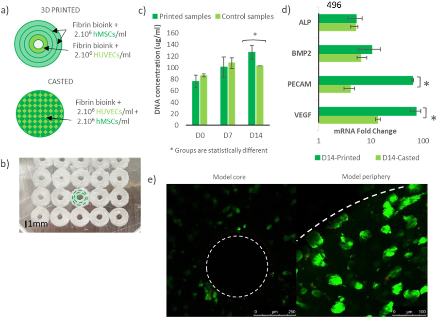
Figure III: Osteon-like scaffolds in-vitro experiment.
(a) Schematic of 3D printed osteon-like scaffolds and casted controls. 3DP scaffolds are composed of four concentric fibrin fibers of 250μm in diameter, containing hMSCs for the three outermost rings, and HUVECs for the innermost ring. Casted samples containing a mixture of MSCs and HUVECs (2.106 cells/ml) were used as controls. (b) Bioplotter picture of 3D printed constructs. The inner channel is 350μm in diameter and the complete scaffold is 2.5 mm in diameter. (c) DNA quantification assay showed significant increase in DNA concentration (p<0.05) over 14 days of in vitro culture in the printed samples compared to the casted samples (n=9). In addition, a significant higher DNA concentration (p<0.05) was shown in the printed samples, while the casted samples showed no significant changes. (d) mRNA expression fold change between day 0 and day 14 of in vitro culture (n=9). Rt-PCR showed no significant difference in gene expression of osteogenic markers (p=0.098), but a significant fold change in mRNA of angiogenic markers (p<0.05). (e) Live/Dead images of 3D printed scaffolds at day 14 (n=3). Live cells are stained green, and dead cells are stained red. Cells were counted using Image J in order to quantify cell viability. All data points represent mean +/− standard deviation.

