

Abstract
Gluteus medius and minimus tears have recently been reported to be very common and the main etiology of lateral sided hip pain. The purpose of this study was to determine whether there is any correlation between the dimensions of the tendon insertions and bare areas (BA) and various bony landmarks. Twenty-seven hemipelvises from adult male hips were included. The bony landmarks [anterior tip (Ta), posterior tip of trochanter, vastus tubercle (VT) and center of BA] were marked. The longitudinal lengths and widths (maximum) of posterosuperior (PS), lateral facets (LF), minimus insertion (Min) and BA and the distance between posterior (Tp) and Ta and between anterior/posterior tips and the VT or center of BA were measured using a digital caliper. A correlation analysis was performed between variables. There was a correlation between LFlength and Minlength (r = 0.4, P = 0.01) and between Ta–BA and PS + LF (r = 0.5, P = 0.003) or Minlength (r = 0.4, P = 0.016). LFwidth was negatively correlated with BAwidth (r = −0.4, P = 0.002). Tp–BA was negatively correlated with BAwidth (r = −0.4, P = 0.01). LFwidth was correlated with Tp–BA, and this nearly reached statistical significance (r = 0.3, P = 0.05). BA can be used intraoperatively as landmarks to estimate the width of the LF and also to determine the length of the longitudinal insertion of the gluteus medius and minimus tendons.
INTRODUCTION
Gluteus and minimus tears are recognized as the main etiology of lateral sided hip pain [1]. Partial and total ruptures of gluteal muscles tendon insertions have been described [2–4]. Open and arthroscopic treatments of gluteal tendon tears have been reported to be associated with pronounced short-term pain relief and gain of function [5, 6]. Recently, bald areas without any tendon attachment between the gluteus medius and minimus tendons have been described in previous works as possible reference points while repairing gluteal tendon tears and as reference points for intramedullary nail entry to reduce soft tissue injury [7, 8]. These areas have been reported to be slightly ellipsoidal with a diameter of 21 mm. Diameters of gluteal tendon insertions [superoposterior facet and lateral facet (LF)] were also analyzed; however, no correlation has been reported between the distance from any bony landmarks and the dimensions of the bare area (BA) [7, 8].
The gluteus minimus insertion (Min) on the trochanter has been described as L-shaped or triangular shaped in one study and shaped like a bowstring or crescent in another; no study has yet given its dimensions [9–11].
The purpose of this study was first to assess the presence of the bald spot and second to determine whether there is a correlation between the dimensions of the BA, width of the trochanter (the distance between anterior and posterior tips) or length of the trochanter [distance between anterior tip (Ta) and vastus tubercle (VT) or BA] and the insertional dimensions of the gluteal tendons. It is here hypothesized that there is a correlation between the dimensions of the bony landmarks and the insertional dimensions of the tendons, which, in turn, may help the surgeon to understand the size of the tear to plan its repair.
MATERIALS AND METHODS
This study included 27 hemipelvises (14 formalin preserved pelvises) from the hips of cadavers of adult Turkish men preserved at the Department of Anatomy, Dokuz Eylül University. Two female pelvises (four hemipelvis) were also measured to allow gross comparison to those of the all-male study group. Exact ages and body dimensions were not known.
Macroscopic examinations performed to assess gross tendon or muscle damage and signs of tears in the gluteal tendon or osteoarthritis (e.g. cartilage damage and/or osteophyte formation), any evidence of peripheral vascular disease, changes in the skin, prior fracture and traumatic or surgical scar were performed. The right hemipelvis of the first pelvis was used for pilot hemi-dissection. Dissection consisted of excision of skin, peritrochanteric muscles and fascia to isolate tendon insertions of the gluteus medius, gluteus minimus, piriformis and vastus lateralis were isolated. Sharp dissections of these tendons were then performed. Bony landmarks (Ta, posterior tip of trochanter, VT and center of BA) were also marked.
Longitudinal lengths and widths (maximum) of posterosuperior (PS),LF, Min, BA, distance between posterior (Tp), Ta, between anterior, posterior tips, VT or center of BA were measured using digital caliper (Figs 1 and 2). The distance from the center of the BA to the center of the trochanter or the most proximal point of the trochanter was also measured. The angle between longitudinal axis of the BA and LF and the axis of the femur shaft were measured using a goniometer. Correlations between the measured parameters were assessed using the Spearman correlation analysis test. Significance was set as P < 0.05.
Fig. 1.

Schematic drawings of the measured parameters (Ta, Tp, BA, Min, Med: medius tendon, P: piriformis and VT).
Fig. 2.
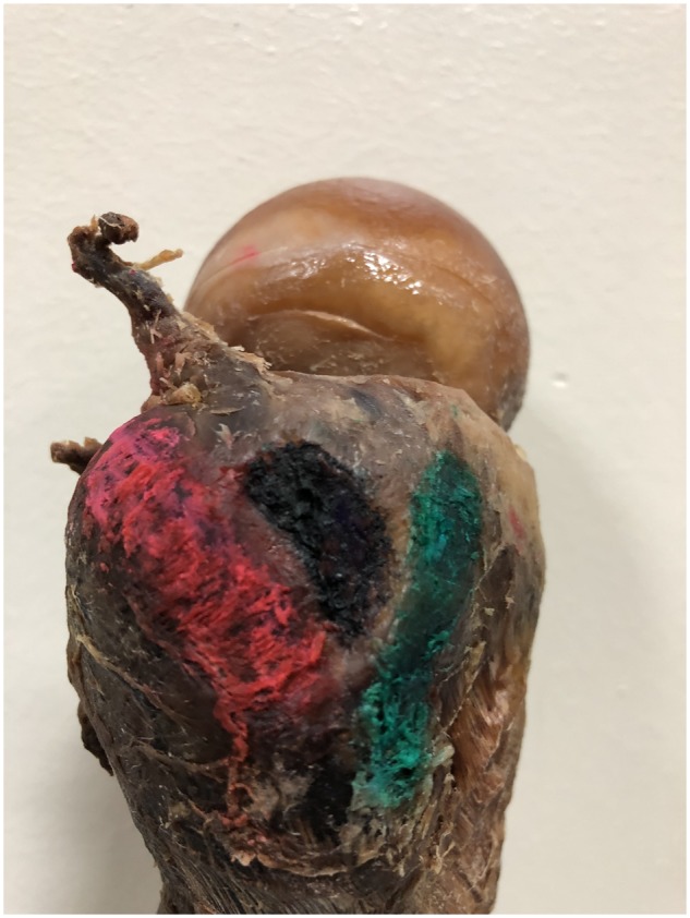
Gluteus minimus and medius insertions on the trochanter and BA in between.
RESULTS
None of the specimens yielded any macroscopic evidence of tendon damage, muscle atrophy, or scarring or any evidence on any scale of peripheral vascular disease, prior fracture or osteoarthritis. The data are presented in Table I. The distance between the most proximal point of the trochanter and the center of the BA was 8 ± 2 mm [9 (7–9)]. The distance between the center of the trochanter and the center of the BA was 13 ± 2 mm [13 (12–14)]. The angle between the longitudinal axis of the femur shaft and the LF was 32 ± 6 [30 (29–35)], while the angle between the shaft and the BA was 23 ± 6 [24 (20–26)].
Table I. .
Dimensions of insertions and distances between bony landmarks (PS, LF, Min, BA, Ta and Tp of greater trochanter and VT)
| Mean (± SD), mm | Median, mm | [% 95 CI], mm | |
|---|---|---|---|
| PS + LF (mid) | 55 ± 6 (a: 58, p: 63) [10] | 57 | [52–57] |
| LF (length × width) | 28 ± 4 × 15 ± 2 (34 ± 4 × 13 ± 2) [7, 8] (34 × 30) [12] | 27 × 15 | [26–30] × [14–16] |
| Min (length × width) | 26 ± 4 × 10 ± 2 (21 mm) [12] | 26 × 10 | [25–28] × [10–11] |
| BA (length × width) | 19 ± 2 × 9 ± 2 (r: 10 mm) [7, 8] | 20 × 9 | [18–20] × [8–10] |
| Ta–Tp | 34 ± 8 | 31 | [31–37] |
| Ta–VT | 37 ± 9 | 38 | [34–41] |
| Ta–BA | 13 ± 4 | 12 | [11–15] |
| Tp–BA | 31 ± 4 | 32 | [29–33] |
The gluteus Min appeared to be shaped like a bow tie in most of the specimens (20 specimens), while the rest were crescent shaped.
There was a correlation between LFlength and Minlength (r = 0.4, P = 0.01, PS + LF versus Minlengthr = 0.3, P = 0.06) and between Ta–BA and PS + LF (r = 0.5, P = 0.003) or Minlength (r = 0.4, P = 0.016) or BAlength (r = −0.3, P = 0.07).
LFwidth was negatively correlated with BAwidth (r = −0.4, P = 0.002). Tp–BA was negatively correlated with BAwidth (r = −0.4, P = 0.01). LFwidth showed a correlation with Tp–BA that almost reached statistical significance (r = 0.3, P = 0.05). Female subjects (n = 4) had lower parameters than their male counterparts (PS + LF = 48 ± 3, LF = 23 ± 4 × 14 ± 1, Min = 24 × 12 ± 2 mm, BA = 15 ± 1 × 10 ± 1, Ta–Tp = 32 ± 1 mm, Ta–VT = 18 ± 1, Ta–BA = 12 ± 1, Tp–BA = 25 ± 4).
DISCUSSION
Greater trochanteric pain syndrome is now used for laterally based hip pain and includes several pathologies, such as trochanteric bursitis, gluteal muscle tendinopathies and external coxa saltans due to iliotibial band thickening. If conservative measures such as rest and anti-inflammatory medications fail injections (‘recently’ platelet-rich plasma) and mini-open, endoscopic treatments have been described [12–15].
The main findings of this study were that the BA between the gluteus medius and minimus tendon insertions was 20 mm × 9 mm in size, the width of which was negatively correlated to that of the LF. The distance between the posterior tip of the trochanter and the center of this particular area was correlated with the width of LF (P = 0.05), and the length of the BA was correlated with the distance from the Ta to the center point; this correlation approached statistical significance (P = 0.07). The lengths of the insertions of the gluteus medius and minimus were positively correlated with the distance from the Ta of the trochanter to the center of the BA.
Robertson et al. [8] were the first to describe the presence of the bald center or area in between the gluteal tendons on the trochanter as approximating a least squares circle with a radius of 10 ± 1 mm. Robertson et al. reported the dimensions of the BA, angle between its longitudinal axis and femoral shaft, coordinates of the localization of the BA (e.g. central point distance to most proximal part of trochanter or central point of trochanter) and dimensions of the LF. It was reported that the shape of this area is slightly ellipsoidal with its major axis in the posterior superior to anterior inferior at an angle of 34° (17–48°) to the femoral shaft axis [similar to this study (20–26°)] with a diameter of 21 mm (17–25) [7]. In this study, it was observed that the same ellipsoid shape of the area with a longitudinal length similar to that shown in the previous study (20 mm); however, the width in this study was 9 mm. In the previous work, the center of the area was 11 mm (7–14 mm) distal to the most proximal point of the trochanter [similar to present study (7–9 mm)] and 5 mm (0–9 mm) anterior to the center of the trochanter [less than this study (12–14 mm)] [7].
Robertson et al. also reported the dimensions of the LF to be 34 ± 4 mm × 13 ± 2 mm. This study showed the LF to be slightly shorter (26–30 mm) with a 14–16-mm width [8]. However, both the previous studies were performed on fresh cadavers of both female and male pelvises with a smaller number of specimens, unlike in this study [7, 8]. Another study reported dimensions of LF to be 34 mm × 30 mm without mentioning about the BA [16].
Most recently, Philippon et al. [10] reported the total anterior length of the PS facet plus LF to be 58 mm and the posterior length to be 63 mm. Philippon et al. [10] also described the distances of footprint centrums to various bony landmarks on the trochanter; however, no report has yet discussed correlations between the dimensions of the footprints and the distances between the landmarks. In this study, the mid longitudinal length of the PS facet plus LF was reported to be 57 mm rather than providing distinct anterior or posterior total lengths.
For the gluteus minimus trochanteric insertion, the first study described its shape to be irregular L-shaped or triangular but did not provide its dimensions [9]. More recent studies described it to be shaped like a crescent (n = 11) or bow tie (n = 3) [9, 10] but still not providing the dimensions of the anterior facet or distances to various bony landmarks [10, 11]. Another study reported the width of the anterior facet to be 21 ± 3 mm [16]. This study showed a correlation between the length of the minimus tendon insertion area and the LF length or the distance from the Ta to the BA center.
The findings of this study may suggest that the BA is present, and it can be used intraoperatively as a landmark to estimate the width of LF or even determine the longitudinal insertion length of the gluteus medius and minimus tendons. The reason why other bony landmarks (e.g. VT) and the distance between the anterior and posterior tips were not correlated with the dimensions of tendon insertion remains unclear. This may be because the distance between the BA and the trochanteric tip is on the same plane or was shorter. These can be reproduced unlike other measurements, which will require further studies for clarification.
This study does have some limitations. First, formalin-fixed cadavers were used, which could affect the soft tissue relationships. Second, only male cadavers were included in correlation analysis, and the exact ages of the cadavers were not known. Due to the small number of female pelvises statistical analysis could not be performed. Most of the studies did not report the sexual differences for the tendinous attachment dimensions [8–11, 17–19]. Only one study reported sexual differences between parameters, such that that LF (34 versus 27 mm; present study 28 versus 23 mm) and tendon length were found to be longer in males than in females [16].
Further studies utilizing fresh cadavers and including the female pelvises and an overall larger number of specimens are needed to validate the correlations found in this study and to further delineate possible other correlations, which, in turn, may help surgeons determine the sizes and configurations of tears to plan treatment.
CONFLICT OF INTEREST
None declared.
REFERENCES
- 1. Karpinski MR, Piggott H.. Greater trochanteric pain syndrome. A report of 15 cases. J Bone Joint Surg Br 1985; 67:762–3. [DOI] [PubMed] [Google Scholar]
- 2. Kingzett-Taylor A, Tirman PF, Feller J. et al. Tendinosis and tears of gluteus medius and minimus muscles as a cause of hip pain: MR imaging findings. AJR Am J Roentgenol 1999; 173:1123–6. [DOI] [PubMed] [Google Scholar]
- 3. Connell DA, Bass C, Sykes CA. et al. Sonographic evaluation of gluteus medius and minimus tendinopathy. Eur Radiol 2003; 13:1339–47. [DOI] [PubMed] [Google Scholar]
- 4. Bunker TD, Esler CN, Leach WJ.. Rotator-cuff tear of the hip. J Bone Joint Surg Br 1997; 79-B: 618–20. [DOI] [PubMed] [Google Scholar]
- 5. Kagan A., II Rotator cuff tears of the hip. Clin Orthop Relat Res 1999; 368:135–40. [PubMed] [Google Scholar]
- 6. Hartigan DE, Perets I, Ho SW. et al. Endoscopic repair of partial-thickness undersurface tears of the abductor tendon: clinical outcomes with minimum 2-year follow-up. Arthroscopy 2018; 34:1193–9. [DOI] [PubMed] [Google Scholar]
- 7. Gardner MJ, Robertson WJ, Boraiah S. et al. Anatomy of the greater trochanteric ‘bald spot’: a potential portal for abductor sparing femoral nailing? Clin Orthop Relat Res 2008; 466:2196–200. [DOI] [PMC free article] [PubMed] [Google Scholar]
- 8. Robertson WJ, Gardner MJ, Barker JU. et al. Anatomy and dimensions of the gluteus medius tendon insertion. Arthroscopy 2008; 24:130–6. [DOI] [PubMed] [Google Scholar]
- 9. Beck M, Sledge JB, Gautier E. et al. The anatomy and function of the gluteus minimus muscle. J Bone Joint Surg Br 2000; 82-B: 358–63. [DOI] [PubMed] [Google Scholar]
- 10. Philippon MJ, Michalski MP, Campbell KJ. et al. Surgically relevant bony and soft tissue anatomy of the proximal femur. Orthop J Sports Med 2014; 2: 2325967114535188. [DOI] [PMC free article] [PubMed] [Google Scholar]
- 11. Flack NA, Nicholson HD, Woodley SJ.. A review of the anatomy of the hip abductor muscles, gluteus medius, gluteus minimus, and tensor fascia lata. Clin Anat 2012; 25:697–708. [DOI] [PubMed] [Google Scholar]
- 12. Byrd JWT, Jones KS.. Endoscopic repair of hip abductor tears: outcomes with two-year follow-up. J Hip Preserv Surg 2017; 94:80–4. [DOI] [PMC free article] [PubMed] [Google Scholar]
- 13. DeFroda S, Silverman A, Quinn M, Tabaddor R.. Mini-open double row gluteus medius repair provides good short-term functional outcomes. J Hip Preserv Surg 2019; 6:271–6. [Google Scholar]
- 14. Khoury AN, Brooke K, Helal A. et al. Proximal iliotibial band thickness as a cause for recalcitrant greater trochanteric pain syndrome. J Hip Preserv Surg 2018; 8; 5:296–300. [DOI] [PMC free article] [PubMed] [Google Scholar]
- 15. Ali M, Oderuth E, Atchia I, Malviya A.. The use of platelet-rich plasma in the treatment of greater trochanteric pain syndrome: a systematic literature review. J Hip Preserv Surg 2018; 5:209–19. 30; [DOI] [PMC free article] [PubMed] [Google Scholar]
- 16. Flack NA, Nicholson HD, Woodley SJ.. The anatomy of the hip abductor muscles. Clin Anat 2014; 27:241–53. [DOI] [PubMed] [Google Scholar]
- 17. Márquez-Arabia WH, Gómez-Hoyos J, Gómez M. et al. Influence of aging on microvascular supply of the gluteus medius tendon: a cadaveric and histologic study. Arthroscopy 2017; 33:1354–136. [DOI] [PubMed] [Google Scholar]
- 18. Al-Hayani A. The functional anatomy of hip abductors. Folia Morphol (Warsz) 2009; 68:98–103. [PubMed] [Google Scholar]
- 19. Márquez WH, Gómez-Hoyos J, Woodcock S. et al. The regional microvascular density of the gluteus medius tendon determined by immunohistochemistry with CD31 staining: a cadaveric study. Hip Int 2015; 25:168–71. [DOI] [PubMed] [Google Scholar]