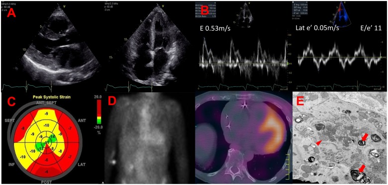
A 70-year old woman with history of long-standing rheumatoid arthritis and recent diagnosis of complete heart block leading to a dual-chamber pacemaker implantation presented to the hospital with several weeks of worsening dyspnoea and lower extremity swelling. On physical exam, she had evidence of both intra- and extravascular fluid accumulation as evidenced by elevation in jugular venous pressure and lower extremity oedema. Electrocardiogram demonstrated normal pacemaker function with atrial-sensed, ventricular-paced rhythm. Echocardiography showed moderate–severe increased wall thickness with moderately reduced left ventricular ejection fraction (Supplementary material online, Videos S1–S4). Diastolic parameters were consistent with a Grade III abnormal filling pattern, low mitral annular tissue velocity, and a global longitudinal strain (GLS) of −9.4% with associated relative apical sparing (ratio of apical/basal segments 1.2). Given concern for an infiltrative cardiomyopathy, she underwent 99mtechnetium pyrophosphate scintigraphy revealing semiquantitative visual Grade 3 and diffuse uptake (Perugini Grade 3) on single-photon emission computed tomography. Serum immunofixation, free light chains, and alpha galactosidase A were normal. Right ventricular biopsy was negative on Congo red staining. Electron microscopy revealed disorganized myofibrils, abundant vacuoles, multiple lamellar inclusion bodies, and curvilinear cytoplasmic bodies, findings that are pathognomonic for hydroxychloroquine-induced restrictive cardiomyopathy (Figure). Hydroxychloroquine was discontinued, and the patient started on guideline directed heart failure therapy. Several months later, repeat echocardiography revealed modest improvement in left ventricular ejection fraction.
Hydroxychloroquine accumulates in lysosomes, inhibiting molecular breakdown with resultant accumulation of phospholipids and glycogen. Clinical presentation includes biventricular hypertrophy, restrictive physiology (late-stage), and conduction system abnormalities. Importantly, in an era when bone scintigraphy is being used with increasing frequency to diagnose transthyretin (ATTR) amyloid cardiomyopathy, we hope to raise awareness for the potential of a false-positive result in patients taking hydroxychloroquine.
Hydroxychloroquine-induced restrictive cardiomyopathy. (Panel A) Parasternal long-axis and apical views showing left ventricular concentric hypertrophy. (Panel B) Pulsed-wave and tissue Doppler showing a pseudo-normal filling pattern and depressed mitral annular velocity with a normal E/e′ ratio. (Panel C) Polar map of the left ventricle showing relative apical sparing and a GLS of −9.4%. (Panel D) Diffuse uptake of 99mtechnetium pyrophosphate on planar imaging and single-photon emission computed tomography. (Panel E) Electron microscopy of a myocardial section obtained from an endomyocardial biopsy showing cytoplasmic changes of the cardiomyocyte including disorganization of myofibrils, lamellar inclusion bodies (red arrows), abundant vacuoles, and curvilinear cytoplasmic bodies (red arrow heads).
A.M. is supported by a competitive investigator-initiated grant from Pfizer (ASPIRE). S.B.H. has received research support and personal compensation for consulting, serving on a scientific advisory board, speaking, or other activities with Akcea, Ionis, Pfizer, and Eidos.
Supplementary material is available at European Heart Journal online.
Supplementary Material
Associated Data
This section collects any data citations, data availability statements, or supplementary materials included in this article.


