Abstract
The frequency and severity of injection-site lesions in the outside round muscles of both beef and dairy cattle were evaluated through a series of audits. Audits were conducted in 2017 on 1,300 rounds from dairy and beef cows from seven locations throughout the United States. Outside round muscles were butterfly cut into 1.25-cm slices and, if present, lesions were counted, measured, and categorized. Rounds from beef (7%) and dairy cattle (15%) had at least one injection-site lesion present. The most common location of injection-site lesions was quadrant 2 and 3, which contained both the biceps femoris and semitendinosus muscles. Injection-site lesions were more frequent (P < 0.05) in the biceps femoris for both beef and dairy rounds. Clear lesions accounted for 57% of injection-sites in both beef and dairy rounds, whereas metallic lesions made up 23% of the total in beef and 25% in dairy. Overall, there was a dramatic decline in the frequency (P < 0.05) of injection-site lesions since the 1998 (24 and 45 percentage units greater in beef and dairy rounds, respectively) and 2000 audits (13 and 20 percentage units greater in beef and dairy rounds, respectively). Educational programs, such as Beef Quality Assurance (BQA) and requirements for BQA training, have resulted in substantial improvements in beef management practices for both the beef and dairy industries.
Keywords: audit, beef, dairy, injection-site lesion
INTRODUCTION
Injection-site lesions represent a major economic loss to the beef industry (Roeber et al., 2001). More specifically, the presence of injection-site lesions in whole muscle cuts limit their value and use, especially for further processors (Roeber et al., 2001). Furthermore, steaks afflicted with visually and palpably mild injection-site lesions have greater Warner–Bratzler shear force values, reduced sensory panel tenderness ratings, and greater palatability variation among steaks within a primal/subprimal cut with lesions than those without injection-site lesions (George et al., 1996).
The National Beef Quality Market Cow and Bull Audit has been conducted four times (1994, 1999, 2007, and 2016) over the past 25 yr. Following the original audit, attention was drawn to injection-site lesions and the need to further assess the lesions. Previous research evaluated injection-site lesions in beef and dairy cattle, especially in the muscles from the top sirloin butt and the round (Dexter et al., 1994; George et al., 1996; Sullivan et al., 2009).
The 2016 National Beef Quality Market Cow and Bull Audit reported a major decline in the presence of surface knots or injection-site lesions compared with previous audits. The presence of lesions was discussed in detail at the strategy workshop held as the third phase of the 2016 National Beef Quality Market Cow and Bull Audit. Further processors expressed extreme concern of loss and the need to once again evaluate lesions as evaluated in 1998, 1999, and 2000. Although previous audits quantified the frequency of injection-site lesions, limited information is currently available on the location of lesions (Harris et al., 2017).
Therefore, the objective of this study was to evaluate the frequency and presence of injection-site lesions in the outside round muscles of cows. Evaluating the frequencies and severity of injection sites allows each segment of the beef and dairy industries to evaluate the progress made since the audit conducted by Roeber et al. (2002), as well as continue to create better management practices for the future.
MATERIALS AND METHODS
Plant and Product Selection
Data were collected in 2017 at seven U.S. beef packing plants selected based on geographic location, as well as size and scale of slaughter and fabrication production. At six plants outside rounds, including both the biceps femoris and semitendinosus muscles, were randomly selected and collected from approximately 100 dairy cows and 100 beef cows, whereas only 50 dairy and 50 beef rounds were evaluated at the seventh plant.
Product Preparation and Evaluation
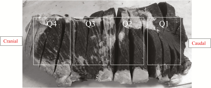
The rounds were trimmed of fat and then butterfly cut into 1.25-cm slices by a trained plant employee. Two trained researchers evaluated the sliced round muscles for the presence of injection-site lesions. If a lesion(s) was present, it was documented based on its location (muscle and quadrant) as well as diameter and depth from the most lateral point. The outside round consisted of four quadrants (Figure 1), with quadrant 1 (Q1) as the most caudal end and closest to the shank, whereas quadrant 4 (Q4) was the most cranial end and only include the biceps femoris. Both quadrants 2 (Q2) and 3 (Q3) were evenly split between Q1 and Q4, and Q1, Q2, and Q3 included both the biceps femoris and semitendinosus muscles. The depth of each lesion was measured from the outside surface (fat trimmed) to the innermost (center) of the lesion. The diameter was measured using the lengths of the lesion throughout the muscles. The type of lesion was classified using five different classifications. The lesions were classified using the 5-point scale described by Dexter et al. (1994), as well as Roeber et al. (2002): 1 = clear, lesion contains primarily clear connective tissue; 2 = woody, lesion contains organized connective tissue and fat; 3 = nodular, lesion contains nodules, a central foci, and granulomatous inflammation; 4 = metallic, lesion contains mineralized remnants of muscle cells, typically bloody color; or 5 = cystic, lesion contains fluid.
Figure 1.
Quadrants of the outside round where injection-site lesions were evaluated. Q1 was identified as the most caudal end and was the closest to the shank, whereas Q4 was identified as the most cranial end and only included the biceps femoris muscle. Q2 and Q3 were evenly split between Q1 and Q4. Q1, Q2, and Q3 included both the biceps femoris and the semitendinosus muscles.
Statistical Analysis
The presence of injection-site lesions, number of lesions per round, depth, and diameter for both dairy and beef cow rounds were analyzed using the general linear models procedure in SAS 9.4 (SAS Inst. Inc., Cary, NC). A pairwise t-test was used to separate differences between the presence, number, depth, and diameter of lesions when the analysis of variance demonstrated differences (P < 0.05). Differences between frequency percentages of lesion presence in the 2017 audit as well as 1998, 1999, and 2000 audits were evaluated using the Chi-square statistic in Excel (Microsoft Office, 2016) by comparing incidence percentage and total observations of each audit.
RESULTS AND DISCUSSION
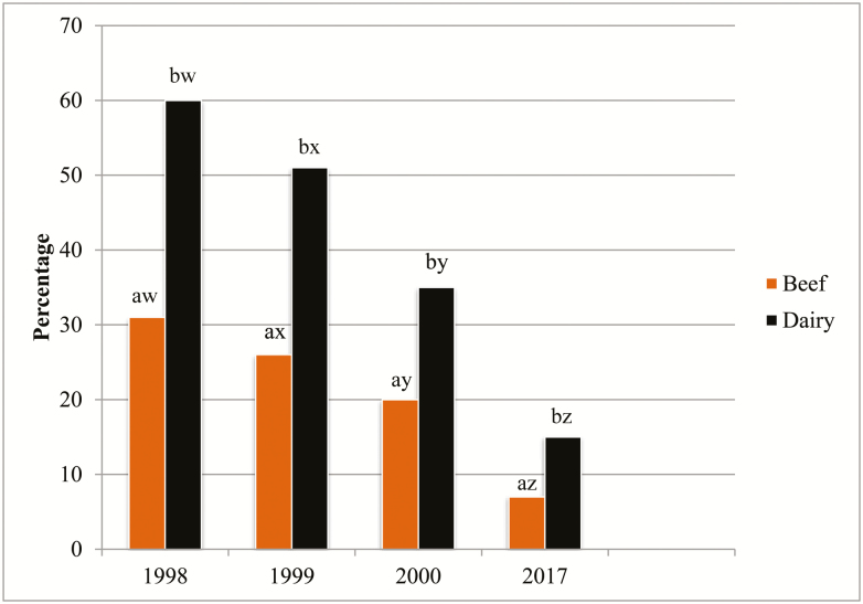
In the 2017 audit, lesions were identified in 6.9% and 14.9% of rounds from beef and dairy carcasses, respectively (Table 1). In the 2017 audit, as well as previous audits, the frequencies of injection-site lesions were greater (P < 0.05) in rounds of dairy cows than beef cows (Table 1 and Figure 2). More importantly, the number of injection-site lesions present has declined greatly from 2000 (20% and 35% of beef and dairy cow rounds, respectively) and 1998 audits (31% and 60% of beef and dairy cow rounds, respectively) (Figure 2). This sharp reduction in injection-site lesions mirrors the results of the 2016 National Beef Quality Market Bull and Cow Audit, which demonstrated a major decrease in the presence of surface knots and injection-site lesions (Harris et al., 2017).
Table 1.
Frequency of injection-site lesions in beef and dairy rounds in 2017 (n = 1,300)
| Beef | Dairy | |
|---|---|---|
| Total pieces audited | 677 | 623 |
| Pieces with lesion(s) | 47 | 93 |
| Percent of rounds with lesion(s) | 6.9a | 14.9b |
| Average number of lesions per pieces with lesion(s) | 1.0 | 1.1 |
| Max lesions in one round | 2 | 4 |
a,bWithin a row, means lacking a common superscript letter differ, P < 0.05.
Figure 2.
Frequency of injection-site lesions in 1998, 1999, 2000, and 2017. a,bWithin each year comparing breed type, with differing superscript letters differ (P < 0.05). w,x,y,zWithin each breed type comparing year, with differing superscript letter differ (P < 0.05).
The location of the injection-site lesion within the outside round was evaluated. Providing the location gives better insight into the administration of injections and ultimately prevention programs for the future. The location of lesions was much more frequent (P < 0.05) in the biceps femoris muscle of the outside round than the semitendinosus muscle for both dairy and beef type (Table 2).
Table 2.
Location of injection-site lesion within the round muscles as a percentage of total lesions pieces audited in 2017
| Location in outside round muscles | Beef (n = 47) | Dairy (n = 93) |
|---|---|---|
| Semitendinosus Q1 | 0 | 5 |
| Semitendinosus Q2 | 3 | 10 |
| Semitendinosus Q3 | 7 | 22 |
| Semitendinosus all quadrants | 9a,x | 35b,x |
| Biceps femoris Q1 | 4 | 8 |
| Biceps femoris Q2 | 10 | 23 |
| Biceps femoris Q3 | 22 | 27 |
| Biceps femoris Q4 | 4 | 12 |
| Biceps femoris all quadrants | 40a,y | 68b,y |
a,bColumn percentages, comparing breed type of all whole muscle (all quadrants), with differing superscript letters differ (P < 0.05).
x,yRow percentages, comparing muscle type (all quadrants) within breed type, with differing superscript letters differ (P < 0.05).
According to Dexter et al. (1994) and Roeber et al. (2002), clear and woody lesions are “older” lesions resulting from an injection administered during earlier stages of the calf’s life, whereas nodular and cystic lesions arise from injections administered more recently in the animal’s life, thereby not having time to fully heal from the trauma of an intramuscular injection. In the present audit, clear lesions were the most prevalent (P < 0.05) in both dairy and beef rounds, and the frequency of nodular lesions was greater (P < 0.05) than the presence of woody, metallic, or cystic lesions (Table 3). There were no differences found between the presence of woody, metallic, or cystic lesions (Table 3).
Table 3.
Frequency data, depth and diameter of injection-site lesions by classification of lesion and breed type for 2017.
| Depth (cm) | Diameter (cm) | |||||
|---|---|---|---|---|---|---|
| Trait | Beef (n = 47) | Dairy (n = 93) | Beef (n = 47) | Dairy (n = 93) | Beef (n = 47) | Dairy (n = 93) |
| Classification of lesion | ||||||
| Clear1 | 57c | 57c | 2.18 ± 0.33 | 2.25 ± 0.23 | 3.60 ± 0.04 | 3.60x ± 0.37 |
| Woody2 | 10a | 12a | 2.28 ± 0.79 | 1.74 ± 0.51 | 5.08 ± 1.16 | 5.40y ± 0.82 |
| Nodular3 | 23b | 25b | 1.21 ± 0.53 | 1.25 ± 0. 35 | 4.39 ± 0.78 | 3.22x ± 0.55 |
| Metallic4 | 8a | 5a | 1.59 ± 0.88 | 2.79 ± 0.79 | 2.86 ± 1.30 | 4.57x ± 1.26 |
| Cystic5 | 2a | 1a | 1.27 ± 1.76 | 1.27 ± 1.76 | 2.54 ± 2.60 | 7.62y ± 2.82 |
a,b,cPercentages in the same column, comparing kind of lesion in a breed type for a given year, with differing superscript letters differ (P < 0.05).
x,yMeans in the same column with differing superscript letters differ (P < 0.05).
1Clear: Lesion contains primarily clear connective tissue.
2Woody: Lesion contains organized connective tissue and fat.
3Nodular: Lesion contains nodules, a central foci, and granulomatous inflammation.
4Metallic: Lesion contains mineralized remnants of muscle cells, typically bloody color.
5Cystic: Lesion contains fluid.
In dairy rounds, lesions diameter varied among the lesion types, with woody and cystic lesions clearly larger (P < 0.05), and damaging more sellable product, than clear, nodular, or metallic lesions (Table 3). Neither diameter nor depth of lesions differed (P > 0.05) between beef and dairy rounds.
The reduction in lesions between 1998 and 2000 showed positive improvement in beef medication administration and producer education. The continued reduction from 2000 to 2017 also supports this—during this time not only did Beef Quality Assurance (BQA) program trainings continue across the country, but a new online certification system was also released, potentially exposing additional cow–calf and dairy producers to the educational materials on proper injection placement. Various programs and animal health companies have worked to reduce injection-site lesions. Imler et al. (2017) outline best management practices that include follow label directions, administer all injections in the neck, keep injection sites and equipment clean, and tent the skin. These types of guidelines that are found through many educational programs provide producers with detailed instructions that help to avoid injection-site lesions. In addition, companies such as Merck Animal Health have created web-based producer educational programs for both dairy (Dairy Care 365) and beef (Creating Connections) cattle (Merck Animal Health, 2018a, 2018b).
Furthermore, many herds have focused on improving nutrition and management as a preventative method to help improving overall herd health (Leblanc et al., 2006). Nordlund (1998) expressed the importance of fixing the production system to ensure a healthy herd. Also, Leblanc et al. (2006) explain the importance of determining health status based on the components of host, agent, and environment that are all affected by the management practices. Finally, the large decrease in injection-site lesions in dairy animals could be due to a significant reduction in bovine somatotropin usage throughout the United States in the past 10 yr (Stepp, 2018).
These improvements show the increased knowledge of BQA and support the education programs targeting the beef and dairy industry have led directly to the dramatic decrease in injection-site lesions in muscles of the round over the past 25 yr. Continued emphasis on these programs and education should equate to further beef quality improvements and greater use of the beef and dairy meat products.
ACKNOWLEDGMENTS
This project was funded by the Cattlemens Beef Board through the National Cattlemen’s Beef Association.
LITERATURE CITED
- Dexter D. R., Cowman G. L., Morgan J. B., Clayton R. P., Tatum J. D., Sofos J. N., Schmidt G. R., Glock R. D., and Smith G. C.. 1994. Frequency of injection-site lesions in beef top sirloin butts. J. Anim. Sci. 72: 824–837. [DOI] [PubMed] [Google Scholar]
- George M. H., Cowman G. L., Tatum J. D., and Smith G. C.. 1996. Incidence and sensory evaluation of injection-site lesions in beef top sirloin butts. J. Anim. Sci. 74:2095–2103. [DOI] [PubMed] [Google Scholar]
- Harris M. K., Eastwood L. C., Boykin C. A., Arnold A. N., Gehring K. B., Hale D. S., Kerth C. R., Griffin D. B., Savell J. W., Belk K. E.,. et al. 2017. National Beef Quality Audit–2016: transportation, mobility, live cattle, and carcass assessments of targeted producer-related characteristics that affect value of market cows and bulls, their carcasses, and associated by-products. Transl. Anim. Sci. 1:570–584. doi:10.1093/tas/txx002 [DOI] [PMC free article] [PubMed] [Google Scholar]
- Imler A., Hersom M., Thrift T., Yelich J., and Irsik M.. 2017. Cull cow beef quality issues: injection sites and abscesses. Department of Animal Sciences, UF/IFAS Extension; Available from http://edis.ifas.ufl.edu/pdffiles/AN/AN30800.pdf [Google Scholar]
- LeBlanc S. J., Lissemore K. D., Kelton D. F., Duffield T. F., and Leslie K. E.. 2006. Major advances in disease prevention in dairy cattle. J. Dairy Sci. 89:1267–1279. doi:10.3168/jds.S0022-0302(06)72195-6 [DOI] [PubMed] [Google Scholar]
- Merck Animal Health 2018a. Dairy care 365 [accessed July 3, 2018] https://www.dairycare365.com/?utm_source= MAH-USA&utm_medium=website.
- Merck Animal Health 2018b. Creating connections [Accessed July 3, 2018] http://www.creatingconnections.info/Training.
- Nordlund K. 1998. Grumpy old vets: the 1960’s practice hits the 21st century. Bovine Pract. 32:58–62. [Google Scholar]
- Roeber D. L., Cannell R. C., Wailes W. R., Belk K. E., Scanga J. A., Sofos J. N., Cowman G. L., and Smith G. C.. 2002. Frequencies of injection-site lesions in muscles from rounds of dairy and beef cow carcasses. J. Dairy Sci. 85:532–536. doi:10.3168/jds.S0022-0302(02)74105-2 [DOI] [PubMed] [Google Scholar]
- Roeber D. L., Mies P. D., Smith C. D., Belk K. E., Field T. G., Tatum J. D., Scanga J. A., and Smith G. C.. 2001. National market cow and bull beef quality audit—1999: a survey of producer-related defects in market cows and bulls. J. Anim. Sci. 79:2615–2618. [DOI] [PubMed] [Google Scholar]
- Stepp E. 2018. Personal communication on behalf of the National Milk Producers Federation. [Google Scholar]
- Sullivan M. M., Vanoverbeke D. L., Kinman L. A., Krehbiel C. R., Hilton G. G., and Morgan J. B.. 2009. Comparison of the biobullet versus traditional pharmaceutical injection techniques on injection-site tissue damage and tenderness in beef subprimals. J. Anim. Sci. 87:716–722. doi:10.2527/jas.2007-0763 [DOI] [PubMed] [Google Scholar]


