

Abstract
Cortical bone loss in revision hip arthroplasty requires an adequate stabilization to achieve a durable implant fixation. This case series shall illustrate possible indications for the use of allogenic grafts in revision hip arthroplasty. Twelve patients with femoral bone loss were treated with allografts. In addition to established clinical scores, the radiological follow-ups were analyzed for hints of implants loosening and the osteointegration of the allografts. After a mean follow-up of 3.0 years the mHHS was 61.3 points and the UCLA 3.8. One patient showed a non-progressive radiolucency around the hip implant. The osteointegration of all allogenic grafts happened on time. Up to the last follow-up no revision surgery of the hip implants and the associated femoral bone graft was observed. Allogenic bone grafts present a method for biological stabilization in situations of large femoral cortical bone defects in revision hip arthroplasty.
Key words: Femoral bone loss, revision hip arthroplasty, allogen bone graft, defect Augmentation, Paprowsky
Introduction
Over the last decades the number of performed primary [total hip arthroplasties (THA)] increased continuously.1 Subsequently, the number of revisions raised. Kurtz et al. estimated for the USA that the volume of revision replacements will enhance up to 96.700 per year in 2030, an increase about 137.0%.2 However, not only the number of revisions overall is increasing, but also more revisions per patient are required.3 With rising number of revisions in the same patient, the surgeon is faced with the problem of achieving adequate stability of the revision implants due to increasing bony defects. For bony defects of the acetabulum different solutions exist for compensation, e.g. autogenous/allogen bone (impaction grafting) with or without combination of revision cups, tantalum augmentation or individual implants.4,5 The number of treatment options for femoral defects are lower. In general, for the first revision of a primary femoral stem a longer revision stem may be used to compensate the loss of spongiosa due to explantation in order to gain good primary stability. An additional option may be to use a modular implant that fills the further cancellous bony defects of the metaphysis as accurately as possible.5 Extensive femoral spongiosa bone loss can also be filled with autogenous / allogenic bone or compensate with cemented implants. For combined cortical and cancellous defects implants with a diaphyseal fixation are used or a proximal femur replacement up to a megaprosthesis (e.g. total femur) are implanted.6 However, isolated cortical defects or periprosthetic fractures still represent a challenge due to limited treatment options. The aim of surgery is to stabilize and bridge the insufficient bone area with an osteosynthesis plate and cerclages.7 For cortical defects the use of an allogenic bone grafting combined with a long revision implant seems to represent an useful stabilization of insufficient bone conditions of the femur.
The aim of this study was to review cases with the use of strut grafts in revision hip arthroplasty. This analysis shall illustrate possible indications for the use of this technique. Along with that, clinical and radiological outcomes in terms of resorption and osseointegration were analyzed. Thus, the survival rate of allogenic femoral bone grafts and further revision treatments were determined.
Materials and Methods
Surgical procedure
During surgery the preparation to the area of bone loss was performed until sufficient depiction. The allografts were fitted and placed in a way as to cover the defect of the host femur and fixed with wires (Figure 1). In every case the correct position was controlled by intraoperative fluoroscopy. Except for one patient, all patients were partial weight mobilized for 6 weeks.
Demographics
The current study is a retrospective case series admitted by the local ethical commission (No. 7743_BO_S_2018). The study included twelve consecutive cases of proximal femoral bone loss, treated with femoral bony allografts between 2010 and 2017. Of the twelve patients, ten were women (83.3%) and two men (16.7%). Three patients were excluded: One patient died before the time of contact, another did not wish to participate, and for one patient no actual contact data was present (25.0%). As a result, the clinical und radiological evaluation based on the data of nine cases (75.0%). Three patients wished to participate only by mail and permitted the evaluation of the last radiological pictures by our institution. Two of them already lived in nursing homes.
Statistical analysis
Several patient- and surgery-specific data were collected, especially the number of previous surgeries, the indication and defect classification according to Paprosky,8 the localization of bone loss, the size of the allogenic graft and the number of wires for fixation. The analyzed allogenic femoral bone grafts had a mean length of 10.6cm (±7.0; 2.5-25.0). In one case the allograft was fixed by 1 wire (11.1%), in 2 cases by 2 wires (22.2%), in 3 cases by 4 (33.3%), in one case by 5 (11.1%) and in 2 cases by 6 (22.2%).
The clinical follow-up was assessed using the [modified Harris Hip Score (mHHS)], the [University of California and Los Angeles activity score (UCLA)] and the [visual analog scale (VAS)] in terms of pain and gait control. Both scores ranged from 0.0 to 10.0, 0.0 being an excellent result and 10.0 the poorest.
The revision-free survival was defined as absence of any surgical intervention of the hip implants and the associated allogenic femoral bone grafts.
The patients received a radiographical follow-up in two planes. The x-rays acquired immediately postoperatively served as reference for comparison. For the evaluation of implant stability, the subsidence and the alignment were measured.9 Subsidence was defined as a change in the distance between the top of the greater trochanter and the lateral border of the femoral stem. A migration about more than 4 mm was diagnosed as a relevant stem subsidence. 10 If the trochanter major could not conduce as reference because of defect situations, other landmarks were chosen (e.g. distal point of trochanter minor). The stem alignment was defined as angle between the anatomical femur axis and the vertical stem axis. A neutral position was present between 0-2°. Alignments greater than 2° were described as “misalignment” of the femoral stem.10 In addition, the periprosthetic bone was analyzed for radiolucency > 1 mm in the femoral zones according to Gruen.11 The osteointegration of the allografts was described with “none” (<50%, neither side osteointegrated), “partial” (<50%, one side osteointegrated) and “complete” (>50%, both sides osteointegrated).9 The radiological images were presented and analyzed with Carestream® (Carestream Solutions, Vue Solutions, Version 11.4.1.1011- sias110). A describing data analysis was performed for the clinical and radiological parameters using IBM® SPSS® Statistics (IBM® SPSS® Statistics, Version 24.0.0.0, 64-Bit-Version). For metric data the mean value and the standard deviation were reported, for ordinal/categorical data absolute values and percentages. With respect to the characteristics of a case series, no comparing statistics were performed due to less power.
Results
Of the nine patients, who were clinically followed-up, seven were females (77.8%) and two males (22.2%). Before surgery the mean age was 69.7 years (±7.8; 58.5-83.1) and the BMI 29.3kg/m² (±6.7; 22.2-42.6). In five cases the indication was the loosening of the previous implants (55.6%), three of them septic in a twostage- revision (33.3%) and two aseptic (22.2%). According to Paprosky,8 four patients showed a femoral grade 3A defect (44.4%) and five a 3B (55.6%). In one case (11.1%) the indication was an osteolysis involving the cortical bone, one pseudarthrosis (11.1%) and two (22.2%) periprosthetic fractures type B/C1 according to Vancouver.12 The number of previous surgeries ranged from 1-4 (1 = 1; 2 =5; 3 = 1; 4 = 2). All revision hip arthroplasty with the aid of allogenic femoral bone grafts were performed through a lateral approach. The mean surgical time took 166.1 min (±53.5; 74.0-248.0) and the mean postoperative length of stay was 17.3 days (±10.2; 10.0-43.0).
Table 1.
Patient-related clinical outcome. The most clinical limitations were observed for the UCLA and the VAS gait control.
| Pat. No. | mHHS | UCLA | VAS - pain | VAS - gait |
|---|---|---|---|---|
| 1 | 86.0 | 3.0 | 0.0 | 7.7 |
| 2 | 87.0 | 4.0 | 0.3 | 9.5 |
| 3 | 49.0 | 5.0 | 7.1 | 6.6 |
| 4 | 77.0 | 7.0 | 0.0 | 9.3 |
| 5 | 37.0 | 2.0 | 4.1 | 3.1 |
| 6 | 52.0 | 3.0 | 3.8 | 3.7 |
| 7 | 43.0 | 4.0 | 6.3 | 1.4 |
| 8 | 69.0 | 4.0 | 1.0 | 3.3 |
| 9 | 52.0 | 2.0 | 1.3 | 3.1 |
Table 2.
Patient-related radiolucency (mm) in zonal distribution according Gruen.11 One patient respectively showed radiolucency around the tip of the femoral stem.
| Pat. No. | Gruen 1 | Gruen 2 | Gruen 3 | Gruen 4 | Gruen 5 | Gruen 6 | Gruen 7 |
|---|---|---|---|---|---|---|---|
| 1 | - | - | - | - | - | - | n |
| 2 | n | n | - | - | - | n | n |
| 3 | n | - | - | - | - | - | n |
| 4 | - | - | - | - | - | - | - |
| 5 | n | - | - | - | - | - | - |
| 6 | - | - | - | - | - | - | - |
| 7 | - | n | n | - | - | - | - |
| 8 | - | n | n | - | - | - | - |
| 9 | n | n | - | - | 2.3 | - | - |
- = no radiolucency; n = not present due to bony defect.
Figure 1.
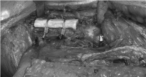
A bone defect about 6 x 2 cm located at the lateral femur covered by allogenic femoral bone graft and two wires.
Clinical evaluation
After a follow-up of 3.0 years (±2.2; 0.3 – 7.5) the mean mHHS was 61.3 (±18.8; 37.0 – 87.0), the UCLA 3.8 (±1.6; 2.0 – 7.0) and the VAS for pain 2.7 (±2.8; 0.0 – 7.1) respectively gait control 5.3 (±3.0; 1.4 – 9.5). The patients with the numbers 6, 7 and 9 already lived in nursing homes and number 4 is still working as a medical doctor (Table 1).
Radiological evaluation
One (11.1%) of the nine participants showed a radiolucency around the hip implants. This non-progressive radiolucency was located at the tip of a long cementless revision stem (Table 2). In no case the femoral stem showed a relevant subsidence or a change of alignment. In 7 cases (77.7%) the allogenic grafts were completely integrated at last radiological follow-up, while in one case (11.1%) a partial osteointegration was observed. Under recognition of a three-month radiological follow-up the result was evaluated as an on-time osteointegration. Patient number 7 (11.1%) did not come to the recommended routine examinations, so no statement concerning the osteointegration of the allogenic femoral bone graft was possible.
Complications
There were two reported revisions, both treated in other clinics. The first case was patient number 1. After 3.5 years, the patient suffered a periprosthetic fracture of the equilateral knee arthroplasty falling three steps down and received osteosynthesis by plate. At time of evaluation, the follow- up treatment had just ended, and the patient just resumed her work as a bookseller. In the most recent follow-up x-rays there were no sign of subsidence, loosening or mechanical failure of the hip implants and the allogenic graft. In the second case, patient number 8 fell in her kitchen after alcohol consumption in the setting of a known dependency. The trauma cropped up 7 months after hip revision replacement. An occurred distal femur fracture (AO 33-B1) was treated with an open reposition and internal fixation (9-hole NCB plate) and a fracture of the tibial shaft (AO 42-A1) was treated with a closed repositioning and stabilized with an internal fixation (315*12mm nail). The radiological follow-up showed no signs of periprosthetic fractures or other mechanical complications of the hip revision arthroplasty or the allografting hip stabilization. Overall the septic and aseptic survival rates of the strut grafts and the associated hip implants were 100.0%.
Discussion
The present study illustrated the indications for the additional application of an allogenic femoral bone graft in hip revision arthroplasty. This technique enables a biological stabilization in case of bone loss with a high risk for or already in case of periprosthetic fracture of the host femur. The bony deficiency or fractur situation were bridged by the allogenic graft, which osteointegrate and reconstruct the cortical ring of the host femur. For the initial osteointegration the most cases were trained with partial weight bearing. In selected cases femoral strut grafting may present a chance for a stable and durable hip revision arthroplasty before the use of total femur replacement becomes necessary, which is associated with a high rate of complications, especially [periprosthetic joint infections (PJI)].13 Allogenic femoral bone grafts generally seem to osteointegrate in about 6-12 months.14-16 The radiological follow-ups of the current study have proven a timely osteointegration of all allografts without a hint of resorption. The implants presented a stable fixation without relevant radiolucencies, sintering or changes in alignment. This was on the contrary to the recent results of Wilke et al., who reported about a small rate of durable bone stock restoration. In 74.0% of the cases with aseptic revision arthroplasty after allograft-prosthetic composites the allogenic bone was completely removed.17 In spite of two revision cases, most of the participants were economically active and self-sufficient. The patients group reached a mean mHHS of 61.3 points, a UCLA of 3.8 and a VAS for pain of 2.7 respectively gait control of 5.3. According to the mHHS categorization the findings represented a poor clinical status at last time of follow-up. However, there is a wide range between the patients. As table I presents, two patients showed good outcomes with 86.0 resp. 87.0 points, and one patient with 77.0 points was still working as a medical doctor without pain (VAS 0). Three patients were already housed in nursing homes with a limited mobilization by using walkers or bedriddenness. These factors might be partly explaining the mean result in mHHS. In addition, due to lack of preoperative values there was no ability to perform a comparison to the baseline status. Previous studies affirmed a good biological restoration of femoral bone loss in hip revision arthroplasty and a satisfiable functional outcome by using allografts.14,18-22 Lim et al. reported about 28 cases of revision hip arthroplasty using cortical allografts combined with cementless modular stems. The authors presented a postoperative mHHS about 71.0 points on average, which is comparable to our results. 3 of the 28 cases had to be revised due to PJI and periprosthetic fracture.9 These results were supported by Kim et al., who found a postoperative mHHS about 86.0 points after a mean follow- up of 16.1 years in 120 patients. The 16-years survival-rate was 91.0% with 6 cases of postoperative infection.22 In general, the infection rate in literature ranged between 5.0-11.8%.18,22,23 There might be a correlation between the infection rate and the long operating time in revision hip replacement with femoral restoration by allografting.9 As a biological stabilization method, resorption stays a relevant, specific complication, which was not observed in our study.24
In spite of limitations, e.g. the small group of patients, different types of hip revision systems and the shortness of follow- up times, the current study supported previously published results in literature by using allogenic femoral bone grafts for bone stock restoration in hip revision arthroplasty. The number of cases in this series was small as the indication for the use of allogenic femoral bone graft was chosen very strictly.
Conclusions
The use of allogenic femoral bone graft for local femoral bone deficiencies combined with revision of the arthroplasty presented good radiological results in a select group of patients. The evaluation of this case series illustrated the indications for cortical bone loss in septic and aseptic loosening, periprosthetic fracture or pseudarthrosis. Due to the fact that allogen grafts consisted of long bone themselves, the surgeon could suitably cut these donor bones especially for meta- and diaphyseal defect situations. Furthermore, shaft pain after long stem fixation and predetermined breaking points in patients after total hip and revision knee arthroplasty were also conceivable indications for allogen grafting.
Funding Statement
Funding: None.
References
- 1.Kurtz S, Mowat F, Ong K, et al. Prevalence of primary and revision total hip and knee arthroplasty in the united states from 1990 through 2002. J Bone Joint Surg Am 2005;87:1487-97. [DOI] [PubMed] [Google Scholar]
- 2.Kurtz S, Ong K, Lau E, et al. Projections of primary and revision hip and knee arthroplasty in the united states from 2005 to 2030. J Bone Joint Surg Am 2007;89:780-5. [DOI] [PubMed] [Google Scholar]
- 3.Kurtz SM, Lau E, Ong K, et al. Future young patient demand for primary and revision joint replacement: National projections from 2010 to 2030. Clin Orthop Relat Res 2009;467:2606-12. [DOI] [PMC free article] [PubMed] [Google Scholar]
- 4.Boscainos PJ, Kellett CF, Maury AC, et al. Management of periacetabular bone loss in revision hip arthroplasty. Clin Orthop Relat Res 2007;465:159-65. [DOI] [PubMed] [Google Scholar]
- 5.Sculco PK, Abdel MP, Lewallen DG. Management of femoral bone loss in revision total hip arthroplasty. Hip Int 2015;25:380-7. [DOI] [PubMed] [Google Scholar]
- 6.Sakellariou VI, Babis GC. Management bone loss of the proximal femur in revision hip arthroplasty: Update on reconstructive options. World J Orthop 2014;5:614-22. [DOI] [PMC free article] [PubMed] [Google Scholar]
- 7.Davenport D, Hutt JR, Mitchell PA, et al. Management of peri-prosthetic fractures around total hip arthroplasty: A contemporary review of surgical options. Ann Joint 2018;3:65. [Google Scholar]
- 8.Paprosky WG, Greidanus NV, Antoniou J. Minimum 10-year-results of extensively porous-coated stems in revision hip arthroplasty. Clin Orthop Relat Res 1999;369:230-42. [DOI] [PubMed] [Google Scholar]
- 9.Lim CT, Amanatullah DF, Huddleston JI, et al. Cortical strut allograft support of modular femoral junctions during revision total hip arthroplasty. J Arthroplasty 2017;32:1586-92. [DOI] [PubMed] [Google Scholar]
- 10.Cinotti G, Della Rocca A, Sessa P, et al. Thigh pain, subsidence and survival using a short cementless femoral stem with pure metaphyseal fixation at minimum 9-year follow-up. Orthop Traumatol Surg Res 2013;99:30-6. [DOI] [PubMed] [Google Scholar]
- 11.Gruen TA, McNeice GM, Amstutz HC. “Modes of failure” of cemented stemtype femoral components: A radiographic analysis of loosening. Clin Orthop Relat Res 1979;141:17-27. [PubMed] [Google Scholar]
- 12.Duncan CP, Masri BA. Fractures of the femur after hip replacement. Instr Course Lect 1995;44:293-304. [PubMed] [Google Scholar]
- 13.Toepfer A, Harrasser N, Petzschner I, et al. Short- to long-term follow-up of total femoral replacement in non-oncologic patients. BMC Musculoskelet Disord 2016;17:498. [DOI] [PMC free article] [PubMed] [Google Scholar]
- 14.Emerson RH, Malinin TI, Cuellar AD, et al. Cortical strut allografts in the reconstruction of the femur in revision total hip arthroplasty. A basic science and clinical study. Clin Orthop Relat Res 1992;285:35-44. [PubMed] [Google Scholar]
- 15.Haddad FS, Duncan CP, Berry DJ, et al. Periprosthetic femoral fractures around well-fixed implants: Use of cortical onlay allografts with or without a plate. J Bone Joint Surg Am 2002;84:945-50. [PubMed] [Google Scholar]
- 16.Head WC, Wagner RA, Emerson RH, Malinin TI. Revision total hip arthroplasty in the deficient femur with a proximal load-bearing prosthesis. Clin Orthop Relat Res 1994;298:119-26. [PubMed] [Google Scholar]
- 17.Wilke BK, Houdek MT, Rose PS, Sim FH. Proximal femoral allograft-prosthetic composites: Do they really restore bone? A retrospective review of revision allograft-prosthetic composites. J Arthroplasty 2019;34:346-51. [DOI] [PubMed] [Google Scholar]
- 18.Allan DG, Lavoie GJ, McDonald S, et al. Proximal femoral allografts in revision hip arthroplasty. J Bone Joint Surg Br 1991;73:235-40. [DOI] [PubMed] [Google Scholar]
- 19.Pak JH, Paprosky WG, Jablonsky WS, Lawrence JM. Femoral strut allografts in cementless revision total hip arthroplasty. Clin Orthop Relat Res 1993;295: 172-8. [PubMed] [Google Scholar]
- 20.Barden B, Fitzek JG, Huttegger C, Loer F. Supportive strut grafts for diaphyseal bone defects in revision hip arthroplasty. Clin Orthop Relat Res 2001;387: 148-55. [DOI] [PubMed] [Google Scholar]
- 21.Head WC, Malinin TI. Results of onlay allografts. Clin Orthop Relat Res 2000;371:108-12. [DOI] [PubMed] [Google Scholar]
- 22.Kim YH, Park JW, Kim JS, Rastogi D. High survivorship with cementless stems and cortical strut allografts for large femoral bone defects in revision tha. Clin Orthop Relat Res 2015;473: 2990-3000. [DOI] [PMC free article] [PubMed] [Google Scholar]
- 23.Canbora K, Kose O, Polat A, et al. Management of vancouver type b2 and b3 femoral periprosthetic fractures using an uncemented extensively porous-coated long femoral stem prosthesis. Eur J Orthop Surg Traumatol 2013;23: 545-52. [DOI] [PubMed] [Google Scholar]
- 24.Perren SM. Evolution of the internal fixation of long bone fractures. The scientific basis of biological internal fixation: Choosing a new balance between stability and biology. J Bone Joint Surg Br 2002;84:1093-110. [DOI] [PubMed] [Google Scholar]