Abstract
Hypertensive retinopathy and choroidopathy have important short- and long-term implications on patients’ overall health and mortality. Eye care professionals should be familiar with the severity staging of these entities and be able to readily recognize and refer patients who are in need of systemic blood pressure control. This paper will review the diagnosis, staging, treatment, and long-term implications for vision and mortality of patients with hypertensive retinopathy and choroidopathy.
Keywords: hypertensive retinopathy, hypertensive choroidopathy, hypertensive chorioretinopathy
Introduction
Hypertension is a risk factor for many systemic conditions that cause serious morbidity and mortality. The World Health Organization defines hypertension as a systolic blood pressure of greater than 140 mmHg and/or a diastolic blood pressure of greater than 90 mmHg, with an estimated 1.13 billion people worldwide affected by this condition.1 Hypertension can affect the eyes in several ways, including the development of retinopathy, choroidopathy, and optic neuropathy. It is also a risk factor for other vision-threatening eye conditions including branch retinal artery occlusion (BRAO), central retinal artery occlusion (CRAO), branch retinal vein occlusion (BRVO), central retinal vein occlusion (CRVO), retinal artery macroaneurysms, and non-arteritic anterior ischemic optic neuropathy (NAION). Hypertension increases the risk for the development and progression of diabetic retinopathy, glaucoma, and age-related macular degeneration.2 Hypertension is also a risk factor for the development of suprachoroidal hemorrhage during ophthalmic surgery.
The eye is the only organ in the body where the vascular changes due to systemic hypertension can be observed in vivo. The most common ocular presentation of hypertension is hypertensive retinopathy. There are several grading systems for hypertensive retinopathy that have been proposed to classify its severity. The most widely cited is the Keith-Wagener-Barker classification proposed in 1939 (Table 1).3 Wong and Mitchell recently proposed a simplified grading system in 2004 (Table 2).4 Hypertensive retinopathy can be the presenting sign of hypertensive emergency, an acute, life-threatening condition resulting from markedly increased blood pressure that leads to acute end-organ damage. Elevated blood pressure with evidence of moderate hypertensive retinopathy has been referred to as “accelerated hypertension,” while elevated blood pressure with evidence of severe hypertensive retinopathy, including optic disc swelling, has been referred to as “malignant hypertension.”
Table 1.
Keith-Wagener-Barker Classification of Hypertensive Retinopathy
| Grade 1 | Mild generalized retinal arteriolar narrowing |
| Grade 2 | Definite focal narrowing and arteriovenous nipping |
| Grade 3 | Signs of grade 2 retinopathy plus retinal hemorrhages, exudates and cotton wool spots |
| Grade 4 | Severe grade 3 retinopathy plus papilledema |
Table 2.
Wong and Mitchell Classification of Hypertensive Retinopathy
| Mild | 1 or more of the following signs: generalized arteriolar narrowing, focal arteriolar narrowing, arteriovenous nicking, arteriolar wall opacity |
| Moderate | 1 or more of the following signs: retinal hemorrhage (blot-, dot-, or flame-shaped), microaneurysm, cotton wool spot, hard exudates |
| Severe | Moderate retinopathy plus optic disc swelling |
Pathophysiology of Hypertensive Retinopathy
The vascular changes associated with systemic elevated blood pressure are visible in the retina as hypertensive retinopathy. Early findings include generalized narrowing of the retinal arteriolar vessels due to vasospasm and increased vascular tone. Chronic hypertension leads to structural changes in the vessel wall such as intimal thickening and hyaline degeneration. These manifest as focal or diffuse areas of vessel wall opacification, called copper or silver wiring.2,4 Thickening of the arterioles leads to compression of the venules where they cross, termed arteriovenous (AV) crossing or nicking (Figure 1A). Microaneurysms may form. Severe hypertension creates focal areas of ischemia of the retinal nerve fiber layer, seen as cotton-wool spots. Breakdown of the blood-retina barrier causes exudation of blood as retinal hemorrhages or exudation of lipids as hard exudates (Figure 1B). Very severe hypertension can lead to increased intracranial pressure, causing optic nerve ischemia and optic disc swelling (Figure 1C).4,5
Figure 1.
Grades of hypertensive retinopathy. (A) Mild hypertensive retinopathy (in an eye with an unrelated chorioretinal lesion) with arteriolar narrowing (white arrow), copper wiring (black star), and AV nicking (black arrow). (B) Moderate hypertensive retinopathy with features of mild hypertensive retinopathy as well as cotton wool spots (yellow arrow) and intraretinal hemorrhages (red arrow). (C) Severe hypertensive retinopathy with features of moderate hypertensive retinopathy and optic disc swelling (white star).
Pathophysiology of Hypertensive Chorioretinopathy
Hypertensive choroidopathy, usually seen in combination with hypertensive retinopathy and termed hypertensive chorioretinopathy, is often seen in younger patients who have acute elevations in blood pressure. Hypertensive choroidopathy is characterized by fibrinoid necrosis of choroidal arterioles, with resultant non-perfusion of the overlying choriocapillaris and focal ischemic damage to the retinal pigment epithelium (RPE).6 Areas of focal ischemia appear as yellowish lesions at the level of the RPE, called Elschnig spots. Over time, these become irregularly pigmented with a depigmented halo. Ischemia along choroidal lobules appear as linear hyperpigmented streaks along the course of underlying choroidal arteries, called Siegrist streaks. Macular edema is often present. When blood pressure is severely elevated, global choroidal dysfunction affects the pumping capacity of the RPE, which leads to serous retinal or RPE detachments.
Hypertensive chorioretinopathy is thought to happen more commonly in younger patients due to the elasticity of their blood vessels. In response to acute systemic hypertension, the flexible choroidal arterioles initially undergo constriction, leading to non-perfusion of the choriocapillaris. In addition, the choroidal arteries supply the choriocapillaris at right angles, so the systemic blood pressure is transmitted more directly to the choriocapillaris.6
Diagnosis of Hypertensive Chorioretinopathy
Hypertensive chorioretinopathy is diagnosed based on its clinical appearance on funduscopic exam as well as coexistent hypertension. The funduscopic exam would show the findings described above, with hypertensive chorioretinopathy demonstrating the findings of hypertensive retinopathy plus choroidal findings.
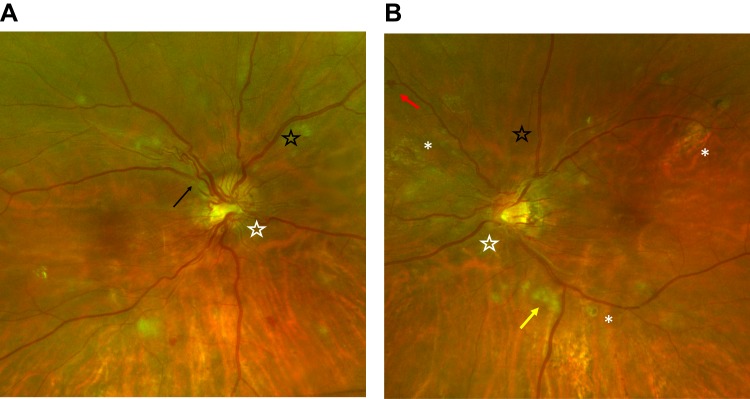
There are many imaging modalities that are helpful for the diagnosis and management of hypertensive chorioretinopathy. Retinal imaging, in particular wide-field color fundus photography, is useful to document the findings seen on funduscopic exam including copper wiring, AV nicking, cotton-wool spots, intraretinal hemorrhages, Elschnig spots, and optic disc swelling (Figure 2). Fundus photography allows for these findings to be followed over time to assess for improvement in end-organ damage from systemic hypertension. New technologies are being developed for analysis of retinal microvasculature in fundus photographs, which may soon allow for automated screening for conditions such as diabetic retinopathy, hypertensive retinopathy, and macular degeneration.7 Poplin et al used deep-learning models on retinal imaging to predict cardiovascular risk factors, such as age, gender, smoking status, systolic blood pressure, and major adverse cardiac events.8
Figure 2.
Optos wide-field color fundus photographs of the right eye (A) and left eye (B) of a patient with hypertensive chorioretinopathy. The photographs demonstrate copper wiring (black star), AV nicking (black arrow), intraretinal hemorrhages (red arrow), cotton wool spots (yellow arrow), Elschnig spots (white asterisk), and optic disc swelling (white star).
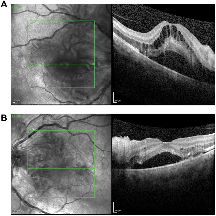
Spectral domain optical coherence tomography (SD-OCT) may demonstrate thinning of the inner retina and focal attenuation of the ellipsoid zone in areas of the Elschnig spots. Findings such as pigmentary epithelial detachments or serous retinal detachments may be visualized on SD-OCT, and their evolution or resolution may be monitored (Figure 3). Swept source optical coherence tomography (SS-OCT) allows deeper penetration into the tissues; one study reported an increase in choroidal thickness in the acute phase of hypertensive choroidopathy.9 SS-OCT angiography may reveal a moth-eaten appearance of the choriocapillaris, signifying areas of choroidal nonperfusion.10 Reperfusion of the choriocapillaris may be seen after antihypertensive treatment is initiated.11
Figure 3.
Optical coherence tomography of the macula of the right eye (A) and left eye (B) of a patient with hypertensive chorioretinopathy shows macular edema and serous retinal detachments. These serous retinal detachments improved after medical management of hypertension.
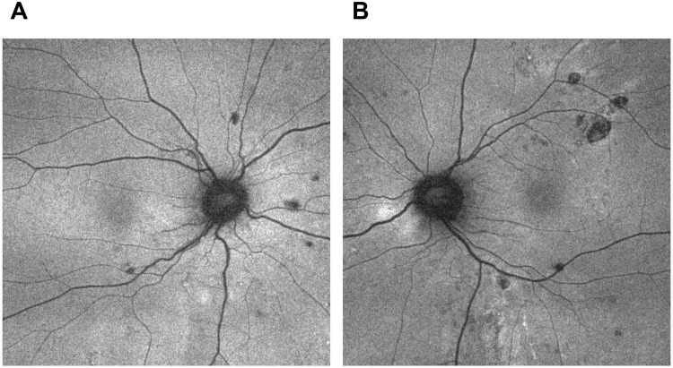
Fundus autofluorescence demonstrates hypoautofluorescence of Elschnig spots.12 There may also be hypoautofluorescence due to intraretinal hemorrhages and cotton wool spots (Figure 4).
Figure 4.
Fundus autofluorescence of the right eye (A) and left eye (B) of a patient with hypertensive chorioretinopathy. Magnification of the posterior pole reveals hypoautofluorescence of Elschnig spots, retinal hemorrhages and cotton wool spots.
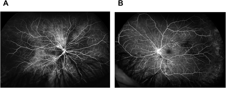
Fluorescein angiography is another helpful modality for investigating hypertensive chorioretinopathy. There is patchy and delayed choroidal filling, and severely delayed retinal arterial filling with areas of retinal capillary nonperfusion. Elschnig spots appear as areas of early hyperfluorescence with late subretinal leakage.12,13 There may also be optic disc leakage and blockage from cotton wool spots and intraretinal hemorrhages (Figure 5). Indocyanine green (ICG) angiography demonstrates hypocyanescence of ischemic areas of the choroid.10
Figure 5.
Fluorescein angiography of the right eye (A) and left eye (B) of a patient with hypertensive chorioretinopathy shows patchy and delayed choroidal filling and areas of retinal capillary nonperfusion. There is also blockage from intraretinal hemorrhages and cotton wool spots, as well as optic disc leakage.
Differential diagnoses for hypertensive chorioretinopathy include diabetic retinopathy, radiation retinopathy, anemia and other blood dyscrasias, ocular ischemic syndrome, and retinal vein occlusion. Clinical history and the presence of elevated blood pressure are valuable for distinguishing hypertensive chorioretinopathy from the aforementioned conditions.
Significance of Hypertensive Chorioretinopathy
Hypertensive retinopathy and choroidopathy are markers of hypertensive changes elsewhere in the body. Signs of mild hypertensive retinopathy, including generalized and focal arteriolar narrowing, copper wiring, and AV nicking, have been associated with coronary artery disease,14 stroke,15 and renal dysfunction.16 The Ibaraki Prefectural Health Study found that mild hypertensive retinopathy was a risk factor for cardiovascular mortality, independent of cardiovascular risk factors.17 For patients with mild hypertensive retinopathy, multivariable hazard ratios for total cardiovascular mortality were 1.23–1.24 for men and 1.12–1.44 for women, while hazard ratios for total stroke mortality were 1.31–1.38 for men and 1.30–1.70 for women.
Signs of moderate hypertensive retinopathy, including retinal hemorrhage, cotton wool spots, hard exudates, and microaneurysms, are even more strongly associated with increased risk of death from cardiovascular causes. The Atherosclerosis Risk in Communities study showed that signs of moderate hypertensive retinopathy were associated with a two- to four-times higher risk of incident stroke, independent of other risk factors including long-term elevations in blood pressure, cigarette smoking, and elevated lipid levels.18 This study also showed that after controlling for pre-existing risk factors, moderate hypertensive retinopathy was associated with a two-fold higher risk of congestive heart failure.19 Habib et al found that a higher grade of hypertensive retinopathy was significantly associated with higher angiographic severity of coronary artery disease by syntax score.20
Hypertensive chorioretinopathy is seen more commonly in younger patients with acute elevations in blood pressure and is associated with a poor prognosis if left untreated. It has been reported in conditions such as malignant hypertension,13,20 pre-eclampsia,21 eclampsia,22 acute or chronic renal failure,23,24 renal artery stenosis,25 and adrenal carcinoma.26 These conditions are all medical emergencies that require immediate treatment.
Management
The treatment of hypertensive retinopathy and choroidopathy is focused on reducing systemic blood pressure and, if indicated, managing the underlying medical condition. The optometrist or ophthalmologist who discovers hypertensive chorioretinopathy in a patient with undiagnosed hypertension is in a position to reduce the patient’s morbidity and mortality. There are no official recommendations for routine screening for hypertensive retinopathy in asymptomatic patients who carry a diagnosis of systemic hypertension. However, if a patient without a diagnosis of hypertension presents with signs of mild hypertensive retinopathy, we recommend referral to a general practitioner within one week. For moderate hypertensive retinopathy, the patient should be evaluated by a general practitioner within one or two days. Patients who present with severe hypertensive retinopathy or hypertensive choroidopathy should have their blood pressure measured immediately and should be referred to the nearest emergency room for urgent blood pressure management. There are no official recommendations for screening women with pregnancy-induced hypertension; however, we recommend that pregnant women who present with hypertensive retinopathy should be referred to their obstetrician for evaluation of pre-eclampsia.
While optometrists and ophthalmologists generally do not manage hypertension, close partnership with general practitioners is needed to ensure that patients receive appropriate treatment. These patients should be followed frequently to assess for resolution of changes due to hypertensive chorioretinopathy.
Conclusion
Eye care professionals should be familiar with the signs and symptoms of hypertensive retinopathy and chorioretinopathy as they have both short- and long-term correlations to the patient’s overall health and mortality. Care should be coordinated with the patient’s primary care physician or, in cases of acutely elevated blood pressure and ocular signs, with an emergency department.
Acknowledgement
A portion of this work was supported by an unrestricted department grant from Research to Prevent Blindness.
Disclosure
The authors report no conflicts of interest in this work.
References
- 1.The World Health Organization. A global brief on hypertension: silent killer, global public health crisis. Available from: http://apps.who.int/iris/bitstream/10665/79059/1/WHO_DCO_WHD_2013.2_eng.pdf. Accessed August 2019.
- 2.Wong TY, Mitchell P. The eye in hypertension. Lancet. 2007;369:425–435. doi: 10.1016/S0140-6736(07)60198-6 [DOI] [PubMed] [Google Scholar]
- 3.Keith NM, Wagener HP, Barker NW. Some different types of essential hypertension: their course and prognosis. Am J Med Sci. 1939;197:332–343. doi: 10.1097/00000441-193903000-00006 [DOI] [PubMed] [Google Scholar]
- 4.Wong TY, Mitchell P. Hypertensive retinopathy. N Engl J Med. 2004;351:2310–2317. doi: 10.1056/NEJMra032865 [DOI] [PubMed] [Google Scholar]
- 5.Fraser-Bell S, Symes R, Vaze A. Hypertensive eye disease: a review. Clin Exp Ophthalmol. 2017;45(1):45–53. doi: 10.1111/ceo.12905 [DOI] [PubMed] [Google Scholar]
- 6.Tso MO, Jampol LM. Pathophysiology of hypertensive retinopathy. Ophthalmol. 1982;89:1132–1145. doi: 10.1016/S0161-6420(82)34663-1 [DOI] [PubMed] [Google Scholar]
- 7.Kipli K, Hoque ME, Lim LT, et al. A review on the extraction of quantitative retinal microvascular image feature. Comput Math Methods Med. 2018;2018:4019538. doi: 10.1155/2018/4019538 [DOI] [PMC free article] [PubMed] [Google Scholar]
- 8.Poplin R, Varadarajan AV, Blumer K, et al. Prediction of cardiovascular risk factors from retinal fundus photographs via deep learning. Nat Biomed Eng. 2018;2(3):158–164. doi: 10.1038/s41551-018-0195-0 [DOI] [PubMed] [Google Scholar]
- 9.Velazquez-Villoria D, Marti Rodrigo P, DeNicola ML, Zapata Vitori MA, Segura García A, García-Arumí J. Swept source optical coherence tomography evaluation of chorioretinal changes in hypertensive choroidopathy related to HELLP syndrome. Retin Cases Brief Rep. 2019;13(1):30–33. doi: 10.1097/ICB.0000000000000524 [DOI] [PubMed] [Google Scholar]
- 10.Rezkallah A, Kodjikian L, Abukhashabah A, Denis P, Mathis T. Hypertensive choroidopathy: multimodal imaging and the contribution of wide-field swept-source oct-angiography. Am J Ophthalmol Case Rep. 2019;13:131–135. doi: 10.1016/j.ajoc.2019.01.001 [DOI] [PMC free article] [PubMed] [Google Scholar]
- 11.Saito M, Ishibazawa A, Kinouchi R, Yoshida A. Reperfusion of the choriocapillaris observed using optical coherence tomography angiography in hypertensive choroidopathy. Int Ophthalmol. 2018;38(5):2205–2210. doi: 10.1007/s10792-017-0705-1 [DOI] [PubMed] [Google Scholar]
- 12.Stryjewski TP, Papakostas TD, Eliott D. Multimodal imaging of elschnig spots: a case of simultaneous hypertensive retinopathy, choroidopathy, and neuropathy. Semin Ophthalmol. 2017;32(4):397–399. doi: 10.3109/08820538.2015.1115091 [DOI] [PubMed] [Google Scholar]
- 13.Verstappen M, Draganova D, Judice L, et al. Hypertensive choroidopathy revealing malignant hypertension in a young patient. Retina. 2019;39(5):e12–e13. doi: 10.1097/IAE.0000000000002509 [DOI] [PubMed] [Google Scholar]
- 14.Duncan BB, Wong TY, Tyroler HA, Davis CE, Fuchs FD. Hypertensive retinopathy and incident coronary heart disease in high risk men. Br J Ophthalmol. 2002;86(9):1002–1006. doi: 10.1136/bjo.86.9.1002 [DOI] [PMC free article] [PubMed] [Google Scholar]
- 15.Ong YT, Wong TY, Klein R, et al. Hypertensive retinopathy and risk of stroke. Hypertension. 2013;62:706–711. doi: 10.1161/HYPERTENSIONAHA.113.01414 [DOI] [PMC free article] [PubMed] [Google Scholar]
- 16.Wong TY, Coresh J, Klein R, et al. Retinal microvascular abnormalities and renal dysfunction: the atherosclerosis risk in communities study. J Am Soc Nephrol. 2004;15:2467–2473. doi: 10.1097/01.ASN.0000136133.28194.E4 [DOI] [PubMed] [Google Scholar]
- 17.Sairenchi T, Iso H, Yamagishi K, et al. Mild retinopathy is a risk factor for cardiovascular mortality in Japanese with and without hypertension: the Ibaraki Prefectural Health Study. Circulation. 2011;124(23):2502–2511. doi: 10.1161/CIRCULATIONAHA.111.049965 [DOI] [PubMed] [Google Scholar]
- 18.Wong TY, Klein R, Couper DJ, et al. Retinal microvascular abnormalities and incident stroke: the atherosclerosis risk in communities study. Lancet. 2001;358(9288):1134–1140. doi: 10.1016/S0140-6736(01)06253-5 [DOI] [PubMed] [Google Scholar]
- 19.Wong TY, Rosamond W, Chang PP, et al. Retinopathy and risk of congestive heart failure. JAMA. 2005;293:63–69. doi: 10.1001/jama.293.1.63 [DOI] [PubMed] [Google Scholar]
- 20.Habib SA, Jibran MS, Khan SB, Gul AM. Association of hypertensive retinopathy with angiographic severity of coronary artery disease determined by syntax score. J Ayub Med Coll Abbottabad. 2019;31(2):189–191. [PubMed] [Google Scholar]
- 21.Lee CS, Choi EY, Lee M, Kim H, Chung H. Serous retinal detachment in preeclampsia and malignant hypertension. Eye (Lond). 2019;33(11):1707–1714. doi: 10.1038/s41433-019-0461-8 [DOI] [PMC free article] [PubMed] [Google Scholar]
- 22.Song YS, Kinouchi R, Ishiko S, Fukui K, Yoshida A. Hypertensive choroidopathy with eclampsia viewed on spectral-domain optical coherence tomography. Graefes Arch Clin Exp Ophthalmol. 2013;251:2647–2650. doi: 10.1007/s00417-013-2462-9 [DOI] [PubMed] [Google Scholar]
- 23.Pociej-Marciak W, Karska-Basta I, Kuźniewski M, Kubicka-Trząska A, Romanowska-Dixon B. Sudden visual deterioration as the first symptom of chronic kidney failure. Case Rep Ophthalmol. 2015;6(3):394–400. doi: 10.1159/000442182 [DOI] [PMC free article] [PubMed] [Google Scholar]
- 24.Kameda Y, Hirose A, Iida T, Uchigata Y, Kitano S. Giant retinal pigment epithelial tear associated with fluid overload due to end-stage diabetic kidney disease. Am J Ophthalmol Case Rep. 2017;5:44–47. doi: 10.1016/j.ajoc.2016.11.004 [DOI] [PMC free article] [PubMed] [Google Scholar]
- 25.Malhotra SK, Gupta R, Sood S, Kaur L, Kochhar S. Bilateral renal artery stenosis presenting as hypertensive retinopathy and choroidopathy. Indian J Ophthalmol. 2002;50(3):221–223. [PubMed] [Google Scholar]
- 26.Rahhal-Ortuño M, Fernández-Santodomingo AS, Barreiro-González A, Díaz-Llopis M. Hypertensive choroidopathy and adrenal carcinoma: a new association. Arch Soc Esp Oftalmol. 2019;94(1):37–40. doi: 10.1016/j.oftal.2018.09.003 [DOI] [PubMed] [Google Scholar]
Associated Data
This section collects any data citations, data availability statements, or supplementary materials included in this article.
Data Citations
- The World Health Organization. A global brief on hypertension: silent killer, global public health crisis. Available from: http://apps.who.int/iris/bitstream/10665/79059/1/WHO_DCO_WHD_2013.2_eng.pdf. Accessed August 2019.


