

Click Video under article title in Contents at ajog.org
The current coronavirus disease 2019 (COVID-19) pandemic is a challenge to every health system worldwide. Unfortunately, it is likely that this emergency will not disappear soon. No health system, with its present resources and work flow, is ready to deal with a full-blown wave of this pandemic. Rapid acquisition of specific new skills may be fundamental in delivering appropriate healthcare for our patients. COVID-19 is classically diagnosed by real-time reverse transcription polymerase chain reaction and radiological investigations (X-ray or high-resolution computerized tomography). These techniques are not without limitations. Ultrasonography has been suggested as a reliable and accurate tool for assessing the lungs in patients with suspected pneumonia. Obstetricians and gynecologists are usually familiar with the use of ultrasound. Lung ultrasound (LUS) findings can show specific signs of interstitial pneumonia, which is a characteristic of COVID-19. We believe that extensive and rapid training of healthcare providers on the application of ultrasound in the detection of characteristic pulmonary signs of COVID-19, in addition to proper care and handling of their ultrasound machines, is feasible and may be critical to provide appropriate management especially to the obstetrical patients in the coming period. We present a systematic approach to lung examination, simplified to encourage its adoption by obstetricians and gynecologists, in addition to an example of a recent pregnant woman with COVID-19, in which LUS was useful in its management.
Letter to the Editors
The current COVID-19 pandemic is a challenge to every healthcare system. Pregnant women and fetuses represent a high-risk population during pandemics.1 LUS is a reliable tool in the assessment of patients with suspected pneumonia.2 , 3 We provide a guide to perform LUS and an example on its implementation in pregnancy.
Transducer
Linear, phased array, or convex probes can all be used for LUS.
Position
LUS is usually performed in the sitting, lateral, and supine positions.
Examined areas ( Figure 1 )
Figure 1.

An illustration showing the 6 areas to be examined in each hemithorax
The anterior and posterior axillary lines are the hallmarks used to determine the 6 regions. Each region should be examined in the axial and sagittal views.
Youssef. Lung ultrasound in the coronavirus disease 2019 pandemic. Am J Obstet Gynecol 2020.
Each hemithorax is divided into 6 regions by the aid of anterior and posterior axillary lines as follows: 2 anterior, 2 lateral, and 2 posterior regions. Each region is examined in sagittal and axial views.
Normal findings ( Table , Video )
-
•
Pleural line: Normally, only the pleural line is visualized, as hyperechoic sliding line, moving forward and backward with ventilation.
-
•
A-lines: Regularly spaced horizontal lines under the pleural line and are normal reverberation artifacts of the pleural line.
Table.
Normal and abnormal findings on lung ultrasound examination
| Description | Ultrasound image |
|---|---|
Normal
|
Figures 3 and 4 |
| Abnormal | |
| Irregular pleural line | Figure 5 |
| Well-separated B-lines | Figure 6 |
| Coalescent B-lines (White lung) | Figure 7 |
| Consolidations (hypoechoic tissue-like lung) with bronchogram (hyperechoic punctiform images) | Figure 8 |
Youssef. Lung ultrasound in the coronavirus disease 2019 pandemic. Am J Obstet Gynecol 2020.
Abnormal findings
-
•
Pleural line: In inflammatory lung diseases, the pleural line is usually irregular and blurred.3
-
•
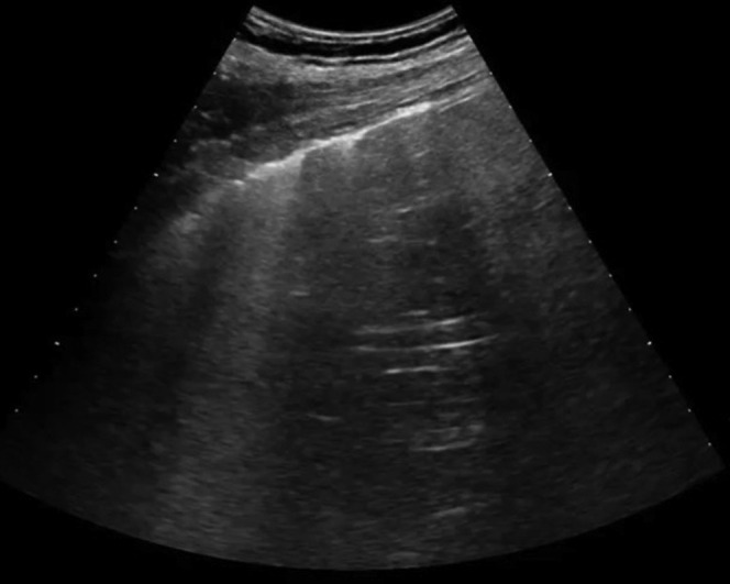
B-lines: These are vertical ultrasound artifacts resulting from abnormal gas-tissue interface. B-lines start from the pleural line and “shine” vertically to erase A-lines.
-
•
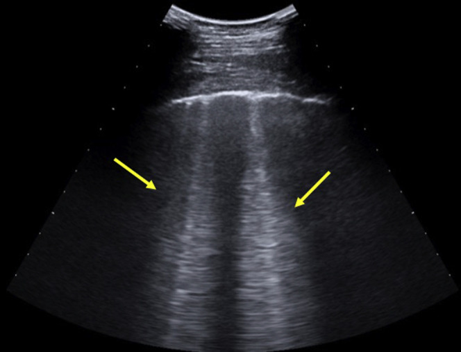
Coalescent B-lines (white lung): B-lines may amalgamate.
-
•
Lung consolidation: This is due to massive aeration loss. Lungs acquire a tissue-like echotexture. Hyperechoic punctiform images may be seen corresponding to the air-filled bronchi.
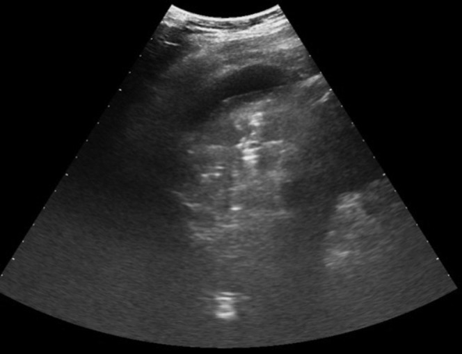
Case Discussion (Figure 2)
Figure 2.

Lung ultrasound image of the pregnant woman at presentation (A) and after 4 days (B)
Youssef. Lung ultrasound in the coronavirus disease 2019 pandemic. Am J Obstet Gynecol 2020.
A 33-year-old primigravida presented to the emergency room of our university hospital at 26 weeks’ gestation with fever and mild chest pain and dyspnea for 3 days. At the emergency room assessment, the lady had no fever with normal oxygen saturation. Obstetrical ultrasound findings were normal. We performed LUS and the results showed pleural thickening and diffuse coalescent B-lines involving both lungs. The patient underwent a nasopharyngeal swab for COVID-19 and was admitted to our hospital. The results from the swab were positive for COVID-19. During the following 4 days of admission, the woman exhibited no symptoms and had normal oxygen saturation. A repeat LUS was performed, which showed improvement in the ultrasonographic aspect with pleural thinning and only some thin B-lines. The woman was discharged. She is now at 29 weeks’ gestation, asymptomatic, with normally progressing pregnancy.
We believe that extensive training of physicians may be considerably helpful in case of an unfortunate but likely continuing increase in the number of COVID-19 cases.
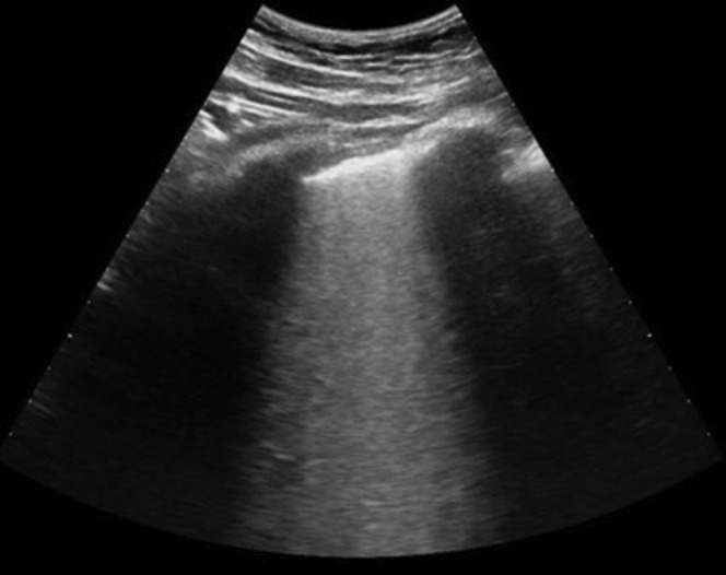
Figure 3.
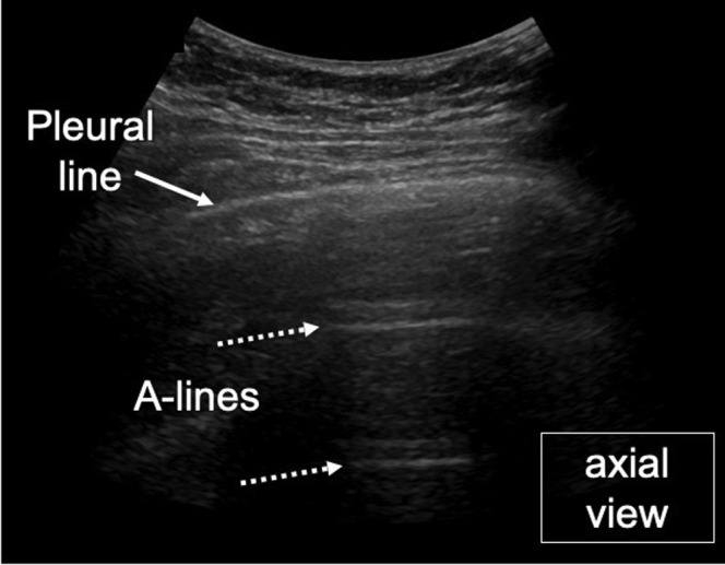
Normal lung, axial view
Youssef. Lung ultrasound in the coronavirus disease 2019 pandemic. Am J Obstet Gynecol 2020.
Figure 4.
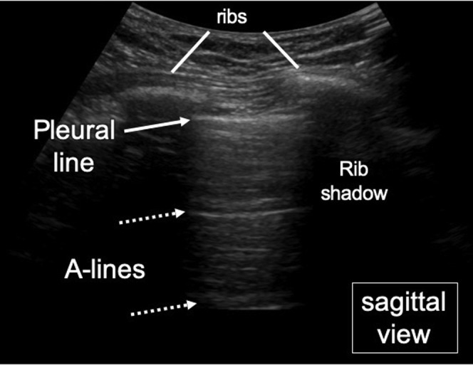
Normal lung sagittal view
Youssef. Lung ultrasound in the coronavirus disease 2019 pandemic. Am J Obstet Gynecol 2020.
Figure 5.
Irregular pleural line
Youssef. Lung ultrasound in the coronavirus disease 2019 pandemic. Am J Obstet Gynecol 2020.
Figure 6.
Well-separated B-lines
Youssef. Lung ultrasound in the coronavirus disease 2019 pandemic. Am J Obstet Gynecol 2020.
Figure 7.
Coalescent B-lines (white lung)
Youssef. Lung ultrasound in the coronavirus disease 2019 pandemic. Am J Obstet Gynecol 2020.
Figure 8.
Lung consolidation with air bronchogram
Youssef. Lung ultrasound in the coronavirus disease 2019 pandemic. Am J Obstet Gynecol 2020.
Footnotes
The authors report no conflict of interest.
Supplementary Data
Normal pleural line with pleural sliding.
A video abstract summarizing the content of the letter.
Youssef. Lung ultrasound in the coronavirus disease 2019 pandemic. Am J Obstet Gynecol 2020.
References
- 1.Dashraath P., Wong J.L.J., Lim M.X.K., et al. Coronavirus disease 2019 (COVID-19) pandemic and pregnancy. Am J Obstet Gynecol. 2020 doi: 10.1016/j.ajog.2020.03.021. [Epub ahead of print] [DOI] [PMC free article] [PubMed] [Google Scholar]
- 2.Ye X., Xiao H., Chen B., Zhang S. Accuracy of lung ultrasonography versus chest radiography for the diagnosis of adult community-acquired pneumonia: review of the literature and meta-analysis. PLoS One. 2015;10 doi: 10.1371/journal.pone.0130066. [DOI] [PMC free article] [PubMed] [Google Scholar]
- 3.Moro F., Buonsenso D., Moruzzi M.C., et al. How to perform lung ultrasound in pregnant women with suspected COVID-19. Ultrasound Obstet Gynecol. 2020;55:593–598. doi: 10.1002/uog.22028. [DOI] [PubMed] [Google Scholar]
Associated Data
This section collects any data citations, data availability statements, or supplementary materials included in this article.
Supplementary Materials
Normal pleural line with pleural sliding.
A video abstract summarizing the content of the letter.
Youssef. Lung ultrasound in the coronavirus disease 2019 pandemic. Am J Obstet Gynecol 2020.