

Abstract
Objective
The purpose of this in vivo study was to confirm the detection of proximal caries using near-infrared light transillumination (NILTI) (DIAGNOcam) device, and to compare the diagnostic performance of the device with other caries detection methods, including visual examination using the International Caries Detection and Assessment System (ICDAS), bitewing radiography (BW), an LED-based device (Midwest Caries I.D.), and a laser fluorescence device (LFpen).
Methods
A total of 974 proximal surface of permanent posterior teeth from 34 patients (19 females and 15 males between the ages of 22–55) were evaluated in the present study. After clinical examination of each proximal surface by an experienced examiner, they were coded according to the ICDAS criteria and subsequently with BW, the NILTI, LED, and LFpen. The proximal dentin caries of 106 were opened based on the combination of visual, NILTI and radiographic assessment and validated; and were treated with restorative materials. The data were analyzed with descriptive statistics and calculation of the sensitivity, specificity, and area under curve (AUC).
Results
The highest sensitivity values were recorded from NILTI readings (99.1%), followed by BW scores (86.8%). The highest specificity values were recorded from ICDAS (100%). The best AUC values were found from the NILTI readings (0.97), followed by BW (0.93) and ICDAS (0.87).
Conclusion
The NILTI device exhibited the best performance in terms of detecting proximal dentin caries.
Keywords: Proximal caries, Near-infrared light transillumination, Laser fluorescence, Caries diagnostics
1. Introduction
It is difficult the detection of caries lesions on the proximal surfaces of teeth with visual examination (VE), because of the interproximal contact. The International Caries Detection and Assessment System (ICDAS; www.icdas.org) is one of the most widely used methods for VE, which was introduced based on consensus and thus represents the most broadly based visual caries detection system (in terms of comparability). ICDAS was also reported to be easily applied by dental students (Diniz et al., 2009, Luz et al., 2015), and to reliably assess the early phase of decalcification (Ismail et al., 2007). The ICDAS definition of dental caries is applicable to the proximal smooth surface (Ekstrand et al., 2011). According to the ICDAS criteria, detection of smooth surface caries requires visual inspection from the occlusal, buccal, and lingual aspects (Shivakumar et al., 2009). The ICDAS criteria for smooth surfaces are classified as follows: Code 0, there is no evidence of caries; 1, when viewed there is no color changes, but after prolonged air drying, a carious opacity (white or brown lesion) are seen from the buccal or lingual surface; 2, carious opacity or discoloration may be seen directly from the buccal or lingual aspects and as a shadow bounded by enamel, seen through the marginal ridge; 3, when dried for five seconds, visible loss of enamel integrity is viewed from the buccal or lingual aspect; 4, in wet condition, the lesion seems as an intrinsic shadow of discolored dentin which appears as gray, blue and brown; 5, there is cavitation in opaque or discolored enamel with visible dentin; 6, there are distinct loss of tooth structure and deep cavity with visible dentin.
Radiography has simplified dental practice and is the most commonly used lesion detection method (Sochtig et al., 2014). The superiority of radiographic techniques over clinical examination for the detection of interproximal carious lesions has been verified (Feldens et al., 2003, Machiulskiene et al., 1999, Stecksen-Blicks and Wahlin, 1983). However, bitewing radiography (BW), which is the most common method used for diagnosis of proximal caries, has some limitations (Sochtig et al., 2014): Achieving acceptable BW in children and teenagers is frequently difficult and occasionally impossible. The detection of dental caries is made more difficult because of overlapping images on radiographs and fixed orthodontic appliances. In addition, dental X-ray usage is greatly limited because of the dangers associated with exposure to ionizing radiation (Bajaj and Offiah, 2015). The BW radiography has some limitations in detecting proximal enamel lesions (Tonkaboni et al., 2019). The other restrictions of x-ray exposure may comprise pregnancy, frequent follow-ups in epidemiological studies or patient's antipathy (Wenzel, 2004).
A caries detection device (DIAGNOcam; KaVo, Biberach an der Riss, Germany) utilizes near-infrared light transillumination (NILTI) of teeth. NILTI, defined first in 1995, uses a photo-optical process for detection of posterior tooth decay (Fried et al., 1995). This method was developed following advances in the digital imaging fiber optic transillumination method (DIFOTI). Visible light was used in the DIFOTI method however, NILTI method uses invisible long-wavelength light. Decreased scattering allows deeper near-infrared light penetration of objects, which is a major benefit of using longer wavelengths (Hall and Girkin, 2004). The camera of DIAGNOcam based on photo-optical principles uses an illuminating wavelength of 780 nm (Kuhnisch et al., 2015). This device consisting of a USB connection to a computer and specific software is used mostly in the detection of proximal caries, and less for occlusal caries and secondary caries related to restorations (Abdelaziz and Krejci, 2015). The light is not transmitted into the proximal space but directly through the alveolar process. The quality of diagnostic imaging seems to be improved by these modifications (Sochtig et al., 2014). The use of this device allows to capture different stages of interproximal enamel and dentine caries lesions (Kuhnisch et al., 2015). Nevertheless, the diagnostic accuracy of DIAGNOcam exhibited the similarity with bitewing regarding the detection of proximal dentin caries (Abdelaziz et al., 2018, Abogazalah et al., 2017, Kuhnisch et al., 2015).
There are many studies that investigate the detection of the occlusal and smooth surface caries with DIAGNOdent device which based on the detecting the emitted fluorescence after application of pulsed red light [laser fluorescence] (Attrill and Ashley, 2001, Diniz et al., 2019, Lussi et al., 2004, Shi et al., 2001). The direct detection of proximal area is not possible, if a device has not a tip that can penetrate the proximal area (Lussi et al., 2006). The tips of DIAGNOdent pen (LFpen; KaVo) device have a smaller diameter than DIAGNOdent which makes them easier to handle (Lussi and Hellwig, 2006). DIAGNOdent pen device is based on the difference fluorescence emission of carious tissue compared to sound tissue. The fluorescence in carious tissue is caused by the porphyrins produced by some bacterial species after excitation by red light (Lussi et al., 2004). The caries-detection ability of this device is based on the greater fluorescence emission of decalcified regions compared to sound dentin when irradiated with a laser with a wavelength of 655 nm (Sundstrom et al., 1985).
LED-based devices have also been developed for detection of decalcified occlusal and proximal surfaces (Aktan et al., 2012, Huth et al., 2010, Lussi and Hellwig, 2006, Sochtig et al., 2014). The hand piece of LED-based device uses light transmitted by optical fibers to detect dental caries. The reflectance and refraction of LED that captured by fiber optics are turned into electrical signal (Lussi and Hellwig, 2006). According to the manufacturer, when the detection is positive, the light at the tip changes to red. The speed of the audible signal is proportional to the amount of decay detected. For occlusal lesions, this device showed promising sensitivity of 0.7 and specificity of 0.9 on the D3 level (Rodrigues et al., 2011).
The present in vivo study was performed to investigate the detection ability of NILTI device for proximal dentine caries in permanent teeth and compare it to other detection methods, such as visual examination (VE) with ICDAS, digital BW, the LED-based device, and LFpen. The working hypothesis was that NILTI device would show better performance in validation of lesion depth, compared to alternative methods
2. Materials and methods
This clinical research was approved by the Ethics Committee of Gaziantep University, Medical Faculty (Decision no: 74/15). A total of 974 proximal surfaces (sound and carious) of permanent molars and premolars, of 34 patients (15 females and 19 males) ranging in age from 22 to 55 years (mean age, 31.81 ± 9.68 years) were investigated. Both sound and carious teeth with proximal contact were included in the study for the statistical comparison. The presence or absence of dentin caries (intact or enamel caries) formed the basis of the comparison. Teeth with restorations, occlusal caries, residual caries, secondary caries, hypoplasia, or orthodontic bands were excluded. After a careful screening for dental caries in all patients, only 174 surface with dentine caries lesions were included in the present investigation. The participants were informed about the aims, procedures, and benefits of this clinical study, and informed consent was obtained prior to their participation. BW was used for the interproximal caries detection and was performed in cases showing clinical symptoms, such as the presence of cavitated or active initial lesions and detectable demineralization between teeth (Sochtig et al., 2014). All examination methods were performed by same examiner (PhD, specialist dentist in Oral and Maxillofacial Radiology).
A dental intraoral X-ray machine (Planmeca Oy, Helsinki, Finland) with Digora Storage Phosphor Plate (SPS) sensors (Soredex; Orion Corp., Helsinki, Finland) was used. All radiographs were obtained using a film plate holder. The exposure time was 0.08 s at 63 kVp and 7 mA. All images were primarily analyzed by same examiner (PhD, specialist dentist). In cases in which a proximal dentin caries lesion (grade 4–5) (4: radiolucency in the external half of the dentin; and 5: radiolucency in the internal half of the dentin) was diagnosed, all associated treatment procedures were discussed with the patient. If an operative intervention was needed, they were asked to patient in the study. Informed consent was obtained before making another appointment for validation or restoration. The indication of dentin caries was validated only when scores of 4 or 5 on BW and/or NILTI (score 4: dentin caries penetrating the dentino-enamel junction linearly; score 5: deep dentin caries) were present. Patients with intact decalcified surfaces, ambiguous radiolucency near the enamel-dentin junction (EDJ) on BW images, or enamel decalcification with an isolated spot reaching the EDJ on NILTI images, were not approved for operative treatment and were advised to receive preventive care (such as resin infiltrant, fluoride or CPP-ACP treatment). The decision for operative treatment was primarily based on combined clinical (visual and NILTI) and radiographic assessment. It was decided to make restoration of 106 of 174 proximal dentin caries. Determination of caries level was used as the reference standard in this study after all caries were removed with a carbide bur. Lesion depths (LDs) were determined as follows: (0) sound; (1) enamel caries; (2) superficial dentin caries; and (3) deep dentin caries. Sound teeth and lesion depth of enamel caries were determined with visual examination (ICDAS) and BW. Lesion depths of superficial and deep dentin caries were determined with combination of BW images and clinical appearance after caries removal.
2.1. Visual examination (VE)
Before examination, all approximal areas were thoroughly cleaned using a rubber cup, prophylactic paste, and dental floss. The patients were examined by an experienced examiner using operating light illumination, a dental mirror, and an exploratory probe. The proximal lesions were classified using the ICDAS as follows: (0) sound surface; (1) first initial change in enamel; (2) distinct visual change in wet enamel surface; (3) initial breakdown in enamel because of decay with no visible dentin; (4) underlying dark shadow from dentin with or without localized enamel breakdown; (5) distinct cavity with visible dentin; and (6) extensive distinct cavity with visible dentin.
2.2. Examination with near-infrared light for transillumination (NILTI)
All of the 974 permanent posterior tooth surfaces were examined using near-infrared light at a wavelength of 780 nm. The DIAGNOcam camera was centered perpendicularly over the region of interest and the images were captured and stored with KiD software (KaVo Integrated Desktop/Ref: 1.004.7700, KaVo, Biberach, Germany). To prevent any light interference, the light source on the dental box was switched off before NILTI imaging.
Following the clinical procedure, NILTI images were first analyzed by experienced (PhD, specialist) dentist independent from the results of the other diagnostic methods. Enamel and dentin caries lesions were categorized according to the degree of demineralization (Sochtig et al., 2014). Score 0: sound surface (Fig. 1a); score 1: first visible signs of enamel caries (Fig. 1b); score 2: established enamel caries (Fig. 1c); score 3: established enamel caries with a spot reaching the dentino-enamel junction (Fig. 1d); score 4: dentin caries penetrating the dentino-enamel junction linearly (Fig. 1e); score 5: deep dentin caries (Fig. 1f)
Fig. 1.

Classification of enamel and dentin caries lesions (Arrows indicate areas the caries.)
2.3. Examination with laser-induced fluorescence device (LFpen)
LFpen measurements of the proximal surfaces were performed by an examiner. Teeth were examined using the type 1 tip of DIAGNOdent pen 2190 (KaVo) for proximal surfaces in accordance with the manufacturer’s recommendations. Calibration of the LFpen was performed with a ceramic standard before all examinations. The calibration was performed on a sound area. After the tooth was dried, the tip was placed under the contact area and moved until the peak score was reached on both buccal and palatal/lingual surfaces (Braga et al., 2009). Whether there was decalcification of the surface was decided using the peak reading (Lussi et al., 2006), and scored as follows: 0 – 9, healthy tooth surface; 9.1 – 22, lesion in the enamel; >22 lesion in the dentin.
2.4. Examination with LED-based device
Before measurement, the LED-based device (Midwest Caries I.D.; Dentsply Professional, York, PA) was calibrated using the ceramic standard. For detection of proximal caries, red LED radiation was transported to the proximal area using the tip of the probe in direct contact with the marginal surfaces and parallel to the long axis of the tooth. The examined teeth must be moist to avoid erroneous readings. The presence of decayed or demineralized structures was determined by red light emission and an audible tone. The green light emission with an inaudible tone indicated that the tested site was intact. Three different tones associated with red light emission indicated the extent of the caries. The cutoff limits provided by the manufacturer were used to assay the performance of the LED-based device: 0 (green light, no beeping noise: sound); 1 (red light, slow beeping noise: enamel caries); 2 (red light, midlevel beeping noise: superficial dentin caries); and 3 (red light, continuous beeping noise: deep dentin caries).
2.5. Radiographic examination (BW)
Posterior radiographs were classified as follows (Huth et al., 2010): (0) no radiolucency; (1) radiolucency in the external half of the enamel; (2) radiolucency in the internal half of the enamel; (3) radiolucency in the external half of the dentin; and (4) radiolucency in the internal half of the dentin.
2.6. Statistical analysis
To compare the results statistically, the scores were classified as sound, enamel caries, or dentin caries. Cohen’s kappa test was performed for determination of interobserver agreement. McNemar test was were calculated in order to compare sensitivity, specificity, accuracy values among different detection methods. The area under curve (AUC) values were calculated for caries detection for each method assessed in the present study. Comparison of the AUC was used to determine the accuracy of the caries detection methods. Statistical analyses were performed using SPSS for Windows software (ver. 16.0; SPSS Inc., Chicago, IL, USA). In all analyses, P < 0.05 was taken to indicate statistical significance.
3. Results
Of the 974 proximal sites of permanent molar and premolar teeth, 106 proximal dentin caries were treated with restorative materials after examination with BW, VE, and NILTI. Substantial inter-observer agreement was achieved for NILTI, BW, LFpen, VE and LED-based caries detection readings in the present study (κ = 0.939, 0.0.801, 0.647, 0.583 and 0.345, respectively).
In the McNemar test, where the depth of the lesion was regarded as the gold standard, it was examined whether there was a significant difference between the other detection methods and lesion depth. According to this, NILTI and LFpen showed statistically similar results with lesion depth (p > 0.05). According to the Kappa test, the highest agreement value was found at NILTI with 0.939 and then at BW (Table 1).
Table 1.
Differences between the lesion depth and the caries detection methods.
| Mc Nemar test p | Measure of agreement | Kappa test p | |
|---|---|---|---|
| LD-NILTI | 0.375 | 0.939 | <0.001 |
| LD-LED | <0.001 | 0.345 | <0.001 |
| LD-VE | <0.001 | 0.583 | <0.001 |
| LD-LFpen | 0.099 | 0.647 | <0.001 |
| LD-BW | 0.013 | 0.801 | <0.001 |
The highest sensitivity value was determined in NILTI, the highest specificity value was found in ICDAS and the highest accuracy was determined in NILTI according to ROC analysis. (Table 2).
Table 2.
Sensitivity, specificity, Accuracy, Area Under Curve (AUC).
| NILTI | LED | VE | LFpen | BW | |
|---|---|---|---|---|---|
| Sensitivity | 99.1% | 56.6% | 64.2% | 81.1% | 86.8% |
| Specificity | 94.1% | 80.9% | 100% | 85.3% | 95.6% |
| Accuracy | 97.1% | 66.1% | 78.2% | 82.8% | 90.2% |
| AUC | 0.97a | 0.72b | 0.87c | 0.86c | 0.93d |
*The different letters on the line of AUC indicate statistical differences.
When the AUC values obtained from ROC analysis were compared, it was observed that NILTI generally had superior caries detection performance compared to other tests. However, it was observed that the best detection method after NILTI was BW. (Table 2, Table 3). The difference between NILTI and BW tests in terms of caries detection performance was nearly significant (p = 0.0489). Other important findings were that VE and LFpen showed similar performance and LED exhibited the lowest performance in detecting caries (Table 2). There were no statistically differences between the AUC values of the LFpen and VE on pairwise comparisons (Fig. 2 and Table 3).
Table 3.
Comparison of AUC’s.
| Difference between areas | Standard error a | 95% Confidence interval | Z statistic | P | |
|---|---|---|---|---|---|
| NILTI- BW | 0.0425 | 0.0216 | 0.000211 to 0.0847 | 1.970 | 0.0489 |
| BW-LED | 0.215 | 0.038 | 0.141 to 0.289 | 5.688 | 0.0001 |
| BW-LFpen | 0.0666 | 0.0344 | −0.000840 to 0.134 | 1.936 | 0.0529 |
| BW-VE | 0.0606 | 0.0278 | 0.00608 to 0.115 | 2.179 | 0.0294 |
| LFpen-NILTI | 0.109 | 0.0296 | 0.0510 to 0.167 | 3.680 | 0.0002 |
| NILTI-VE | 0.103 | 0.0274 | 0.0494 to 0.157 | 3.767 | 0.0002 |
| LED-LFpen | 0.148 | 0.0437 | 0.0624 to 0.234 | 3.387 | 0.0007 |
| LED-VE | 0.154 | 0.0395 | 0.0766 to 0.231 | 3.897 | 0.0001 |
| LFpen-VE | 0.00597 | 0.0378 | −0.0682 to 0.0801 | 0.158 | 0.8747 |
| NILTI-LED | 0.257 | 0.0352 | 0.188 to 0.326 | 7.301 | 0.0001 |
Fig. 2.
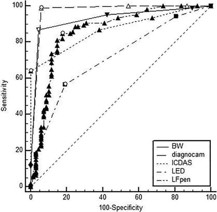
Comparison of ROC curves.
4. Discussion
This study focused on validating proximal caries detection by the near-infrared light for transillumination (NILTI) method, and compared its effectiveness to that of other diagnostic methods, such as VE with ICDAS, BW, an LED-based device, and the LFpen. There are studies that test the performance of NILTI device in the diagnosis of proximal, occlusal and secondary caries (Elhennawy et al., 2018, Schaefer et al., 2018, Sochtig et al., 2014). There have been no studies to date using the LED- and laser fluorescence-based devices together with NILTI to determine the best device for detecting proximal dentin caries. Enamel caries could not be verified because ethical regulations prohibited in vivo validation. Under in vitro conditions, transillumination of the teeth is insufficient because the different photo-optical properties of the embedding materials are not comparable with the periodontal anatomy (Kuhnisch et al., 2015). Reassessing the sound surfaces or enamel lesions after a long period would be an alternative procedure to exclude false-negative results (Huth et al., 2010).
Regardless of the limitations, considerable clinical information was provided in the present study. Our working hypothesis that this near-infrared (NIR) light for transillumination device would have better performance for detection of proximal dentin decalcification compared to other techniques, was supported. Even if the diagnostic performance of VE, laser fluorescence, LED, and BW corresponded to earlier reports (Bader and Shugars, 2004, Huth et al., 2010, Lussi and Hellwig, 2006, Lussi et al., 2004, Rutjes et al., 2007, Sochtig et al., 2014), the observation that NILTI performed nearly better than digital radiography is notable, and may result in a reduction in the usage of BW. The detection of hidden proximal and occlusal caries is difficult on VE. The results of the present study indicated that the best AUC values were obtained from NILTI followed by BW. Considering these results, this device can reduce X-ray exposure in such situations. Kunisch et al. and Sochtig et al. supported this suggestion in their study of NILTI. In a recent study, it was emphasized that proximal caries of the posterior teeth can cause to be overlooked with only visual examination, and near-infrared light transillumination may be an additional method for detection of caries (Kunisch et al., 2019). Furthermore, NILTI can be repeated as often as necessary, and occlusal and proximal surfaces can be evaluated simultaneously with this method.
In daily clinical practice, the use of NILTI cannot replace radiographic examination in all respects—it does not provide information about the pulp and periodontal tissues, and it is difficult to detect a carious lesion beneath restorative materials such as amalgam, ceramic, composite, and gold (Sochtig et al., 2014). If the dental caries is next to the filling, it is possible to detect it with NILTI. In a recent study, NILTI was found to be successful in detecting the caries adjacent to composite restorations (Elhennawy et al., 2018).
Several methods have been used for visual examination, such as ICDAS, Nyvad criteria, and Universal Visual Scoring System (UniViSS). The VE and BW were predominantly used as standard method for deciding process (Kuhnisch et al., 2015). Many studies support this conclusion (Bussaneli et al., 2015, Huth et al., 2010, Jallad et al., 2015, Sochtig et al., 2014). In the present study, ICDAS and the LFpen yielded more accurate results than the LED-based device for detection of proximal dentin caries, with the exception of NILTI and BW, consistent with the results of other studies. As the reflection, absorption, and scattering of signals may prevent suitable readings, Midwest Caries I.D. may not be useful for diagnosis of approximal caries (Sochtig et al., 2014). The LED-based device provides both optical and acoustic feedback. However, the examiners had difficulty in clearly discriminating between the midlevel and continuous beeping noise. It was suggested that understanding of four different colors would be easier rather than three different tones (Sochtig et al., 2014). Bozdemir et al. studied in vivo performance of the LED-based device and LFpen for occlusal caries detection and suggested that these devices may be valuable adjuncts to VE. In one of the related studies, the researchers used the LED-based device and LFpen for occlusal caries diagnosis in vitro and concluded that the LFpen provides more accurate diagnostic results in validation of non-carious occlusal surfaces than the LED-based device, which was more accurate in terms of uncovering the presence of caries compared to the LFpen (Aktan et al., 2012). In another in vitro study, it was concluded that the LED-based device and LFpen were effective for evaluation of proximal surfaces next to restored teeth. In vitro, it is quite difficult to accurately simulate the conditions in the mouth. As mentioned above, because of the signal reflection, absorption, and scattering (Sochtig et al., 2014), the results obtained with the LED-based device may be different from those in vitro. The LFpen device has some disadvantages. The tip of the LFpen can be broken very easily because it is placed underneath the contact area of the tooth, accurate data cannot be obtained from measurements made with broken tips, and the tips must be replaced. In addition, the tips are expensive and therefore increase the cost of caries diagnosis. The limitation of this study was that we only utilized single examiner. Despite this limitation, the large sample size makes the statistical results strong.
5. Conclusions
Taking into consideration the limitations of this in vivo study, it was concluded that NILTI device exhibited the best performance for detection of proximal dentin caries among the methods examined. This device can be used easily in daily dental practice and may lead to a reduction in the use of BW for detecting proximal dentin caries. LFpen and LED-based devices can be adjunct tools for proximal dentin caries detection. Further studies on detection of early enamel lesion, secondary caries and occlusal caries are needed.
Declaration of Competing Interest
The authors declare that they have no conflict of interest and no competing financial interests exist.
Footnotes
Peer review under responsibility of King Saud University.
References
- Abdelaziz M., Krejci I. DIAGNOcam–a Near Infrared Digital Imaging Transillumination (NIDIT) technology. Int. J. Esthet. Dent. 2015;10(1):158–165. [PubMed] [Google Scholar]
- Abdelaziz M., Krejci I., Perneger T., Feilzer A., Vazquez L. Near infrared transillumination compared with radiography to detect and monitor proximal caries: A clinical retrospective study. J. Dent. 2018;70:40–45. doi: 10.1016/j.jdent.2017.12.008. [DOI] [PubMed] [Google Scholar]
- Abogazalah N., Eckert G.J., Ando M. In vitro performance of near infrared light transillumination at 780-nm and digital radiography for detection of non-cavitated approximal caries. J. Dent. 2017;63:44–50. doi: 10.1016/j.jdent.2017.05.018. [DOI] [PubMed] [Google Scholar]
- Aktan A.M., Cebe M.A., Ciftci M.E., Sirin Karaarslan E. A novel LED-based device for occlusal caries detection. Lasers Med. Sci. 2012;27(6):1157–1163. doi: 10.1007/s10103-011-1020-0. [DOI] [PubMed] [Google Scholar]
- Attrill D.C., Ashley P.F. Occlusal caries detection in primary teeth: a comparison of DIAGNOdent with conventional methods. Br. Dent. J. 2001;190(8):440–443. doi: 10.1038/sj.bdj.4800998. [DOI] [PubMed] [Google Scholar]
- Bader J.D., Shugars D.A. A systematic review of the performance of a laser fluorescence device for detecting caries. J. Am. Dent. Assoc. 2004;135(10):1413–1426. doi: 10.14219/jada.archive.2004.0051. [DOI] [PubMed] [Google Scholar]
- Bajaj M., Offiah A.C. Imaging in suspected child abuse: necessity or radiation hazard? Arch. Dis. Child. 2015;100(12):1163–1168. doi: 10.1136/archdischild-2015-308418. [DOI] [PubMed] [Google Scholar]
- Bozdemir E., Karaarslan E.S., Ozsevik A.S., Ata Cebe M., Aktan A.M. In vivo performance of two devices for occlusal caries detection. Photomed. Laser Surg. 2013;31(7):322–327. doi: 10.1089/pho.2012.3458. [DOI] [PubMed] [Google Scholar]
- Braga M.M., Morais C.C., Nakama R.C.S., Leamari V.M., Siqueira W.L., Mendes F.M. In vitro performance of methods of approximal caries detection in primary molars. Oral Surg. Oral Med. Oral Pathol. Oral Radiol. Endod. 2009;108(4):e35–e41. doi: 10.1016/j.tripleo.2009.05.017. [DOI] [PubMed] [Google Scholar]
- Bussaneli D.G., Restrepo M., Boldieri T., Albertoni T.H., Santos-Pinto L., Cordeiro R.C. Proximal caries lesion detection in primary teeth: does this justify the association of diagnostic methods? Lasers Med. Sci. 2015;30:2239–2244. doi: 10.1007/s10103-015-1798-2. [DOI] [PubMed] [Google Scholar]
- Diniz M.B., Campos P.H., Wilde S., Cordeiro R.C.L., Zandona A.G.F. Performance of light-emitting diode device in detecting occlusal caries in the primary molars. Lasers Med. Sci. 2019 doi: 10.1007/s10103-019-02717-4. [DOI] [PubMed] [Google Scholar]
- Diniz M.B., Rodrigues J.A., Hug I., De Cássia Loiola Cordeiro R., Lussi A. Reproducibility and accuracy of the ICDAS-II for occlusal caries detection. Community. Dent. Oral Epidemiol. 2009;37(5):399–404. doi: 10.1111/j.1600-0528.2009.00487.x. [DOI] [PubMed] [Google Scholar]
- Ekstrand K.R., Luna L.E., Promisiero L., Cortes A., Cuevas S., Reyes J.F., Torres C.E., Martignon S. The reliability and accuracy of two methods for proximal caries detection and depth on directly visible proximal surfaces: an in vitro study. Caries Res. 2011;45(2):93–99. doi: 10.1159/000324439. [DOI] [PubMed] [Google Scholar]
- Elhennawy K., Askar H., Jost-Brinkmann P.G., Reda S., Al-Abdi A., Paris S., Schwendicke F. In vitro performance of the DIAGNOcam for detecting proximal carious lesions adjacent to composite restorations. J. Dent. 2018;72:39–43. doi: 10.1016/j.jdent.2018.03.002. [DOI] [PubMed] [Google Scholar]
- Feldens C.A., Tovo M.F., Kramer P.F., Feldens E.G., Ferreira S.H., Finkler M. An in vitro study of the correlation between clinical and radiographic examinations of proximal carious lesions in primary molars. J. Clin. Pediatr. Dent. 2003;27(2):143–147. doi: 10.17796/jcpd.27.2.858m05m461q2k613. [DOI] [PubMed] [Google Scholar]
- Fried D., Glena R.E., Featherstone J.D., Seka W. Nature of light scattering in dental enamel and dentin at visible and near-infrared wavelengths. Appl. Opt. 1995;34(7):1278–1285. doi: 10.1364/AO.34.001278. [DOI] [PubMed] [Google Scholar]
- Hall A., Girkin J.M. A review of potential new diagnostic modalities for caries lesions. J. Dent. Res. 2004;83(Spec No C):C89–C94. doi: 10.1177/154405910408301s18. [DOI] [PubMed] [Google Scholar]
- Huth K.C., Lussi A., Gygax M., Thum M., Crispin A., Paschos E., Hickel R., Neuhaus K.W. In vivo performance of a laser fluorescence device for the approximal detection of caries in permanent molars. J. Dent. 2010;38(12):1019–1026. doi: 10.1016/j.jdent.2010.09.001. [DOI] [PubMed] [Google Scholar]
- Ismail A.I., Sohn W., Tellez M., Amaya A., Sen A., Hasson H., Pitts N.B. The International Caries Detection and Assessment System (ICDAS): an integrated system for measuring dental caries. Community Dent. Oral Epidemiol. 2007;35(3):170–178. doi: 10.1111/j.1600-0528.2007.00347.x. [DOI] [PubMed] [Google Scholar]
- Jallad M., Zero D., Eckert G., Ferreira Zandona A. In vitro detection of occlusal caries on permanent teeth by a visual, light-induced fluorescence and photothermal radiometry and modulated luminescence methods. Caries Res. 2015;49(5):523–530. doi: 10.1159/000437214. [DOI] [PubMed] [Google Scholar]
- Kuhnisch J., Sochtig F., Pitchika V., Laubender R., Neuhaus K.W., Lussi A., Hickel R. In vivo validation of near-infrared light transillumination for interproximal dentin caries detection. Clin. Oral Investig. 2015;20(4):821–829. doi: 10.1007/s00784-015-1559-4. [DOI] [PubMed] [Google Scholar]
- Kunisch J., Schaefer G., Pitchika V., Garcia-Godoy F., Hickel R. Evaluation of detecting proximal caries in posterior teeth via visual inspection, digital bitewing radiography and near-infrared light transillumination. Am. J. Dent. 2019;32(2):74–80. [PubMed] [Google Scholar]
- Lussi A., Hack A., Hug I., Heckenberger H., Megert B., Stich H. Detection of approximal caries with a new laser fluorescence device. Caries Res. 2006;40(2):97–103. doi: 10.1159/000091054. [DOI] [PubMed] [Google Scholar]
- Lussi A., Hellwig E. Performance of a new laser fluorescence device for the detection of occlusal caries in vitro. J. Dent. 2006;34(7):467–471. doi: 10.1016/j.jdent.2005.11.002. [DOI] [PubMed] [Google Scholar]
- Lussi A., Hibst R., Paulus R. DIAGNOdent: an optical method for caries detection. J. Dent. Res. 2004;83(Spec No C):C80–C83. doi: 10.1177/154405910408301s16. [DOI] [PubMed] [Google Scholar]
- Luz P.B., Stringhini C.H., Otto B.R., Port A.L., Zaleski V., Oliveira R.S., Pereira J.T., Lussi A., Rodrigues J.A. Performance of undergraduate dental students on ICDAS clinical caries detection after different learning strategies. Eur. J. Dent. Educ. 2015;19(4):235–241. doi: 10.1111/eje.12131. [DOI] [PubMed] [Google Scholar]
- Machiulskiene V., Nyvad B., Baelum V. A comparison of clinical and radiographic caries diagnoses in posterior teeth of 12-year-old Lithuanian children. Caries Res. 1999;33(5):340–348. doi: 10.1159/000016532. [DOI] [PubMed] [Google Scholar]
- Rodrigues J.A., Hug I., Neuhaus K.W., Lussi A. Light-emitting diode and laser fluorescence-based devices in detecting occlusal caries. J. Biomed. Opt. 2011;16(10):107003. doi: 10.1117/1.3631796. [DOI] [PubMed] [Google Scholar]
- Rutjes A.W., Reitsma J.B., Coomarasamy A., Khan K.S., Bossuyt P.M. Evaluation of diagnostic tests when there is no gold standard. A review of methods. Health Technol. Assess. 2007;11(50):iii. doi: 10.3310/hta11500. ix-51. [DOI] [PubMed] [Google Scholar]
- Schaefer G., Pitchika V., Litzenburger F., Hickel R., Kühnisch J. Evaluation of occlusal caries detection and assessment by visual inspection, digital bitewing radiography and near-infrared light transillumination. Clin. Oral. Investig. 2018;22(7):2431–2438. doi: 10.1007/s00784-018-2512-0. [DOI] [PubMed] [Google Scholar]
- Shi X.Q., Tranaeus S., Angmar-Mansson B. Validation of DIAGNOdent for quantification of smooth-surface caries: an in vitro study. Acta Odontol. Scand. 2001;59(2):74–78. doi: 10.1080/000163501750157153. [DOI] [PubMed] [Google Scholar]
- Shivakumar K., Prasad S., Chandu G. International Caries Detection and Assessment System: A new paradigm in detection of dental caries. J. Conserv. Dent. 2009;12(1):10–16. doi: 10.4103/0972-0707.53335. [DOI] [PMC free article] [PubMed] [Google Scholar]
- Sochtig F., Hickel R., Kuhnisch J. Caries detection and diagnostics with near-infrared light transillumination: clinical experiences. Quintessence Int. 2014;45(6):531–538. doi: 10.3290/j.qi.a31533. [DOI] [PubMed] [Google Scholar]
- Stecksen-Blicks C., Wahlin Y.B. Diagnosis of approximal caries in pre-school children. Swed. Dent. J. 1983;7(5):179–184. [PubMed] [Google Scholar]
- Sundstrom F., Fredriksson K., Montan S., Hafstrom-Bjorkman U., Strom J. Laser-induced fluorescence from sound and carious tooth substance: spectroscopic studies. Swed. Dent. J. 1985;9(2):71–80. [PubMed] [Google Scholar]
- Tonkaboni A., Saffarpour A., Aghapourzangeneh F., Fard M.J.K. Comparison of diagnostic effects of infrared imaging and bitewing radiography in proximal caries of permanent teeth. Lasers Med. Sci. 2019;34(5):873–879. doi: 10.1007/s10103-018-2663-x. [DOI] [PubMed] [Google Scholar]
- Wenzel A. Bitewing and digital bitewing radiography for detection of caries lesions. J. Dent. Res. 2004;83(Spec No C):C72–C75. doi: 10.1177/154405910408301s14. [DOI] [PubMed] [Google Scholar]