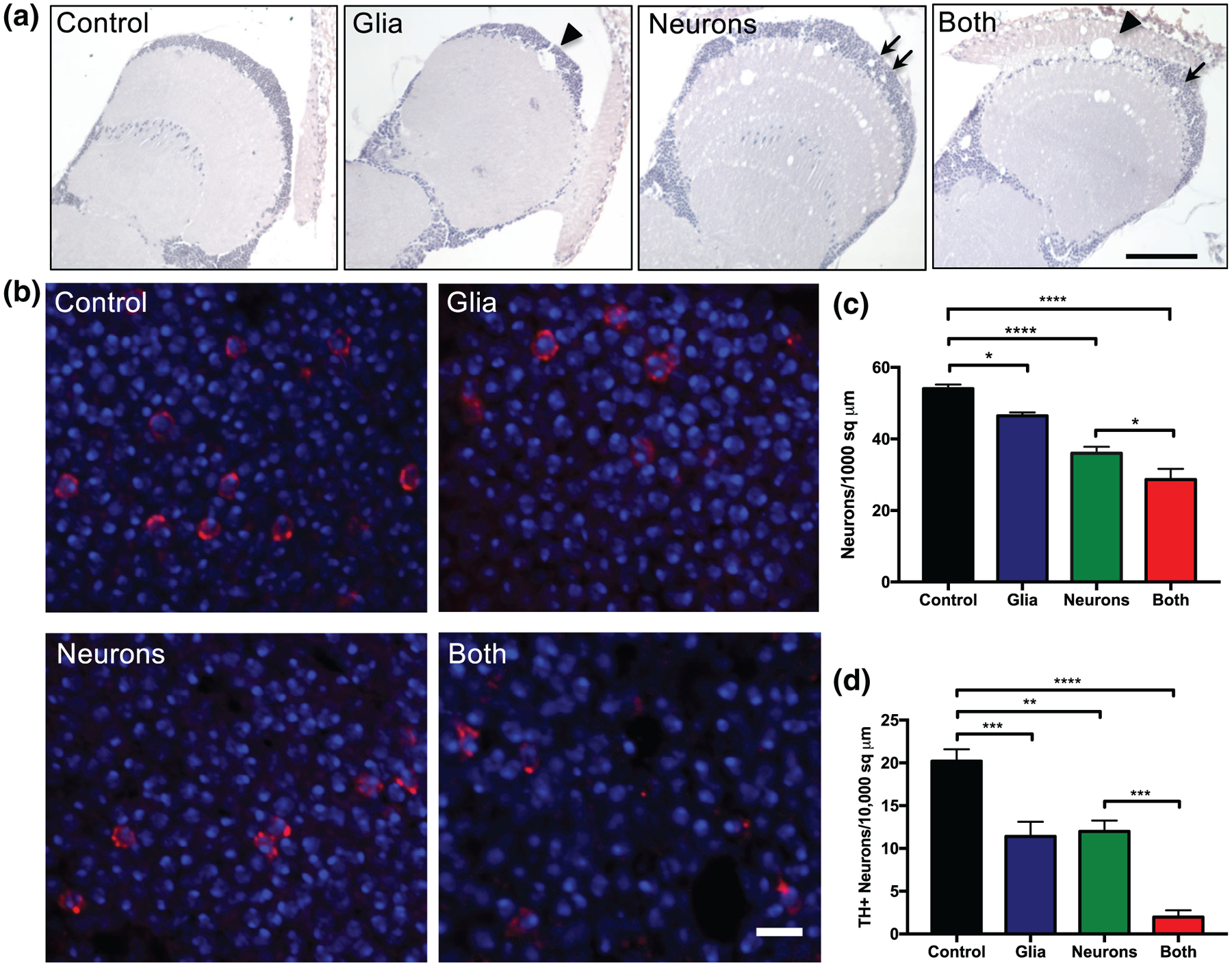
FIGURE 3.

Glial α-synuclein causes neurodegeneration. (a) Optic lobe sections stained with hematoxylin demonstrating vacuolization, an indicator of neurodegeneration. Glial α-synuclein caused infrequent large vacuoles (arrowhead) whereas neuronal α-synuclein caused frequent small vacuoles (arrows). Scale bar = 100 μm. (b) Representative anterior medulla sections stained with DAPI (blue) and tyrosine hydroxylase antibody (red, mouse, 1:200, Immunostar) to indicate dopaminergic neurons. Scale bar = 5 μm. (c) Quantification of total neurons from hematoxylin stained slides of anterior medulla (not shown), n = 6 replicates per genotype. (d) Quantification of dopaminergic neurons from anterior medulla, n = 6 replicates per genotype. *p < .05, **p < .01, ***p < .005, ****p < .001, determined with one-way ANOVA
