
Figure 13.
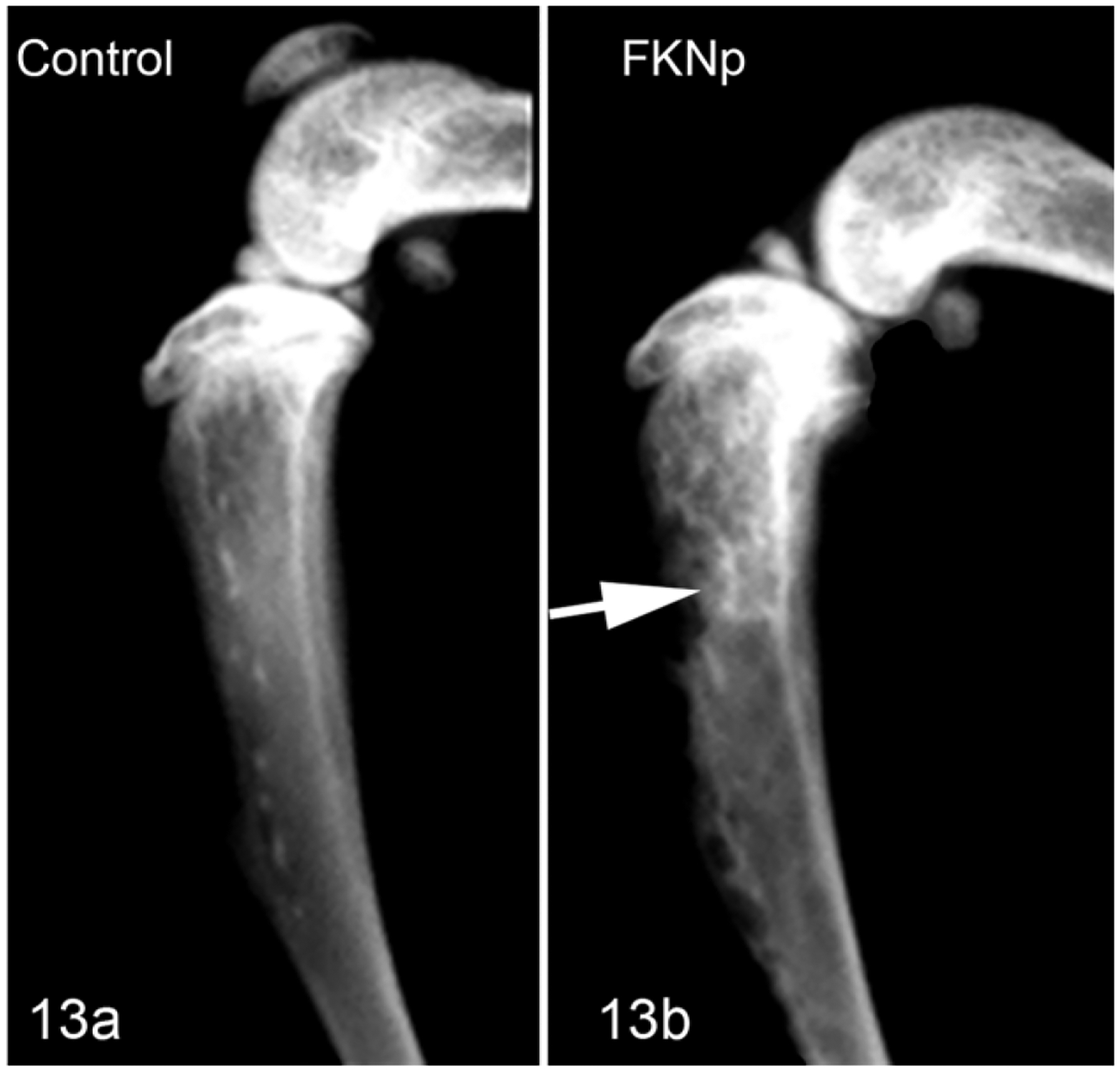
Tibia, Nude mouse. Radiograph. (a) Tibia without injection used as control. (b) The FKNp Luc cells in the tibia marrow space induced mixed osteolytic/osteoblastic bone metastases in the metaphysis and diaphysis (arrow).
Official websites use .gov
A
.gov website belongs to an official
government organization in the United States.
Secure .gov websites use HTTPS
A lock (
) or https:// means you've safely
connected to the .gov website. Share sensitive
information only on official, secure websites.
Tibia, Nude mouse. Radiograph. (a) Tibia without injection used as control. (b) The FKNp Luc cells in the tibia marrow space induced mixed osteolytic/osteoblastic bone metastases in the metaphysis and diaphysis (arrow).