Abstract
This study aimed to reveal the correlation between the radiolucency area around the crown of impacted maxillary canines and dentigerous cysts using cone beam CT (CBCT). CBCT data were obtained from patients with impacted maxillary canines. Three points of five areas (tooth cusp area and buccal, lingual, mesial and distal areas of the crown) were randomly selected, and the distance between the point and the surrounding hard tissue was measured respectively. The mean values were recorded as the radiolucency area. These results were compared with the occurrence of dentigerous cysts during surgery. 58 patients with 76 impacted maxillary canines were included. 14 of the 76 impacted canines were accompanied by cysts (18.42%). With the increase in the thickness of the radiolucency area, the incidence of cysts was significantly increased (p < 0.05). No cysts were found in the compacted canines with 0–1 mm thickness of the radiolucency area. The highest incidence (71.43%) was observed in canines with 3–4 mm thickness of the radiolucency area. This study found that the thickness of the radiolucency area around the crown of the maxillary impacted canine was closely related to the occurrence of dentigerous cysts. CBCT can be used to estimate the occurrence possibility of dentigerous cyst and guide surgical operations.
Keywords: Impacted maxillary canine, Dentigerous cyst, CBCT
Introduction
Impacted teeth may not only interfere with the function of the stomatognathic system but also act as a source of many pathological lesions such as odontogenic cysts and tumours. One of the most prevalent types of odontogenic cysts associated with the impacted tooth is the dentigerous cyst.1 The occurrence of dentigerous cysts is due to the accumulation of cystic fluid between the crown of the impacted tooth and the reduced enamel epithelium. Moreover, dentigerous cysts are extremely prone to occur in the impacted maxillary canines.2 However, it is not that all of the impacted maxillary canines will cause the dentigerous cysts.
In clinical examinations, dentists often find that asymptomatic impacted canines are embedded in the maxilla. It is difficult to make a choice between performing the interventional treatment and keeping continuous observation both for doctors and patients. This is the responsibility of the dentists to find a theoretical basis for judging whether early intervention treatment is necessary. Therefore, to evaluate the occurrence probability of dentigerous cysts around the impacted canines based on the imaging data might be helpful to guide the dentist to make a decision on the clinical treatment time and methods.
Cone beam CT (CBCT) is more accurate than conventional X-ray tomography and cranial positioning lateral slices.3 Compared with traditional CT, CBCT has the advantages of the high pixel, high definition, high resolution, fast imaging and low radiation, and can observe tomographic images and three-dimensional images at any angle. Therefore, it has been widely used in dental science. We can perform many measurements such as distance, angle, and so on in the images as needed by directly using the functions accompanied by the software of CBCT, without the aid of other measuring tools.4,5 The measurement results are consistent and reliable, the selection of reference points is reproducible, and the data studied are comparable.6
In this study, CBCT was used to measure the distance between the crown of an impacted maxillary canine and the surrounding hard tissue, and then the thickness of the surrounding radiolucency image was deduced. The aim of the present study was to reveal the correlation between the radiolucency area around the crown of impacted maxillary canines and dentigerous cysts and to provide a reference for clinical diagnosis and treatment.
Methods and materials
This study was approved by the Institutional Ethics Committee of the Affiliated Stomatology Hospital of Southwest Medical University (certificate number, 2015005) and conducted in accordance with the Declaration of Helsinki. All procedures were carried out with the adequate understanding and written consent of the subjects.
Patients selection
From January 2016 to December 2017, patients with impacted maxillary canines and who needed surgery for assisting teeth eruption or removing the teeth were recruited from the Department of Oral and Maxillofacial Surgery, the Affiliated Stomatology Hospital of Southwest Medical University (Luzhou, China). Informed consent was obtained from all included patients. The inclusion criteria were: (1) patients with completely intraosseous impacted maxillary canines and (2) impacted maxillary canines which received surgery for assisting teeth eruption or removing the teeth. The exclusion criteria were patients with uncontrolled systemic bone disease and cleft palate. The following information was recorded: (1) basic information of the patient, including gender, age etc. (2) The number and location (left, right) of the impacted maxillary canines.
Radiographic examination
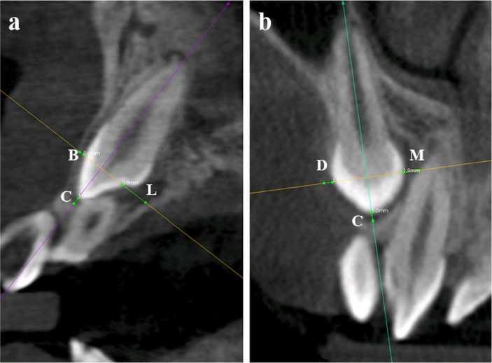
CBCT scans were performed at 86 kVp, 10 mAs and 10.8 s exposure, with a resolution of 0.20 mm per slice by a KODAk 9500 (Carestream Health, Rochester, America). The three-dimensional reconstruction images were obtained by multiplanar reconstruction (MPR). Image data were evaluated using the built-in CS 3D Imaging Software 3.2.9. (Carestream Health, Rochester, America). In detail, five areas were selected for measurement, including the tooth cusp area and buccal, lingual, mesial and distal areas that were located at the middle of the crown. The reason for selecting these five areas was that the cusp and the middle of the crown could be easily detected on the CBCT images, ensuring the repeatability, accuracy and stability of the measurement. In the measurement, the three-dimensional sections of the tooth were selected as the datum (Figure 1).
Figure 1.
The distance between the crown of the impacted maxillary canine and the surrounding hard tissue was measured in five areas. (a) Buccolingual section of the canine. (b) Mesiodistal section of the canine. C, cusp. B, buccal. L, lingual. M, mesial. D, distal.
On the section obtained from longitudinal cutting paralleled to the long axis of the tooth through the cusp, the cusp point and the buccal, lingual, mesial and distal points at the middle of the crown were chosen. Then, the distance between these points and the surrounding hard tissue was measured (Figure 1). These steps were performed in triplicate. The mean values were calculated and recorded as the radiolucency area around the crown of the impacted canine. Later on, the values were grouped according to the size and divided into 0–1 mm group, 1–2 mm group, 2–3 mm group and 3–4 mm group. All the measurements were performed by the same investigator.
Surgical procedure
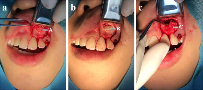
There were two types of surgery for the patients in this study, fenestration and dental extraction. Surgeons consulted with orthodontists to decide which surgery to perform. Thereafter, local anaesthesia was induced with 2% lidocaine with 1:100,000 epinephrine. After local anaesthesia, the gingiva was incised and elevated, and the bone was removed as required. In the cases where a dentigerous cyst was found, the cyst wall was removed with a scalpel. Then, the surgical fenestration proceeded, or the minimally invasive technology was used to extract the teeth. During the operation, the presence of a cystic wall around the crown and cystic fluid between the crown and the cyst wall was diagnosed as a dentigerous cyst (Figure 2).
Figure 2.
An example of the extraction of the impacted maxillary canine and the detection during operation. (a) cystic wall after deboning. (b) removing the cystic wall. (c) cyst.
Statistical analysis
Data were statistically analysed using the IBM SPSS statistical package 22.0 (IBM Co., Chicago, IL). In the study, the incidence of dentigerous cysts was calculated by the ratio of the number of impacted canines with dentigerous cysts to the total number of corresponding impacted canines and recorded as percentage. The incidence of dentigerous cysts in the variations of impacted maxillary canines was analysed by the χ2 test to evaluate the association between impacted canines and dentigerous cysts. A significance level of 0.05 was chosen.
Results
Characteristics of patients
58 patients were included in this study, including 21 males and 37 females. Ages ranged from 10 to 20 years old, with a mean value of 12.5 ± 2.8 years old. Among the 58 patients, 40 patients (68.97%) presented with a unilateral impact, and 18 patients (31.03%) presented with a bilateral impact. In total, there were 76 impacted maxillary canines (Table 1).
Table 1.
Characteristics of the included patients
| Number | Impacted canines | Dentigerous cysts | ||||
|---|---|---|---|---|---|---|
| Left | Right | Bilaterally | Total | |||
| Male | 21 (36.21%) | 7 | 9 | 14 | 30 | 5 |
| Female | 37 (63.79%) | 14 | 10 | 22 | 46 | 9 |
| Total | 58 | 21 | 19 | 36 | 76 | 14 |
Results of radiographical measurement
Measurements showed that there were 33 teeth with a pericoronal radiolucency that was less than 1 mm thick, 19 teeth with a radiolucency area of 1–2 mm thick, 17 teeth with a radiolucency area of 2–3 mm thick and seven teeth with a radiolucency area of 3–4 mm thick (Table 2).
Table 2.
Incidence of dentigerous cyst according to the thickness of the pericoronal radiolucency area (mm)
| Thickness of the radiolucency area (mm) | Cysts | Incidence | |
|---|---|---|---|
| Yes | No | ||
| 0–1 | 0 | 33 | 0.00% |
| 1–2 | 2 | 17 | 10.53% |
| 2–3 | 7 | 10 | 41.18% |
| 3–4 | 5 | 2 | 71.43% |
| Total | 14 | 62 | 18.42% |
With the increase in thickness of the radiolucency area, the incidence of tooth cysts was significantly increased (χ2 = 27.186, p < 0.05)
Results of intraoperative observation
Intraoperative observation found that 14 of the 76 impacted canines were accompanied by cysts, resulting in an incidence of 18.42%. There were 5 (incidence: 16.67%) and 9 (incidence: 19.57%) dentigerous cysts were respectively detected from males and females. There were no statistically significant differences between the cyst probabilities in male and female (χ2 = 0.102, p > 0.05).
With the increase in the thickness of the radiolucency area, the incidence of tooth cysts was significantly increased (χ2 = 27.186, p < 0.05). No cysts were found around the crown in the compacted maxillary canines with a 0–1 mm thickness radiolucency area. For canines with 1–2 mm and 2–3 mm thickness radiolucency area, the incidence of cysts was 10.53 and 41.18%, respectively. The highest incidence (71.43%) was observed in compacted canines with 3–4 mm thickness pericoronal radiolucency area (Table 2). Furthermore, we found that in the cases with 0–1 mm and 1–2 mm radiolucency area, the boundary between the crown of the impacted maxillary canines and the alveolar bone was difficult to distinguish during the surgery.
Discussion
Impacted teeth are a common disease in the clinic and might be due to various systemic and local factors. It may occur in any part of the dental arch. The impacted canine is the most likely to occur in the dentition except for the third molar.7–9 As located in the front of the dental arch, impacted maxillary canines often cause disorder of the anterior teeth, which is a common disease in dentistry and influences the appearance and function of the maxillofacial region. Furthermore, impacted maxillary canines might cause dentigerous cysts.10,11 With the passage of time, the dentigerous cyst may gradually expand as the fluid accumulates between the cyst wall and tooth crown. The compression on the jaw and adjacent teeth from cyst enlargement can cause jaw enlargement and results in irregular dentition and facial deformity and other serious consequences.12,13 Therefore, impacted maxillary canines should be closely observed and need to be managed as early as possible once dentigerous cysts were formed.
Dentigerous cysts go together with the crown of an impacted tooth or partially erupted tooth, connecting at the cervical portion of the tooth (the cemento-enamel junction). These cysts represent the most common developmental cysts. Permanent third molars and maxillary canines are the teeth most likely to fail to erupt and consequently are the most common region for the occurrence of dentigerous cysts.14 Dentigerous cysts generally occur after the formation of the crown and around the crowns. The fluid between the reduced enamel epithelium and the crown surface accumulates into a cyst. The osmotic gradient then pulls the fluid from the surrounding tissue to enhance cystic enlargement.15–17 As the maxillary canine has a great influence on the facial feature, it is relatively easy to detect the related diseases caused by the impacted canine. Therefore, in this study, the relationship between the impacted maxillary canine and the dentigerous cyst was studied by combining the imaging and clinical method.
It is difficult to distinguish a small dentigerous cyst and a large dental follicle despite the availability of both radiographic and histologic information and it seems that identifying the cyst during surgery may be the only reliable way to obtain a definitive diagnosis while radiological and histological features are not sufficient to distinguish between small dentigerous cysts and large dental follicles.18,19 Due to the harmfulness of dentigerous cysts, it is not related to histological classification, but the compression and damage from the expansively developing of the cyst. In this study, we took the presence of a cystic wall and cystic fluid rather than the histological section as criteria for the diagnosis of dentigerous cysts.
As the dentigerous cyst is asymptomatic in general, it is usually detected when a tooth fails to erupt, and an X-ray examination is taken. The typical X-ray feature of a dentigerous cyst is a transmission image that surrounds an unexposed crown.20–22 A three-dimensional CT can stereoscopically and accurately reflect the characteristics of the transmitted image. It has a high isotropic spatial resolution, can perform 1:1 three-dimensional reconstruction on the two-dimensional images obtained from the initial reconstruction and display the dentition and related tissues in a ratio of 1:1. The CBCT can help us to get an accurate understanding of the shape, location and relationship of the surrounding alveolar bone, which provides accurate information for clinicians to develop treatment decisions and select surgical approaches.23–25 Therefore, we can make an accurate and comprehensive measurement of the distance between the crown of the impacted canine and the surrounding alveolar bone in three-dimension space. In this study, 37 females and 21 males with an impacted maxillary canine were included, with a gender ratio of 1.76:1. That may be related to the fact that females care more about their appearance and tend to visit dentists more to inquire about the treatment of an impacted maxillary canine. The results showed that the incidence of dentigerous cysts was 18.42%. These results were closely agreement with the study of Shear and Bernick, whose research results indicated that the percentage of dentigerous cysts associated with a permanent canine were 19.7 and 14.7%, respectively.18
Moreover, the results of the present study indicated that the incidence of dentigerous cysts significantly increased with the increase in the thickness of the pericoronal radiolucency area (p < 0.05). When the measured radiolucency area reached up to 3–4 mm, the incidence of the dentigerous cyst was as high as 71.43%. The possible reason was that the cystic fluid presented between the wall of the cyst and the crown would gradually accumulate, resulting in the compression and absorption of the alveolar bone. Accordingly, the distance between the crown and the surrounding alveolar bone were increased, and the radiolucency area as measured in this study enlarged as well. Therefore, clinical intervention might be necessary to prevent further damage to the alveolar bone and adjacent teeth if the measured radiolucency area were found more than 3 mm.
The treatment of impacted maxillary canines is mainly accomplished through surgical extraction or a combination of surgery and orthodontic traction. Pre-operative estimation of the occurrence of dentigerous cysts is conducive to the surgical operation and useful to the location and operation of the impacted tooth.26,27 From the clinical treatment process in this study, we found the following results and experiences. In the cases with 0–1 and 1–2 mm radiolucency area, the boundary between the crown of the impacted maxillary canines and the alveolar bone was difficult to distinguish during the surgery, since the clue from the cystic wall could not be detected. In these cases, the prediction can be given that there will not be dentigerous cysts. Correspondingly, much care has to be taken in the operation of exposing the crown, and the bones should be carefully removed to avoid damage to the crown. On the contrary, in the cases with more than 3 mm radiolucency area, the prediction could be given that there are dentigerous cysts. The deboning approach can be selected at a thicker location of the radiolucency area on CBCT image; then the bone can be removed more easily. The cystic wall was first exposed, and the crown tissue was exposed after the cystic wall was removed as completely as possible. These procedures are able to reduce the surgical trauma, improve the prognosis, shorten the treatment time, improve the success rate and reduce the patient's pain.
To our knowledge, this is the first study to investigate the correlation between the radiolucency area around the crown of maxillary impacted canines and dentigerous cysts by using CBCT. There were several limitations to the present study. First, as a pilot study, the sample size was relatively limited, and 58 patients with 76 impacted canines were included. Second, histological analyses were not performed in this study, since histological classification was not the aim of this study. In our research, cyst fluid was the key factor to distinguishing cysts from dental follicles. Further research with larger sample size is required to verify the results.
Within the limitations of this study, the following can be concluded. The thickness of the radiolucency area around the crown of the maxillary impacted canine is closely related to the incidence of dentigerous cysts. The thicker the thickness, the higher the incidence of the cyst. Using CBCT to accurately determine the position of the impacted maxillary canine in three-dimensional space and measuring the distance between the crown and the surrounding hard tissue can predict the incidence of dentigerous cysts, which is conducive to make a treatment decision.
Footnotes
Funding: This study was funded by a grant from the National Natural Science Foundation of China (No. 11702231), Sichuan Medical Association (Q15026) and the Cooperation Program of Luzhou Government and Southwest Medical University (2019LZXNYDJ14).
Competing Interests: All of the authors declare that they have no conflicts of interests.
Ethical Approval: This study was approved by the Institutional Ethics Committee of the Affiliated Stomatology Hospital of Southwest Medical University (certificate number, 2015005).
Patient consent: All patients consented to the treatment.
REFERENCES
- 1.Moturi K, Kaila V. Management of non-syndromic multiple impacted teeth with dentigerous cysts: a case report. Cureus 2018; 10: e3323. doi: 10.7759/cureus.3323 [DOI] [PMC free article] [PubMed] [Google Scholar]
- 2.Hasan S, Ahmed S, Reddy LBhaskar, Reddy LB. Dentigerous cyst in association with impacted inverted mesiodens: report of a rare case with a brief review of literature. Int J App Basic Med Res 2014; 4: 61–4. doi: 10.4103/2229-516X.140748 [DOI] [PMC free article] [PubMed] [Google Scholar]
- 3.Shah N, Bansal N, Logani A. Recent advances in imaging technologies in dentistry. World J Radiol 2014; 6: 794–807. doi: 10.4329/wjr.v6.i10.794 [DOI] [PMC free article] [PubMed] [Google Scholar]
- 4.Pauwels R, Nackaerts O, Bellaiche N, Stamatakis H, Tsiklakis K, Walker A, et al. . SEDENTEXCT project Consortium: variability of dental cone beam CT grey values for density estimations. Br J Radiol 2013; 86: 20120135. [DOI] [PMC free article] [PubMed] [Google Scholar]
- 5.Shukla S, Chug A, Afrashtehfar KI. Role of cone beam computed tomography in diagnosis and treatment planning in dentistry: an update. J Int Soc Prev Community Dent 2017; 7(Suppl 3): S125–36. doi: 10.4103/jispcd.JISPCD_516_16 [DOI] [PMC free article] [PubMed] [Google Scholar]
- 6.Lisboa CdeO, Masterson D, da Motta AFJ, Motta AT. Reliability and reproducibility of three-dimensional cephalometric landmarks using CBCT: a systematic review. J Appl Oral Sci 2015; 23: 112–9. doi: 10.1590/1678-775720140336 [DOI] [PMC free article] [PubMed] [Google Scholar]
- 7.Manne R, Gandikota C, Juvvadi SR, Rama HRM, Anche S. Impacted canines: etiology, diagnosis, and orthodontic management. J Pharm Bioallied Sci 2012; 4(Suppl 2): S234–8. doi: 10.4103/0975-7406.100216 [DOI] [PMC free article] [PubMed] [Google Scholar]
- 8.Al-Zoubi H, Alharbi AA, Ferguson DJ, Zafar MS. Frequency of impacted teeth and categorization of impacted canines: a retrospective radiographic study using orthopantomograms. Eur J Dent 2017; 11: 117–21. doi: 10.4103/ejd.ejd_308_16 [DOI] [PMC free article] [PubMed] [Google Scholar]
- 9.Chawla S, Goyal M, Marya K, Jhamb A, Bhatia HP. Impacted canines: our clinical experience. Int J Clin Pediatr Dent 2011; 4: 207–12. doi: 10.5005/jp-journals-10005-1111 [DOI] [PMC free article] [PubMed] [Google Scholar]
- 10.Taffarel IP, Saga AY, Locks LL, Ribeiro GL, Tanaka OM. Clinical outcome of an impacted maxillary canine: from exposition to occlusion. J Contemp Dent Pract 2018; 19: 1552–7. [PubMed] [Google Scholar]
- 11.Arriola-Guillén LE, Ruíz-Mora GA, Rodríguez-Cárdenas YA, Aliaga-Del Castillo A, Boessio-Vizzotto M, Dias-Da Silveira HL, et al. . Influence of impacted maxillary canine orthodontic traction complexity on root resorption of incisors: a retrospective longitudinal study. Am J Orthod Dentofacial Orthop 2019; 155: 28–39. doi: 10.1016/j.ajodo.2018.02.011 [DOI] [PubMed] [Google Scholar]
- 12.Santos BZ, Beltrame AP, Bolan M, Grando LJ, Cordeiro MMR. Dentigerous cyst of inflammatory origin. J Dent Child 2014; 81: 112–6. [PubMed] [Google Scholar]
- 13.Lizio G, Tomaselli L, Landini L, Marchetti C. Dentigerous cysts associated with impacted third molars in adults after decompression: a prospective survey of reduction in volume using computerised analysis of cone-beam computed tomographic images. Br J Oral Maxillofac Surg 2017; 55: 691–6. doi: 10.1016/j.bjoms.2017.04.018 [DOI] [PubMed] [Google Scholar]
- 14.Bilodeau EA, Collins BM. Odontogenic cysts and neoplasms. Surg Pathol Clin 2017; 10: 177–222. doi: 10.1016/j.path.2016.10.006 [DOI] [PubMed] [Google Scholar]
- 15.Shah KM, Karagir A, Adaki S, Pattanshetti C. Dentigerous cyst associated with an impacted anterior maxillary supernumerary tooth. BMJ Case Rep 2013; 31: 1–3. [DOI] [PMC free article] [PubMed] [Google Scholar]
- 16.Choi HJ, Lee JB. Obliteration of recurrent large dentigerous cyst using bilateral buccal fat pad sling flaps. J Craniofac Surg 2016; 27: e465–8. doi: 10.1097/SCS.0000000000002780 [DOI] [PubMed] [Google Scholar]
- 17.Jain N, Gaur G, Chaturvedy V, Verma A. Dentigerous cyst associated with impacted maxillary Premolar: a rare site occurrence and a rare coincidence. Int J Clin Pediatr Dent 2018; 11: 50–2. doi: 10.5005/jp-journals-10005-1483 [DOI] [PMC free article] [PubMed] [Google Scholar]
- 18.Daley TD, Wysocki GP. The small dentigerous cyst. A diagnostic dilemma. Oral Surg Oral Med Oral Pathol Oral Radiol Endod 1995; 79: 77–81. doi: 10.1016/s1079-2104(05)80078-2 [DOI] [PubMed] [Google Scholar]
- 19.Mishra R, Tripathi AM, Rathore M. Dentigerous cyst associated with horizontally impacted mandibular second Premolar. Int J Clin Pediatr Dent 2014; 7: 54–7. doi: 10.5005/jp-journals-10005-1235 [DOI] [PMC free article] [PubMed] [Google Scholar]
- 20.Becker A, Chaushu S. Etiology of maxillary canine impaction: a review. Am J Orthod Dentofacial Orthop 2015; 148: 557–67. doi: 10.1016/j.ajodo.2015.06.013 [DOI] [PubMed] [Google Scholar]
- 21.Mortazavi H, Baharvand M. Jaw lesions associated with impacted tooth: a radiographic diagnostic guide. Imaging Sci Dent 2016; 46: 147–57. doi: 10.5624/isd.2016.46.3.147 [DOI] [PMC free article] [PubMed] [Google Scholar]
- 22.Paul R, Paul G, Prasad RK, Singh S, Agarwal N, Sinha A. Appearance can be deceptive: dentigerous cyst crossing the midline. Natl J Maxillofac Surg 2013; 4: 100–3. doi: 10.4103/0975-5950.117823 [DOI] [PMC free article] [PubMed] [Google Scholar]
- 23.Scarfe WC, Li Z, Aboelmaaty W, Scott SA, Farman AG. Maxillofacial cone beam computed tomography: essence, elements and steps to interpretation. Aust Dent J 2012; 57 Suppl 1(Suppl 1): 46–60. doi: 10.1111/j.1834-7819.2011.01657.x [DOI] [PubMed] [Google Scholar]
- 24.Dağsuyu İlhan Metin, Kahraman F, Okşayan R. Three-Dimensional evaluation of angular, linear, and resorption features of maxillary impacted canines on cone-beam computed tomography. Oral Radiol 2018; 34: 66–72. doi: 10.1007/s11282-017-0289-5 [DOI] [PubMed] [Google Scholar]
- 25.Cetmili H, Tassoker M, Sener S. Comparison of cone-beam computed tomography with bitewing radiography for detection of periodontal bone loss and assessment of effects of different voxel resolutions: an in vitro study. Oral Radio 2018; 18: 75. [DOI] [PubMed] [Google Scholar]
- 26.Spallarossa M, Canevello C, Silvestrini Biavati F, Laffi N. Surgical orthodontic treatment of an impacted canine in the presence of dens invaginatus and follicular cyst. Case Rep Dent 2014; 2014: 1–7. doi: 10.1155/2014/643082 [DOI] [PMC free article] [PubMed] [Google Scholar]
- 27.Dehis HM, Fayed MS. Management of maxillary impacted teeth and complex odontome: a review of literature and case report. Open Access Maced J Med Sci 2018; 6: 1882–7. doi: 10.3889/oamjms.2018.395 [DOI] [PMC free article] [PubMed] [Google Scholar]


