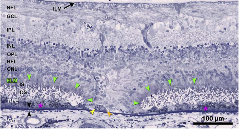
Figure 6: Descent of the external limiting membrane (ELM) towards Bruch’s membrane.
The histology supports the clinical imaging in Figure 2,3, bottom row. The ELM descends in two curved lines on either side of a narrow isthmus of atrophy typically seen in iRORA. The ONL, HFL, OPL, and INL subside in parallel to the ELM, creating a funnel. The ONL is discontinuous, and the HFL is disordered. Where these ELM descents curve, surviving cone photoreceptors lack outer segments and have short inner segments. Layers: ELM, external limiting membrane (green arrowheads); ILM, inner limiting membrane; NFL, nerve fiber layer; GCL, ganglion cell layer; IPL, inner plexiform layer; INL, inner nuclear layer; OPL, outer plexiform layer; HFL, Henle fiber layer; ONL, outer nuclear layer; ChC, choriocapillaris; black arrowheads, Bruch’s Membrane. 87-year-old white male donor. Prepared by M. Li MD PhD and J.D. Messinger DC from the Project MACULA AMD histopathology resource: http://projectmacula.cis.uab.edu/

