Abstract
An outbreak of a bacterial contagion is a critical threat for human health worldwide. Recently, light-activated heterostructured nanomaterials (LAHNs) have shown potential as antibacterial agents, owing to their unique structural and optical properties. Many investigations have revealed that heterostructured nanomaterials are potential antibacterial agents under light irradiation. In this review, we summarize recent developments of light-activated antibacterial agents using heterostructured nanomaterials and specifically categorized those agents based on their various light harvesters. The detailed antibacterial mechanisms are also addressed. With the achievements of LAHNs as antibacterial agents, we further discuss the challenges and opportunities for their future clinical applications.
Keywords: heterostructured nanomaterial, antibacterial agent, antibacterial mechanism, reactive oxygen species, light-activated, synergistic effect
1. Introduction
Antibacterial studies have become more significant because of the increased of multidrug-resistant (MDR) bacteria in this era [1,2,3,4,5,6,7,8,9,10]. Various mechanisms can stimulate antibacterial resistance, including overuse of antibiotics and the delivery of bacteria by different routes [11,12,13]. A recent report indicates that by 2050, antibacterial resistance will be a tremendous public health threat with the potential to cause the death of ten million people each year [14]. Therefore, development of new types of antibacterial agents against MDR bacteria is an urgent task. The development of new antibacterial agents inspired the researchers for the investigation of nanomaterials which can eliminate MDR bacteria without the help of antibiotics [15,16,17,18,19].
Nanomaterials—including metals, metal oxide, semiconductors and polymers—have been extensively studied for applications in nanoscience and nanotechnology, due to their superior chemical and physical properties [20,21,22,23,24,25,26,27,28,29,30,31,32,33,34,35]. Among nanomaterials, the heterostructured nanomaterials have shown unique optical properties, including the increase of light absorption and the extension of absorption region [36,37,38,39,40,41]. Recently, great advancements have been reported in the application of LAHNs as antibacterial agents. With the rise of light absorption and the extension of absorption region, heterostructured nanomaterials have shown superior antibacterial activity, based on reactive oxygen species (ROS) generation under light illumination [42,43,44,45,46,47,48]. The emphasis of this review is on the mechanism study of light-induced ROS generation from heterostructured nanomaterials by synergistic effect. Recent achievements with the uses of LAHNs as antibacterial agents are classified by their various light harvesters. Finally, challenges and perspectives for LAHNs as antibacterial agents are also provided.
2. Antibacterial Nanomaterials Based on Light-induced ROS Generation
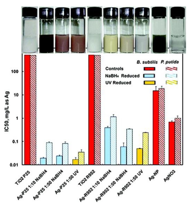
Titanium dioxide (TiO2) materials have been found to possess remarkable biocompatibility and low cell toxicity with significant antibacterial activity [49,50,51,52,53,54,55,56,57,58]. Recently, TiO2-based nanomaterials were popular materials with substantial amount of antibacterial activities, and their antibacterial activities could be further enhanced by light irradiations with various wavelengths, including ultraviolet (UV) light, visible light and near-infrared (NIR) light due to the increase of ROS generation [59,60,61,62,63,64,65,66,67]. For example, with UV light irradiation, heterostructured Ag-TiO2 nanoparticles have displayed higher bactericidal activity compared to that of only UV irradiation, Ag nanoparticles under UV irradiation or TiO2 nanoparticles under UV irradiation (Figure 1) [68]. With UV light illumination, the augmentation of bactericidal activity of hybrid Ag-TiO2 nanoparticles indicated that Ag nanoparticles loaded onto TiO2 nanoparticles served as electron traps to prevent recombination of electron and hole in hybrid Ag-TiO2 nanoparticles. The photo-exited electrons were generated and then transferred to Ag nanoparticles to extend the life of electron-hole pairs. In the system of hybrid Ag-TiO2 nanoparticles, the increases of electron-hole pairs enhanced ROS generation for the destruction of bacterial membrane or DNA [69,70,71,72,73,74]. In the work, hybrid Ag-TiO2 nanoparticles provided more effective bactericides with light irradiations; however, the uses of hybrid Ag-TiO2 nanoparticles as an antibacterial agent were challenging in practical application due to their high cytotoxicity and facile accumulation in tissues and organs.
Figure 1.
IC50 values of different hybrid Ag-TiO2 nanoparticles with and without UV light irradiations. TiO2 P25 (Degussa P25) and TiO2 R-902 (DuPont Ti-Pure R-902) were two commercial TiO2 nanoparticles. Two different molar ratios of AgNO3 and TiO2 including 1:10 and 1:50 were respectively employed to prepare Ag nanoparticles deposited onto the TiO2 nanoparticles by NaBH4 reduction and UV photoreduction to form the samples of Ag-P25 1:10 NaBH4, Ag-P25 1:50 NaBH4, Ag-P25 1:50 UV, Ag-R902 1:10 NaBH4, Ag-R902 1:50 NaBH4 and Ag-R902 1:50 UV. Ag nanoparticles and AgNO3 were purchased from QuantumSphere, Inc. Reproduced with permission from ref. [68]. Copyright © 2011, American Chemical Society.
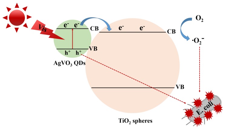
Nanocomposites of AgVO3 quantum dots (QDs) deposited onto TiO2 nanospheres (TiO2/AgVO3) have shown high-performance photocatalytic capability with visible light irradiation [75,76,77]. TiO2/AgVO3 nanocomposites incubated with E. coli have been investigated to study their light-induced bacterial inactivation with illumination of visible light. In the control experiment, after visible light irradiation over 120 min, TiO2 nanospheres were inactivated only 0.13 log of E. coli. With the use of TiO2/AgVO3 nanocomposites as photocatalysts, all of E. coli were killed under visible light irradiation over 120 min. After TiO2/AgVO3 nanocomposites incubated with E. coli, with visible light irradiation; the shape of E. coli was destroyed. Moreover, there were no obvious changes of bacterial inactivity of TiO2/AgVO3 nanocomposites after three cycling antibacterial tests under visible light irradiation indicating that TiO2/AgVO3 nanocomposites were good photocatalysts with superior stability. For the photocatalytic disinfection mechanism, AgVO3 QDs were excited to offer light-induced pairs of electrons and holes with visible light illumination and then the light-induced electrons located in the conduction band of AgVO3 QDs were easily delivered to the conduction band of TiO2 (Figure 2). The light-induced electrons in the conduction band of TiO2 could react with oxygen in medium to form ⋅O2−. Moreover, the holes located in the valence band of AgVO3 QDs could be delivered to their surface to inhibit the growth of E. coli. To sum up, the light-induced electron-hole pairs in TiO2/AgVO3 nanocomposites were efficiently separated to improve the light-induce antibacterial activity of E. coli.
Figure 2.
Antibacterial mechanism of TiO2/AgVO3 nanocomposites with visible light illumination. Reproduced with permission from ref. [75]. Copyright © 2019, Elsevier.
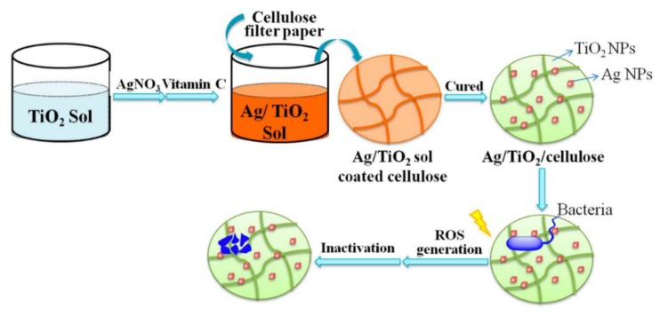
Natural polymers of cellulose are intensively used in daily life [78,79,80]. However, the application of cellulose has been limited, due to its susceptibility to microorganism growth. In recent advancements, a simple sol-gel approach has been used to conjugate the cellulose scaffold with Ag/TiO2 nanoparticles (Ag/TiO2/cellulose) against bacteria (Figure 3) [81]. The antibacterial activities of nanocomposite film of Ag/TiO2/cellulose, film of pristine cellulose and nanocomposite film of TiO2/cellulose have been investigated with E. coli with and without UV light irradiation. The bactericidal performance of the nanocomposite film of Ag/TiO2/cellulose with Ag content of 0.030 wt% has shown the best 99.9% inactivation of E. coli with UV light illumination. The results suggest that the nanocomposite film of Ag/TiO2/cellulose exhibit superior bactericidal performance against E. coli because of the synergistic effect of Ag nanoparticles and anatase TiO2 nanoparticles. Under UV light irradiation, TiO2 nanoparticles may generate ROS—including ⋅OH, ⋅O2− and H2O2—prompting bacterial death. Moreover, Ag nanoparticles may capture electrons, restraining the recombination of photon-induced electron/hole pairs for the increase of ROS formation. Overall, the bactericidal performance of the nanocomposite film of Ag/TiO2/cellulose was significantly enhanced under UV light irradiation.
Figure 3.
Schematic illustration of preparation of the nanocomposite film of Ag/TiO2/cellulose and its antibacterial mechanism. Reproduced with permission from ref. [81]. Copyright © 2018, MDPI.
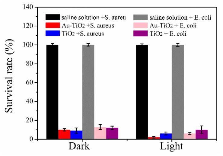
Au-TiO2 nanocomposites embedded into a degradable and antibacterial sodium alginate films have been developed by food packaging industries against bacteria [82]. Sodium alginate/Au-TiO2 nanocomposite (SAT) films were enabled to absorb light form UV to visible wavelength and to improve their hydrophilicity and shape stability. As shown in Figure 4, both sodium alginate/TiO2 nanocomposite (ST) films and SAT films exhibit distinct antibacterial activities for S. aureus and E. coli in dark. With the use of SAT films, 90% of S. aureus and 97.1% E. coli were killed without light illumination, respectively. However, after light illumination for 20 min, the antibacterial abilities of SAT films were respectively improved ~60% and ~50% for S. aureus and E. coli. The improvement of antibacterial abilities of SAT films may be attributed to that the nanocomposites of Au-TiO2 increased light absorption and transfer capability due to their plasmonic effect. The plasmonic Au nanoparticles in SAT films may harvest light to produce light-induced photons for the increase of ROS to kill bacteria.
Figure 4.
Antibacterial activities of saline solution, sodium alginate/TiO2 nanocomposite (ST) film and sodium alginate/Au-TiO2 nanocomposite (SAT) film for S. aureus and E. coli with and without visible light illumination, respectively. Reproduced with permission from ref. [82]. Copyright © 2018, MDPI.
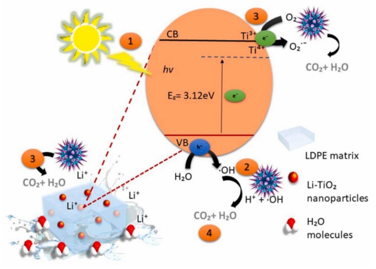
The nanocomposites of lithium-titanate (Li-TiO2) in the low-density polyethylene (LDPE) matrix have shown a significant increase in killing efficiency for S. aureus with visible light illumination [83]. In the heterostructured nanocomposites of Li-TiO2/LDPE, the oxygen vacancies of Ti3+ and interaction of Li-O-Ti bond reduced the band gap of TiO2 nanoparticles, resulting in their response to visible light (Figure 5). LDPE alone did not show any antibacterial activity for S. aureus. With the dopant of Li-TiO2 of 1 wt%, the heterostructured nanocomposites of Li-TiO2/LDPE inhibited the growth of S. aureus by 94% after visible light irradiation for 6 h, then raised the inhibition rate to 99% within 12 h under visible light irradiation. From the results of the scavenger test, the intensified bactericidal effect of the heterostructured nanocomposites of Li-TiO2/LDPE were ascribed to the productions of powerful oxidants including ⋅OH and ⋅O2− under visible light irradiation. These powerful oxidants may particularly react with the polyunsaturated phospholipid composition of the bacterial membrane to form water and carbon dioxide.
Figure 5.
The antimicrobial mechanism of the heterostructured nanocomposites of Li-TiO2/LDPE against S. aureus. Abbreviation: valence band: VB; conduction band: CB. Reproduced with permission from ref. [83]. Copyright © 2019, Elsevier.
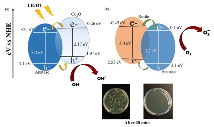
The heterostructured nanomaterials of copper-doped TiO2 (Cu-TiO2) have been applied as an antibacterial agent against E. coli and S. aureus with visible light illumination [84]. By utilizing dopant of oxidizing copper, Cu-TiO2 nanomaterials revealed the increase of absorption in visible region and the band gap of Cu-TiO2 nanomaterials was reduced to 2.8 eV. The photo-induced bactericidal activity of Cu-TiO2 heterostructured nanomaterial have been respectively executed with and without visible light illumination (Figure 6). Without photocatalysts, the E. coli and S. aureus bacteria continued growing under light and dark conditions. Moreover, in the dark, the Cu-TiO2 nanomaterials, anatase TiO2 nanoparticles and rutile TiO2 nanoparticles had no significant bacterial growth after culture for 90 min. After visible light irradiation for 30 min, the Cu-TiO2 nanomaterials had achieved 99.9999% bacterial reduction. However, to obtain 99.9999% bacterial reduction, both anatase and rutile TiO2 nanoparticles required light irradiation for 60 and 90 min, respectively. In comparison with anatase and rutile TiO2 nanoparticles, Cu-TiO2 nanomaterials showed better antibacterial activity. The results indicate that the dopant of Cu in Cu-TiO2 nanomaterials may improve the visible absorption efficiency to enhance bacterial inactivation. After the absorption of light, the charge carriers formed on p-n junction of Cu2O-TiO2 nanomaterials and then reacted with the oxygen and water incorporated on the surface of Cu2O-TiO2 nanomaterials to become radicals to disrupt bacterial membrane and cause bacterial gene alteration to kill bacteria.
Figure 6.
Mechanisms of photocatalytic antibacterial activity (a) p-n junction by heterostructured Cu2O and TiO2 (b) Type-2 heterostructures of anatase and rutile TiO2 nanoparticles. Images of S. aureus colonies in the agar plates with (right) and without (left) Cu-TiO2 nanomaterials after 30 min illumination. Reproduced with permission from ref. [84]. Copyright © 2018, MDPI.
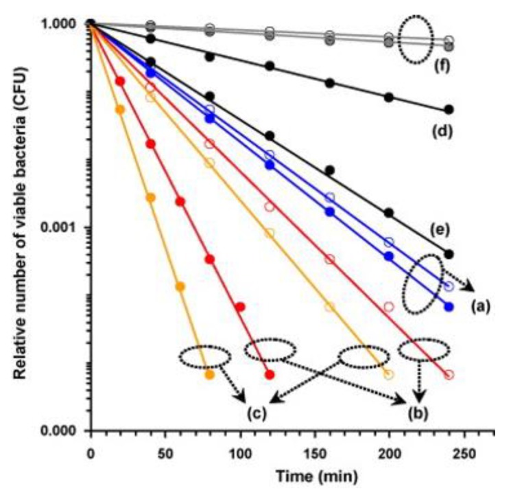
The thin film of photocatalyst of α-Fe2O3 nanograin chains incorporated with anatase TiO2 nanolayer (TiO2/Fe2O3) exhibited bactericidal activity against E. coli with visible light illumination [85]. The bacterial inactivation of the anatase TiO2, α-Fe2O3 and TiO2/α-Fe2O3 thin films has been studied in the bacterial model of E. coli under visible light illumination, with and without H2O2 (Figure 7). As shown in Figure 7, TiO2/α-Fe2O3 thin film reveals the reduction rate of 2.6 × 10⁻² and 6.5 × 10⁻² min−1 without and with H2O2 for E. coil, reflecting a better antibacterial activity than anatase TiO2 and bare α-Fe2O3 thin film. The better bacterial inactivity of TiO2/α-Fe2O3 thin film may be ascribed to the heterostructure of TiO2/Fe2O3. Through visible light irradiation onto TiO2/Fe2O3 heterojunction, electrons located in the valence band of Fe2O3 may be excited to the conduction band—and then, electrons located in the valence band of TiO2 may be driven into the valance band of Fe2O3. Therefore, the anatase TiO2 nanolayer coated with TiO2/α-Fe2O3 thin film could be applied to enhance of the photocatalytic capability of the coated Fe2O3 nanograins. The anatase TiO2 nanolayer could increase the production of Fe2+ to react with the H2O2 for the generation of OH radicals resulted from the reactions of Fenton and photo-Fenton. Furthermore, water or hydroxyl groups adsorbed onto anatase TiO2 nanolayer may be used to trap the holes to produce ROS of hydroxyl radicals for the bactericidal activity enhancement to bacteria.
Figure 7.
Relative number of viable E. coli on surface of (a) the anatase TiO2 (b) the α-Fe2O3 and (c) the TiO2/Fe2O3 thin films under visible light irradiation. (d) The α-Fe2O3 and (e) the TiO2/Fe2O3 thin films without light illumination. (f) Control samples containing the solution without H2O2 (○) and with H2O2 (●) with light illumination. The black circles indicated the samples of a, b, c and f containing the solution without H2O2 (○) and with H2O2 (●) with light illumination. Reproduced with permission from ref. [85]. Copyright © 2009, Elsevier.
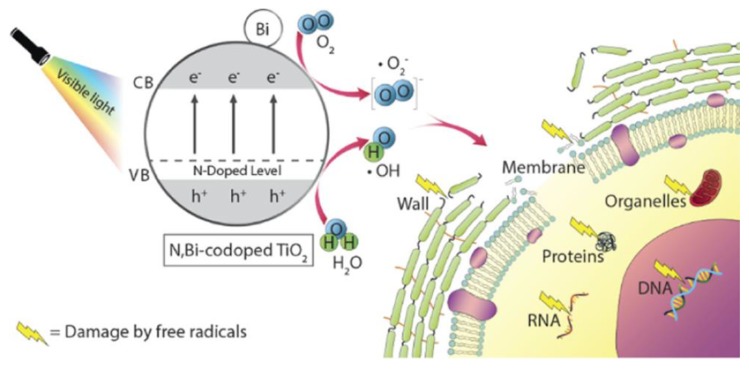
Recent developments of implants have focused on the design of their surface in terms of bacterial inactivation and reusable feasibility. The dopants nitrogen (N) and bismuth (Bi) in TiO2— incorporated with the plasma electrolytic oxidation (PEO)—revealed light-induced antibacterial activity and re-activation potential [86]. With the dopant Bi, the band gap energy of TiO2 was shifted to the visible light region. To confirm the in vitro antibacterial effect, the photocatalysts of TiO2 coated with urea (Urea-TiO2 group), TiO2 doped with Bi (Bi-TiO2 group) and TiO2 co-doped with urea and Bi (Urea, Bi-TiO2 group) were applied as antibacterial agents for two biofilms, including Streptococcus sanguinis and Actinomyces naeslundii. The results of the in vitro antibacterial tests indicated that the urea, Bi-TiO2 group presented the best performance in bactericidal activity with visible light illumination. With a synergistic effect of N and Bi under visible light illumination, the narrowed band gap of Bi2O3 combined with TiO2 could facilitate electrons migration from the valence band to the conduction band located in TiO2 and Ti3+ sites to react with O2 to form ⋅O2− and also ⋅OH radical could be produced according to the reaction of holes located in the valence band of TiO2 with H2O (Figure 8). The ROS of ⋅O2− and ⋅OH may induce damage of bacterial wall, membrane, organelles, proteins and genetic materials (DNA, RNA) to cause death of bacteria.
Figure 8.
Schematic illustration of light-induced antibacterial mechanism of N, Bi-codoped TiO2 photocatalysts. Bacterial DNA should locate in cytoplasm. Reproduced with permission from ref. [86]. Copyright © 2019, American Chemical Society.
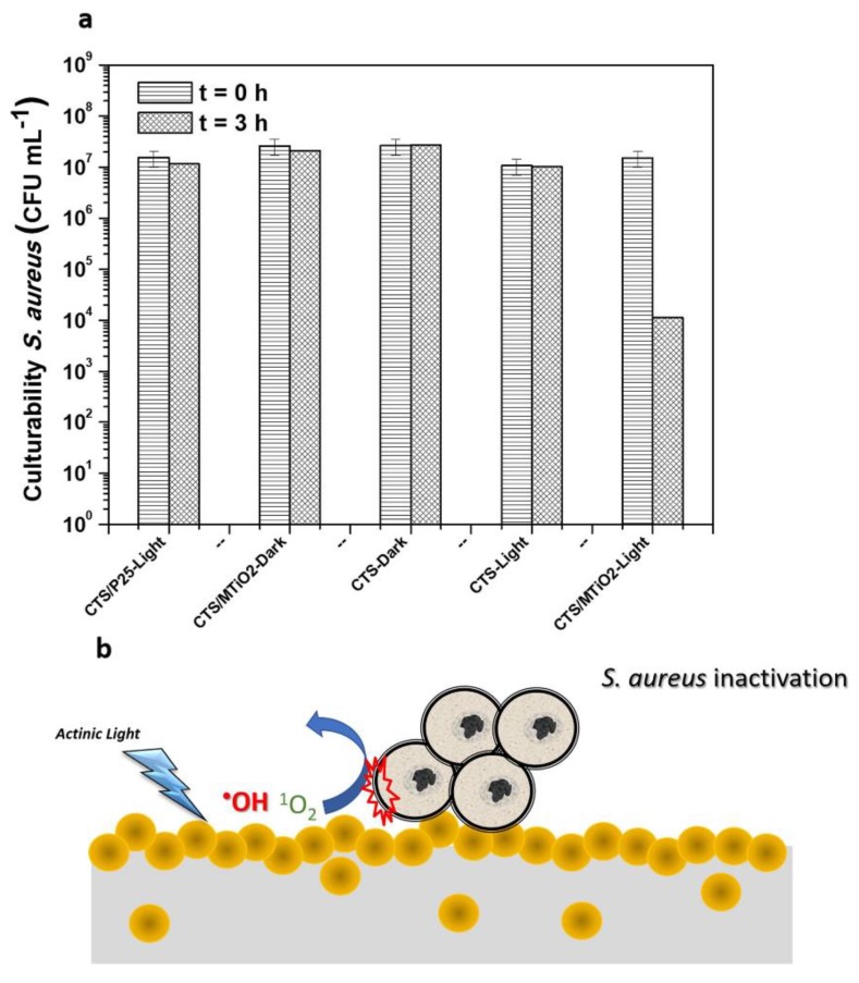
In recent times, nanocomposites of chitosan films containing melon/TiO2 (CTS/MTiO2) have been applied for self-cleaning of malachite green and light-induced antibacterial surfaces of S. aureus under light irradiation [87]. As the most common graphitic carbon nitride material (g-C3N4), melon is a visible-light harvesting molecule. The bacterial inactivation of CTS/MTiO2 nanocomposites was assessed against S. aureus with light illumination (Figure 9). After incubated with S. aureus for 3 h, CTS/P25, CTS/MTiO2 and CTS have revealed different antibacterial activities with and without actinic light irradiations as shown in Figure 9a. Furthermore, in Figure 9b, antibacterial mechanism of CTS/MTiO2 films has been explained under actinic light irradiation. For the control experiments, chitosan and CTS/P25 films showed no significant bacterial inactivation with and without actinic light irradiation for 3 h. On the other hand, CTS/MTiO2 films exhibited an antibacterial rate 99% of S. aureus after 3 h of actinic light irradiation. Previous studies indicated that chitosan films combined neat TiO2 nanoparticles exhibited a bactericidal efficiency of 99.9% for E. coli under light irradiation for 4 h. However, under actinic light irradiation for 4 h, chitosan films combined neat TiO2 nanoparticles had no antibacterial activity for S. aureus. In this aspect, CTS/MTiO2 films had superior antibacterial activity for S. aureus. For the antibacterial mechanism, CTS/MTiO2 films were induced for the formation of ROS such as ⋅OH, 1O2, ⋅O2− and H2O2 to damage bacterial cell membrane with actinic light irradiation.
Figure 9.
(a) Bacterial culturability of S. aureus with and without actinic light irradiation. (b) Antibacterial mechanism of CTS/MTiO2 films under actinic light irradiation. Reproduced with permission from ref. [87]. Copyright © 2019, Elsevier.
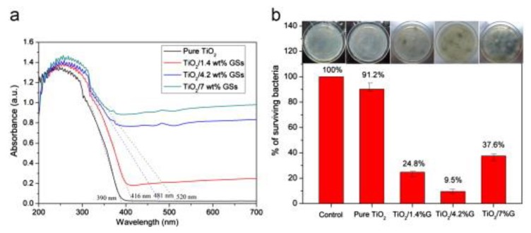
Nanocomposites of uniform TiO2 nanoparticles and graphene sheets (TiO2/GSs) have been fabricated via a facile redox reaction for antibacterial applications [88,89,90,91,92,93]. TiO2 nanoparticles were anchored on the surfaces of GSs by chemical bonds. Moreover, nanocomposites of TiO2/GSs exhibited broad absorption region from UV light to visible light. The photocatalysts of TiO2/GSs were used to kill E. coli under visible light illumination. Uniform TiO2 nanoparticles showed lower antibacterial capability in comparison with TiO2/GSs nanocomposites due to their large band gap, resulting in very low light absorption in the visible region (Figure 10). UV-Vis absorption spectra of pure TiO2 nanoparticles, TiO2/GSs with TiO2/1.4 wt%, TiO2/GSs with TiO2/4.2 wt% and TiO2/GSs with TiO2/7 wt% have shown in Figure 10a. Under visible light illumination for 12 h, viabilities of E. coli incubated with various TiO2-based nanocomposites have been respectively calculated as shown in Figure 10b. Under visible light illumination, the nanocomposites of TiO2/GSs with TiO2/4.2 wt% revealed the best antibacterial activity in comparison with TiO2/GSs with various weight ratios, including TiO2/1.4 wt% and TiO2/7 wt%. The superior antibacterial activity of TiO2/GSs nanocomposites may be explained by the increase of light absorption in visible region and effective separation of photo-generated electron-hole pairs because graphene could be applied as an electron acceptor and transporter. With the effective separation of light-generated pairs of electrons and holes, TiO2/GSs nanocomposites may produce more ROS, including ⋅OH and ⋅O2− for disinfection activity of E. coli. For the nanocomposites of TiO2/GSs with TiO2/7 wt%, the decrease of antibacterial activity may be ascribed to that the active TiO2 nanoparticles were covered by a large number of GSs, forming a shield of active sites on TiO2 nanoparticles.
Figure 10.
(a) UV-Vis absorption spectra of pure TiO2 nanoparticles, TiO2/GSs with TiO2/1.4 wt%, TiO2/GSs with TiO2/4.2 wt% and TiO2/GSs with TiO2/7 wt%. (b) Viabilities of E. coli incubated with various TiO2-based nanocomposites under visible light illumination for 12 h. The insets are the corresponding photographs of agar plates. Reproduced with permission from ref. [88]. Copyright © 2013, Elsevier.
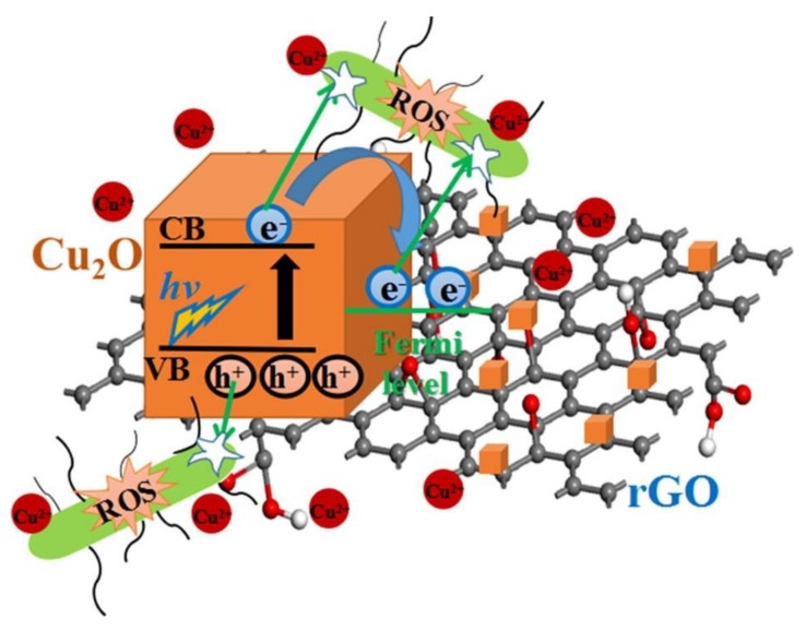
Heterostructured nanocomposites of reduced graphene oxide and cuprous oxide (rGO-Cu2O) have been prepared by the reduction of copper sulfate on graphene oxide for long-term antibacterial activities [94]. The P-type semiconductor of Cu2O may easily separate its electron-hole pairs under light condition. In this study, the fresh rGO-Cu2O nanocomposites generated more ROS compared to that of fresh rGO and Cu2O (Figure 11). The results indicated that the separation performance of photo-excited charges of Cu2O may be distinctly increased by the combination of Cu2O and rGO due to the improvement of interfacial charge transfer between Cu2O and rGO. The ROS antibacterial mechanism of rGO-Cu2O nanocomposites may be attributed to that the light-induced electrons were delivered from Cu2O to rGO to eliminate the recombination of the pairs of electrons and holes [95]. The photo-excited electrons and holes from Cu2O may be used for disinfection of bacteria [96]. Furthermore, rGO have played an important role for the acceptance of light-induced electrons from Cu2O to provide efficient charge transfer between the heterostructured nanocomposites of rGO-Cu2O and bacteria. The photo-excited electrons and holes may create intracellular ROS such as H2O2, ⋅OH and ⋅O2−. Eventually, the active substances of ROS could cause damage of nucleic acids, inactivation of intracellular protein, disability of the mitochondria and destruction of bacterial membrane.
Figure 11.
Reactive oxygen species (ROS) production caused by the heterostructured nanocomposites rGO-Cu2O under light illumination. Reproduced with permission from ref. [94]. Copyright © 2019, Elsevier.
Zinc oxide-selenium (ZnO-Se) heterojunction nanocomposites have been fabricated as antibacterial agents for S. aureus [97]. For the control, ZnO nanoparticles showed the zone of inhibition as 3.0 cm for S. aureus with visible light illumination. In the contrast, ZnO-Se nanocomposites revealed outstanding antibacterial activity with a zone of inhibition as 5 cm for S. aureus with visible light illumination. Furthermore, the bactericidal property of ZnO-Se nanocomposites remained for few days to inhibit the growth of S. aureus. The enhancement of antibacterial activity of ZnO-Se nanocomposites could be attributed to the increase of light-harvesting capability for sustainable production of ROS to kill S. aureus (Figure 12). To investigate the role of ROS in the antibacterial mechanism, electron spin resonance (ESR) was utilized to assess the types of ROS production from ZnO-Se nanocomposites. The results of ESR experiments demonstrated that ZnO-Se nanocomposite may produce ROS, including singlet oxygen and reactive OH species. Overall, the ZnO-Se heterojunction nanocomposites had long term enhancement of their antibacterial activity with visible light illumination and therefore, may be a potential antibacterial agent.
Figure 12.
Antibacterial mechanisms of ZnO-Se nanocomposites with visible light illumination. Reproduced with permission from ref. [97]. Copyright © 2020, Elsevier.
3. Challenges and Opportunities
In this review, we have compiled recent investigations of heterostructured nanomaterials as light-activated antibacterial agents in applications such as medicine, food safety, water sterilization and the textile industry (Table 1). These investigations have demonstrated that heterostructured nanomaterials may be high-performance antibacterial agents due to their synergistic effects under light irradiation. With synergistic effects, heterostructured nanomaterials exhibited excellent antibacterial activities based on the increase of ROS generation under light irradiation. Furthermore, the heterostructures of nanomaterials also inhibited the recombination of photon-induced electron/hole pairs for the increase of ROS formation to kill bacteria. Although various heterostructured nanomaterials have been shown to be light-activated antimicrobial agents, their antibacterial activities still need to be improved. For real clinical applications, the antibacterial efficiency of LAHNs should fit the requirement with a 4-log (99.99%) reduction in bacterial viability, which is one of the leading causes of nosocomial infections in the world. The first challenge to enhance the antimicrobial activity of LAHNs is to increase their synergistic effects. The development of LAHNs with novel compositions would be recommended. The second challenge to improve antibacterial activity for light-activated antimicrobial agents is to combine with other optical properties. For example, the photothermal effect is also used to kill bacteria with light irradiation. Therefore, with ROS generation and photothermal effects, LAHNs may provide better antimicrobial activities. The third challenge for light-activated antimicrobial agents is to increase their biocompatibility for the clinic application. To date, the cytotoxicity of LAHNs is still too high for clinic tests. However, in vitro and in vivo studies of light-activated antimicrobial agents of heterostructured nanomaterials are a critical step for future clinic application. Overall, to realize the antibacterial agents of LAHNs, great efforts are still needed for the improvement of their antibacterial activities. With intensive studies, we believe that antibacterial agents of LAHNs can be utilized as the important antibacterial agents in the near future.
Table 1.
Light-activated nanomaterials for antibacterial application in this review.
| Nanomaterials | Light harvester | Light Source | Bacteria | References |
|---|---|---|---|---|
| Ag-TiO2 | Ag | UV light | B. subtilis and P. putida | [68] |
| TiO2/AgVO3 | AgVO3 | visible light | E. coli | [75] |
| Ag/TiO2/cellulose | Ag | UV light | E. coli | [81] |
| Alginate/Au-TiO2 | Au | visible light | S. aureus and E. coli | [82] |
| Li-TiO2/LDPE | Li | visible light | S. aureus | [83] |
| Cu-TiO2 | Cu | visible light | E. coli | [84] |
| TiO2/α-Fe2O3 | α-Fe2O3 | visible light | E. coli | [85] |
| U,Bi-TiO2 | N and Bi | visible light | S. sanguinis and A. naeslundii | [86] |
| CTS/MTiO2 | melon | visible light | S. aureus | [87] |
| TiO2/GSs | TiO2 | visible light | E. coli | [88] |
| rGO-Cu2O | Cu2O | sunlight | S. aureus and E. coli | [94] |
| ZnO-Se | Se | visible light | S. aureus | [97] |
Funding
This work was supported by MOST 108-2113-M-038-003 and Taipei Medical University.
Conflicts of Interest
The authors declare no conflict of interest.
References
- 1.Luo J., Deng W., Yang F., Wu Z., Huang M., Gu M. Gold nanoparticles decorated graphene oxide/nanocellulose paper for nir laser-induced photothermal ablation of pathogenic bacteria. Carbohydr. Polym. 2018;198:206–214. doi: 10.1016/j.carbpol.2018.06.074. [DOI] [PubMed] [Google Scholar]
- 2.Teng C.P., Zhou T., Ye E., Liu S., Koh L.D., Low M., Loh X.J., Win K.Y., Zhang L., Han M.Y. Effective targeted photothermal ablation of multidrug resistant bacteria and their biofilms with nir-absorbing gold nanocrosses. Adv. Healthc. Mater. 2016;5:2122–2130. doi: 10.1002/adhm.201600346. [DOI] [PubMed] [Google Scholar]
- 3.Wang Y.-W., Fu Y.-Y., Wu L.-J., Li J., Yang H.-H., Chen G.-N. Targeted photothermal ablation of pathogenic bacterium, staphylococcus aureus, with nanoscale reduced graphene oxide. J. Mater. Chem. B. 2013;1:2496–2501. doi: 10.1039/c3tb20144a. [DOI] [PubMed] [Google Scholar]
- 4.Khan S.A., Singh A.K., Senapati D., Fan Z., Ray P.C. Bio-conjugated popcorn shaped gold nanoparticles for targeted photothermal killing of multiple drug resistant salmonella dt104. J. Mater. Chem. 2011;21:17705–17709. doi: 10.1039/c1jm13320a. [DOI] [Google Scholar]
- 5.Magiorakos A.P., Srinivasan A., Carey R.B., Carmeli Y., Falagas M.E., Giske C.G., Harbarth S., Hindler J.F., Kahlmeter G., Olsson-Liljequist B., et al. Multidrug-resistant, extensively drug-resistant and pandrug-resistant bacteria: An international expert proposal for interim standard definitions for acquired resistance. Clin. Microbiol. Infect. 2012;18:268–281. doi: 10.1111/j.1469-0691.2011.03570.x. [DOI] [PubMed] [Google Scholar]
- 6.O’Connell K.M.G., Hodgkinson J.T., Sore H.F., Welch M., Salmond G.P.C., Spring D.R. Combating multidrug-resistant bacteria: Current strategies for the discovery of novel antibacterials. Angew. Chem. Int. Ed. 2013;52:10706–10733. doi: 10.1002/anie.201209979. [DOI] [PubMed] [Google Scholar]
- 7.Simoes M., Bennett R.N., Rosa E.A.S. Understanding antimicrobial activities of phytochemicals against multidrug resistant bacteria and biofilms. Nat. Prod. Rep. 2009;26:746–757. doi: 10.1039/b821648g. [DOI] [PubMed] [Google Scholar]
- 8.Tacconelli E., Cataldo M.A., Dancer S.J., De Angelis G., Falcone M., Frank U., Kahlmeter G., Pan A., Petrosillo N., Rodriguez-Bano J., et al. Escmid guidelines for the management of the infection control measures to reduce transmission of multidrug-resistant gram-negative bacteria in hospitalized patients. Clin. Microbiol. Infect. 2014;20:1–55. doi: 10.1111/1469-0691.12427. [DOI] [PubMed] [Google Scholar]
- 9.Worthington R.J., Melander C. Combination approaches to combat multidrug-resistant bacteria. Trends Biotechnol. 2013;31:177–184. doi: 10.1016/j.tibtech.2012.12.006. [DOI] [PMC free article] [PubMed] [Google Scholar]
- 10.Lam S.J., O’Brien-Simpson N.M., Pantarat N., Sulistio A., Wong E.H.H., Chen Y.Y., Lenzo J.C., Holden J.A., Blencowe A., Reynolds E.C., et al. Combating multidrug-resistant gram-negative bacteria with structurally nanoengineered antimicrobial peptide polymers. Nat. Microbiol. 2016;1:16162. doi: 10.1038/nmicrobiol.2016.162. [DOI] [PubMed] [Google Scholar]
- 11.Hopkins K.L., Davies R.H., Threlfall E.J. Mechanisms of quinolone resistance in escherichia coli and salmonella: Recent developments. Int. J. Antimicrob. Agents. 2005;25:358–373. doi: 10.1016/j.ijantimicag.2005.02.006. [DOI] [PubMed] [Google Scholar]
- 12.Percival S.L., Bowler P.G., Russell D. Bacterial resistance to silver in wound care. J. Hosp. Infect. 2005;60:1–7. doi: 10.1016/j.jhin.2004.11.014. [DOI] [PubMed] [Google Scholar]
- 13.Sabuncu E., David J., Bernède-Bauduin C., Pépin S., Leroy M., Boëlle P.-Y., Watier L., Guillemot D. Significant reduction of antibiotic use in the community after a nationwide campaign in france, 2002–2007. PLoS Med. 2009;6:e1000084. doi: 10.1371/journal.pmed.1000084. [DOI] [PMC free article] [PubMed] [Google Scholar]
- 14.de Kraker M.E., Stewardson A.J., Harbarth S. Will 10 million people die a year due to antimicrobial resistance by 2050? PLoS Med. 2016;13:e1002184. doi: 10.1371/journal.pmed.1002184. [DOI] [PMC free article] [PubMed] [Google Scholar]
- 15.Li X.N., Robinson S.M., Gupta A., Saha K., Jiang Z.W., Moyano D.F., Sahar A., Riley M.A., Rotello V.M. Functional gold nanoparticles as potent antimicrobial agents against multi-drug-resistant bacteria. Acs Nano. 2014;8:10682–10686. doi: 10.1021/nn5042625. [DOI] [PMC free article] [PubMed] [Google Scholar]
- 16.Rai M.K., Deshmukh S.D., Ingle A.P., Gade A.K. Silver nanoparticles: The powerful nanoweapon against multidrug-resistant bacteria. J. Appl. Microbiol. 2012;112:841–852. doi: 10.1111/j.1365-2672.2012.05253.x. [DOI] [PubMed] [Google Scholar]
- 17.Priyadarshini S., Gopinath V., Priyadharsshini N.M., MubarakAli D., Velusamy P. Synthesis of anisotropic silver nanoparticles using novel strain, bacillus flexus and its biomedical application. Colloids Surf. B. 2013;102:232–237. doi: 10.1016/j.colsurfb.2012.08.018. [DOI] [PubMed] [Google Scholar]
- 18.Morones J.R., Elechiguerra J.L., Camacho A., Holt K., Kouri J.B., Ramirez J.T., Yacaman M.J. The bactericidal effect of silver nanoparticles. Nanotechnology. 2005;16:2346–2353. doi: 10.1088/0957-4484/16/10/059. [DOI] [PubMed] [Google Scholar]
- 19.Marambio-Jones C., Hoek E.M.V. A review of the antibacterial effects of silver nanomaterials and potential implications for human health and the environment. J. Nanopart. Res. 2010;12:1531–1551. doi: 10.1007/s11051-010-9900-y. [DOI] [Google Scholar]
- 20.Zuckerman J.E., Gritli I., Tolcher A., Heidel J.D., Lim D., Morgan R., Chmielowski B., Ribas A., Davis M.E., Yen Y. Correlating animal and human phase ia/ib clinical data with calaa-01, a targeted, polymer-based nanoparticle containing sirna. Proc. Natl. Acad. Sci. USA. 2014;111:11449–11454. doi: 10.1073/pnas.1411393111. [DOI] [PMC free article] [PubMed] [Google Scholar]
- 21.Gonavelli G., Chang C.C., Ling Y.C. Facile synthesis of smart magnetic graphene for safe drinking water: Heavy metal removal and disinfection control. ACS Sustain. Chem. Eng. 2013;1:462–472. doi: 10.1021/sc300112z. [DOI] [Google Scholar]
- 22.Lian H.Y., Hu M., Liu C.H., Yamauchi Y., Wu K.C.W. Highly biocompatible, hollow coordination polymer nanoparticles as cisplatin carriers for efficient intracellular drug delivery. Chem. Commun. 2012;48:5151–5153. doi: 10.1039/c2cc31708g. [DOI] [PubMed] [Google Scholar]
- 23.Yang Y.H., Liu C.H., Liang Y.H., Lin F.H., Wu K.C.W. Hollow mesoporous hydroxyapatite nanoparticles (hmhanps) with enhanced drug loading and ph-responsive release properties for intracellular drug delivery. J. Mater. Chem. B. 2013;1:2447–2450. doi: 10.1039/c3tb20365d. [DOI] [PubMed] [Google Scholar]
- 24.Fang J.Y., Fang C.L., Liu C.H., Su Y.H. Lipid nanoparticles as vehicles for topical psoralen delivery: Solid lipid nanoparticles (sln) versus nanostructured lipid carriers (NLC) Eur. J. Pharm. Biopharm. 2008;70:633–640. doi: 10.1016/j.ejpb.2008.05.008. [DOI] [PubMed] [Google Scholar]
- 25.Fang C.L., Al-Suwayeh S.A., Fang J.Y. Nanostructured lipid carriers (NLCS) for drug delivery and targeting. Recent Pat. Nanotechnol. 2013;7:41–55. doi: 10.2174/187221013804484827. [DOI] [PubMed] [Google Scholar]
- 26.Chen Y.P., Chen C.T., Hung Y., Chou C.M., Liu T.P., Liang M.R., Chen C.T., Mou C.Y. A new strategy for intracellular delivery of enzyme using mesoporous silica nanoparticles: Superoxide dismutase. J. Am. Chem. Soc. 2013;135:1516–1523. doi: 10.1021/ja3105208. [DOI] [PubMed] [Google Scholar]
- 27.Li C.H., Shen C.C., Cheng Y.W., Huang S.H., Wu C.C., Kao C.C., Liao J.W., Kang J.J. Organ biodistribution, clearance, and genotoxicity of orally administered zinc oxide nanoparticles in mice. Nanotoxicology. 2012;6:746–756. doi: 10.3109/17435390.2011.620717. [DOI] [PubMed] [Google Scholar]
- 28.Chen M.C., Mi F.L., Liao Z.X., Hsiao C.W., Sonaje K., Chung M.F., Hsu L.W., Sung H.W. Recent advances in chitosan-based nanoparticles for oral delivery of macromolecules. Adv. Drug Deliv. Rev. 2013;65:865–879. doi: 10.1016/j.addr.2012.10.010. [DOI] [PubMed] [Google Scholar]
- 29.Cho E.C., Chang-Jian C.W., Chen H.C., Chuang K.S., Zheng J.H., Hsiao Y.S., Lee K.C., Huang J.H. Robust multifunctional superhydrophobic coatings with enhanced water/oil separation, self-cleaning, anti-corrosion, and anti-biological adhesion. Chem. Eng. J. 2017;314:347–357. doi: 10.1016/j.cej.2016.11.145. [DOI] [Google Scholar]
- 30.Moon R.J., Martini A., Nairn J., Simonsen J., Youngblood J. Cellulose nanomaterials review: Structure, properties and nanocomposites. Chem. Soc. Rev. 2011;40:3941–3994. doi: 10.1039/c0cs00108b. [DOI] [PubMed] [Google Scholar]
- 31.Martin C.R. Nanomaterials - a membrane-based synthetic approach. Science. 1994;266:1961–1966. doi: 10.1126/science.266.5193.1961. [DOI] [PubMed] [Google Scholar]
- 32.Bruce P.G., Scrosati B., Tarascon J.M. Nanomaterials for rechargeable lithium batteries. Angew. Chem. Int. Ed. 2008;47:2930–2946. doi: 10.1002/anie.200702505. [DOI] [PubMed] [Google Scholar]
- 33.Chena S.H., Wu C.H., Fang A., Lin C.K. Effects of adding different morphological carbon nanomaterials on supercapacitive performance of sol-gel manganese oxide films. Ceram. Int. 2016;42:4797–4805. doi: 10.1016/j.ceramint.2015.11.165. [DOI] [Google Scholar]
- 34.Lin C.A.J., Chuang W.K., Huang Z.Y., Kang S.T., Chang C.Y., Chen C.T., Li J.L., Li J.K., Wang H.H., Kung F.C., et al. Rapid transformation of protein-caged nanomaterials into microbubbles as bimodal imaging agents. Acs Nano. 2012;6:5111–5121. doi: 10.1021/nn300768d. [DOI] [PubMed] [Google Scholar]
- 35.Le D., Wagner F., Takamiya M., Hsiao I.L., Alvaradejo G.G., Strahle U., Weiss C., Delaittre G. Straightforward access to biocompatible poly(2-oxazoline)-coated nanomaterials by polymerization-induced self-assembly. Chem. Commun. 2019;55:3741–3744. doi: 10.1039/C9CC00407F. [DOI] [PubMed] [Google Scholar]
- 36.Tian J., Leng Y.H., Zhao Z.H., Xia Y., Sang Y.H., Hao P., Zhan J., Li M.C., Liu H. Carbon quantum dots/hydrogenated TiO2 nanobelt heterostructures and their broad spectrum photocatalytic properties under uv, visible, and near-infrared irradiation. Nano Energy. 2015;11:419–427. doi: 10.1016/j.nanoen.2014.10.025. [DOI] [Google Scholar]
- 37.Carbone L., Cozzoli P.D. Colloidal heterostructured nanocrystals: Synthesis and growth mechanisms. Nano Today. 2010;5:449–493. doi: 10.1016/j.nantod.2010.08.006. [DOI] [Google Scholar]
- 38.Mai L.Q., Yang F., Zhao Y.L., Xu X., Xu L., Luo Y.Z. Hierarchical MnMoO4/CoMoO4 heterostructured nanowires with enhanced supercapacitor performance. Nat. Commun. 2011;2:381. doi: 10.1038/ncomms1387. [DOI] [PubMed] [Google Scholar]
- 39.Liu R., Duay J., Lee S.B. Redox exchange induced MnO2 nanoparticle enrichment in poly(3,4-ethylenedioxythiophene) nanowires for electrochemical energy storage. Acs Nano. 2010;4:4299–4307. doi: 10.1021/nn1010182. [DOI] [PubMed] [Google Scholar]
- 40.Donega C.D. Synthesis and properties of colloidal heteronanocrystals. Chem. Soc. Rev. 2011;40:1512–1546. doi: 10.1039/C0CS00055H. [DOI] [PubMed] [Google Scholar]
- 41.Wang Y.J., Wang Q.S., Zhan X.Y., Wang F.M., Safdar M., He J. Visible light driven type II heterostructures and their enhanced photocatalysis properties: A review. Nanoscale. 2013;5:8326–8339. doi: 10.1039/c3nr01577g. [DOI] [PubMed] [Google Scholar]
- 42.Kőrösi L., Bognár B., Horváth M., Schneider G., Kovács J., Scarpellini A., Castelli A., Colombo M., Prato M.J.A.C.B.E. Hydrothermal evolution of pf-co-doped TiO2 nanoparticles and their antibacterial activity against carbapenem-resistant klebsiella pneumoniae. Appl. Catal. B Environ. 2018;231:115–122. doi: 10.1016/j.apcatb.2018.03.012. [DOI] [Google Scholar]
- 43.Hu C., Lan Y.Q., Qu J.H., Hu X.X., Wang A.M. Ag/AgBr/TiO2 visible light photocatalyst for destruction of azodyes and bacteria. J. Phys. Chem. B. 2006;110:4066–4072. doi: 10.1021/jp0564400. [DOI] [PubMed] [Google Scholar]
- 44.O’Donovan P., Perrett C.M., Zhang X.H., Montaner B., Xu Y.Z., Harwood C.A., McGregor J.M., Walker S.L., Hanaoka F., Karran P. Azathioprine and uva light generate mutagenic oxidative DNA damage. Science. 2005;309:1871–1874. doi: 10.1126/science.1114233. [DOI] [PMC free article] [PubMed] [Google Scholar]
- 45.Hopkinson M.N., Sahoo B., Li J.L., Glorius F. Dual catalysis sees the light: Combining photoredox with organo-, acid, and transition-metal catalysis. Chem. Eur. J. 2014;20:3874–3886. doi: 10.1002/chem.201304823. [DOI] [PubMed] [Google Scholar]
- 46.Takahashi S., Badger M.R. Photoprotection in plants: A new light on photosystem ii damage. Trends Plant Sci. 2011;16:53–60. doi: 10.1016/j.tplants.2010.10.001. [DOI] [PubMed] [Google Scholar]
- 47.Pelaez M., Nolan N.T., Pillai S.C., Seery M.K., Falaras P., Kontos A.G., Dunlop P.S.M., Hamilton J.W.J., Byrne J.A., O’Shea K., et al. A review on the visible light active titanium dioxide photocatalysts for environmental applications. Appl. Catal. B Environ. 2012;125:331–349. doi: 10.1016/j.apcatb.2012.05.036. [DOI] [Google Scholar]
- 48.Li Z.R., Wakao S., Fischer B.B., Niyogi K.K. Sensing and responding to excess light. Annu. Rev. Plant Biol. 2009;60:239–260. doi: 10.1146/annurev.arplant.58.032806.103844. [DOI] [PubMed] [Google Scholar]
- 49.Sivaranjani V., Philominathan P. Synthesize of titanium dioxide nanoparticles using moringa oleifera leaves and evaluation of wound healing activity. Wound Med. 2016;12:1–5. doi: 10.1016/j.wndm.2015.11.002. [DOI] [Google Scholar]
- 50.Lee W.S., Park Y.-S., Cho Y.-K. Significantly enhanced antibacterial activity of TiO2 nanofibers with hierarchical nanostructures and controlled crystallinity. Analyst. 2015;140:616–622. doi: 10.1039/C4AN01682C. [DOI] [PubMed] [Google Scholar]
- 51.Jatoi A.W., Kim I.S., Ni Q.-Q. Cellulose acetate nanofibers embedded with agnps anchored TiO2 nanoparticles for long term excellent antibacterial applications. Carbohydr. Polym. 2019;207:640–649. doi: 10.1016/j.carbpol.2018.12.029. [DOI] [PubMed] [Google Scholar]
- 52.Hyde G.K., Stewart S.M., Scarel G., Parsons G.N., Shih C.C., Shih C.M., Lin S.J., Su Y.Y., Monteiro-Riviere N.A., Narayan R.J. Atomic layer deposition of titanium dioxide on cellulose acetate for enhanced hemostasis. Biotechnol. J. 2011;6:213–223. doi: 10.1002/biot.201000342. [DOI] [PMC free article] [PubMed] [Google Scholar]
- 53.Yang K.C., Zheng J.H., Chen Y.L., Lee K.C., Cho E.C. Carboxyfullerene decorated titanium dioxide nanomaterials for reactive oxygen species scavenging activities. RSC Adv. 2016;6:53025–53033. doi: 10.1039/C6RA09414G. [DOI] [Google Scholar]
- 54.Chen X.B., Liu L., Yu P.Y., Mao S.S. Increasing solar absorption for photocatalysis with black hydrogenated titanium dioxide nanocrystals. Science. 2011;331:746–750. doi: 10.1126/science.1200448. [DOI] [PubMed] [Google Scholar]
- 55.Carp O., Huisman C.L., Reller A. Photoinduced reactivity of titanium dioxide. Prog. Solid State Chem. 2004;32:33–177. doi: 10.1016/j.progsolidstchem.2004.08.001. [DOI] [Google Scholar]
- 56.Matsumoto Y., Murakami M., Shono T., Hasegawa T., Fukumura T., Kawasaki M., Ahmet P., Chikyow T., Koshihara S., Koinuma H. Room-temperature ferromagnetism in transparent transition metal-doped titanium dioxide. Science. 2001;291:854–856. doi: 10.1126/science.1056186. [DOI] [PubMed] [Google Scholar]
- 57.Diebold U. The surface science of titanium dioxide. Surf. Sci. Rep. 2003;48:53–229. doi: 10.1016/S0167-5729(02)00100-0. [DOI] [Google Scholar]
- 58.Chen X., Mao S.S. Titanium dioxide nanomaterials: Synthesis, properties, modifications, and applications. Chem. Rev. 2007;107:2891–2959. doi: 10.1021/cr0500535. [DOI] [PubMed] [Google Scholar]
- 59.Wodka D., Bielanska E., Socha R.P., Elzbieciak-Wodka M., Gurgul J., Nowak P., Warszyński P., Kumakiri I. Photocatalytic activity of titanium dioxide modified by silver nanoparticles. ACS Appl. Mater. Interfaces. 2010;2:1945–1953. doi: 10.1021/am1002684. [DOI] [PubMed] [Google Scholar]
- 60.Gupta K., Singh R., Pandey A., Pandey A. Photocatalytic antibacterial performance of TiO2 and Ag-doped TiO2 against s. Aureus. P. Aeruginosa and e. Coli. Beilstein J. Nanotechnol. 2013;4:345–351. doi: 10.3762/bjnano.4.40. [DOI] [PMC free article] [PubMed] [Google Scholar]
- 61.Adams L.K., Lyon D.Y., Alvarez P.J.J. Comparative eco-toxicity of nanoscale TiO2, SiO2, and ZnO water suspensions. Water Res. 2006;40:3527–3532. doi: 10.1016/j.watres.2006.08.004. [DOI] [PubMed] [Google Scholar]
- 62.Wang J., Guo Y.W., Liu B., Jin X.D., Liu L.J., Xu R., Kong Y.M., Wang B.X. Detection and analysis of reactive oxygen species (ros) generated by nano-sized TiO2 powder under ultrasonic irradiation and application in sonocatalytic degradation of organic dyes. Ultrason. Sonochem. 2011;18:177–183. doi: 10.1016/j.ultsonch.2010.05.002. [DOI] [PubMed] [Google Scholar]
- 63.Cho M., Chung H.M., Choi W.Y., Yoon J.Y. Different inactivation behaviors of ms-2 phage and escherichia coli in TiO2 photocatalytic disinfection. Appl. Environ. Microbiol. 2005;71:270–275. doi: 10.1128/AEM.71.1.270-275.2005. [DOI] [PMC free article] [PubMed] [Google Scholar]
- 64.Xiong D.W., Fang T., Yu L.P., Sima X.F., Zhu W.T. Effects of nano-scale TiO2, ZnO and their bulk counterparts on zebrafish: Acute toxicity, oxidative stress and oxidative damage. Sci. Total Environ. 2011;409:1444–1452. doi: 10.1016/j.scitotenv.2011.01.015. [DOI] [PubMed] [Google Scholar]
- 65.Battin T.J., Kammer F.V.D., Weilhartner A., Ottofuelling S., Hofmann T. Nanostructured TiO2: Transport behavior and effects on aquatic microbial communities under environmental conditions. Environ. Sci. Technol. 2009;43:8098–8104. doi: 10.1021/es9017046. [DOI] [PubMed] [Google Scholar]
- 66.Kim K.T., Klaine S.J., Cho J., Kim S.H., Kim S.D. Oxidative stress responses of daphnia magna exposed to TiO2 nanoparticles according to size fraction. Sci. Total Environ. 2010;408:2268–2272. doi: 10.1016/j.scitotenv.2010.01.041. [DOI] [PubMed] [Google Scholar]
- 67.George S., Pokhrel S., Ji Z.X., Henderson B.L., Xia T., Li L.J., Zink J.I., Nel A.E., Madler L. Role of fe doping in tuning the band gap of TiO2 for the photo-oxidation-induced cytotoxicity paradigm. J. Am. Chem. Soc. 2011;133:11270–11278. doi: 10.1021/ja202836s. [DOI] [PMC free article] [PubMed] [Google Scholar]
- 68.Li M., Noriega-Trevino M.E., Nino-Martinez N., Marambio-Jones C., Wang J., Damoiseaux R., Ruiz F., Hoek E.M. Synergistic bactericidal activity of Ag-TiO2 nanoparticles in both light and dark conditions. Environ. Sci. Technol. 2011;45:8989–8995. doi: 10.1021/es201675m. [DOI] [PubMed] [Google Scholar]
- 69.Billings E.A., Lee C.S., Owen K.A., D’Souza R.S., Ravichandran K.S., Casanova J.E. The adhesion gpcr bai1 mediates macrophage ros production and microbicidal activity against gram-negative bacteria. Sci. Signal. 2016;9:ra14. doi: 10.1126/scisignal.aac6250. [DOI] [PMC free article] [PubMed] [Google Scholar]
- 70.Dharmaraja A.T. Role of reactive oxygen species (ROS) in therapeutics and drug resistance in cancer and bacteria. J. Med. Chem. 2017;60:3221–3240. doi: 10.1021/acs.jmedchem.6b01243. [DOI] [PubMed] [Google Scholar]
- 71.Waldeck W., Heidenreich E., Mueller G., Wiessler M., Toth K., Braun K. Ros-mediated killing efficiency with visible light of bacteria carrying different red fluorochrome proteins. J. Photochem. Photobiol. B. 2012;109:28–33. doi: 10.1016/j.jphotobiol.2012.01.002. [DOI] [PubMed] [Google Scholar]
- 72.Ma H.B., Williams P.L., Diamond S.A. Ecotoxicity of manufactured zno nanoparticles - a review. Environ. Pollut. 2013;172:76–85. doi: 10.1016/j.envpol.2012.08.011. [DOI] [PubMed] [Google Scholar]
- 73.Choi O., Hu Z.Q. Size dependent and reactive oxygen species related nanosilver toxicity to nitrifying bacteria. Environ. Sci. Technol. 2008;42:4583–4588. doi: 10.1021/es703238h. [DOI] [PubMed] [Google Scholar]
- 74.Raghupathi K.R., Koodali R.T., Manna A.C. Size-dependent bacterial growth inhibition and mechanism of antibacterial activity of zinc oxide nanoparticles. Langmuir. 2011;27:4020–4028. doi: 10.1021/la104825u. [DOI] [PubMed] [Google Scholar]
- 75.Liu B., Han X., Mu L., Zhang J., Shi H. TiO2 nanospheres/AgVO3 quantum dots composite with enhanced visible light photocatalytic antibacterial activity. Mater. Lett. 2019;253:148–151. doi: 10.1016/j.matlet.2019.06.057. [DOI] [Google Scholar]
- 76.Ye M.Y., Zhao Z.H., Hu Z.F., Liu L.Q., Ji H.M., Shen Z.R., Ma T.Y. 0d/2d heterojunctions of vanadate quantum dots/graphitic carbon nitride nanosheets for enhanced visible-light-driven photocatalysis. Angew. Chem. Int. Ed. 2017;56:8407–8411. doi: 10.1002/anie.201611127. [DOI] [PubMed] [Google Scholar]
- 77.Lin X., Xu D., Jiang S.S., Xie F., Song M.S., Zhai H.J., Zhao L.N., Che G.B., Chang L.M. Graphitic carbon nitride nanocrystals decorated AgVO3 nanowires with enhanced visible-light photocatalytic activity. Catal. Commun. 2017;89:96–99. doi: 10.1016/j.catcom.2016.10.021. [DOI] [Google Scholar]
- 78.Klemm D., Heublein B., Fink H.P., Bohn A. Cellulose: Fascinating biopolymer and sustainable raw material. Angew. Chem. Int. Ed. 2005;44:3358–3393. doi: 10.1002/anie.200460587. [DOI] [PubMed] [Google Scholar]
- 79.Yang H.P., Yan R., Chen H.P., Lee D.H., Zheng C.G. Characteristics of hemicellulose, cellulose and lignin pyrolysis. Fuel. 2007;86:1781–1788. doi: 10.1016/j.fuel.2006.12.013. [DOI] [Google Scholar]
- 80.Bledzki A.K., Gassan J. Composites reinforced with cellulose based fibres. Prog. Polym. Sci. 1999;24:221–274. doi: 10.1016/S0079-6700(98)00018-5. [DOI] [Google Scholar]
- 81.Li Y., Tian J., Yang C., Hsiao B. Nanocomposite film containing fibrous cellulose scaffold and Ag/ TiO2 nanoparticles and its antibacterial activity. Polymers. 2018;10:1052. doi: 10.3390/polym10101052. [DOI] [PMC free article] [PubMed] [Google Scholar]
- 82.Tang S., Wang Z., Li P., Li W., Li C., Wang Y., Chu P. Degradable and photocatalytic antibacterial Au- TiO2/sodium alginate nanocomposite films for active food packaging. Nanomaterials. 2018;8:930. doi: 10.3390/nano8110930. [DOI] [PMC free article] [PubMed] [Google Scholar]
- 83.Basiron N., Sreekantan S., Akil H.M., Saharudin K.A., Harun N.H., Mydin R.B.S., Seeni A., Rahman N.R.A., Adam F., Iqbal A. Effect of Li- TiO2 nanoparticles incorporation in ldpe polymer nanocomposites for biocidal activity. Nano Struct. Nano Objects. 2019;19:100359. doi: 10.1016/j.nanoso.2019.100359. [DOI] [Google Scholar]
- 84.Mathew S., Ganguly P., Rhatigan S., Kumaravel V., Byrne C., Hinder S., Bartlett J., Nolan M., Pillai S. Cu-doped TiO2: Visible light assisted photocatalytic antimicrobial activity. Appl. Sci. 2018;8:2067. doi: 10.3390/app8112067. [DOI] [Google Scholar]
- 85.Akhavan O., Azimirad R. Photocatalytic property of Fe2O3 nanograin chains coated by TiO2 nanolayer in visible light irradiation. Appl. Catal. A Gen. 2009;369:77–82. doi: 10.1016/j.apcata.2009.09.001. [DOI] [Google Scholar]
- 86.Nagay B.E., Dini C., Cordeiro J.M., Ricomini-Filho A.P., de Avila E.D., Rangel E.C., da Cruz N.C., Barao V.A.R. Visible-light-induced photocatalytic and antibacterial activity of TiO2 codoped with nitrogen and bismuth: New perspective to control implant-biofilm-related disease. ACS Appl. Mater. Interfaces. 2019;11:18186–18202. doi: 10.1021/acsami.9b03311. [DOI] [PubMed] [Google Scholar]
- 87.Vélez-Peña E., Pérez-Obando J., Pais-Ospina D., Marín-Silva D.A., Pinotti A., Cánneva A., Donadelli J.A., Damonte L., Pizzio L.R., Osorio-Vargas P. Self-cleaning and antimicrobial photo-induced properties under indoor lighting irradiation of chitosan films containing melon/ TiO2 composites. Appl. Surf. Sci. 2019;508:144895. doi: 10.1016/j.apsusc.2019.144895. [DOI] [Google Scholar]
- 88.Cao B., Cao S., Dong P., Gao J., Wang J. High antibacterial activity of ultrafine TiO2/graphene sheets nanocomposites under visible light irradiation. Mater. Lett. 2013;93:349–352. doi: 10.1016/j.matlet.2012.11.136. [DOI] [Google Scholar]
- 89.Kamat P.V. Graphene-based nanoarchitectures. Anchoring semiconductor and metal nanoparticles on a two-dimensional carbon support. J. Phys. Chem. Lett. 2010;1:520–527. doi: 10.1021/jz900265j. [DOI] [Google Scholar]
- 90.Xiang Q.J., Yu J.G., Jaroniec M. Preparation and enhanced visible-light photocatalytic H-2-production activity of graphene/C3N4 composites. J. Phys. Chem. C. 2011;115:7355–7363. doi: 10.1021/jp200953k. [DOI] [Google Scholar]
- 91.Zhou W.J., Yin Z.Y., Du Y.P., Huang X., Zeng Z.Y., Fan Z.X., Liu H., Wang J.Y., Zhang H. Synthesis of few-layer MoS2 nanosheet-coated TiO2 nanobelt heterostructures for enhanced photocatalytic activities. Small. 2013;9:140–147. doi: 10.1002/smll.201201161. [DOI] [PubMed] [Google Scholar]
- 92.Zhang Y.H., Tang Z.R., Fu X.Z., Xu Y.J. TiO2-graphene nanocomposites for gas-phase photocatalytic degradation of volatile aromatic pollutant: Is tio2-graphene truly different from other TiO2-carbon composite materials? Acs Nano. 2010;4:7303–7314. doi: 10.1021/nn1024219. [DOI] [PubMed] [Google Scholar]
- 93.Williams G., Seger B., Kamat P.V. Tio2-graphene nanocomposites. Uv-assisted photocatalytic reduction of graphene oxide. Acs Nano. 2008;2:1487–1491. doi: 10.1021/nn800251f. [DOI] [PubMed] [Google Scholar]
- 94.Yang Z., Hao X., Chen S., Ma Z., Wang W., Wang C., Yue L., Sun H., Shao Q., Murugadoss V. Long-term antibacterial stable reduced graphene oxide nanocomposites loaded with cuprous oxide nanoparticles. J. Colloid Interface Sci. 2019;533:13–23. doi: 10.1016/j.jcis.2018.08.053. [DOI] [PubMed] [Google Scholar]
- 95.Wei Q., Wang Y., Qin H., Wu J., Lu Y., Chi H., Yang F., Zhou B., Yu H., Liu J. Construction of rGO wrapping octahedral Ag-Cu2O heterostructure for enhanced visible light photocatalytic activity. Appl. Catal. B Environ. 2018;227:132–144. doi: 10.1016/j.apcatb.2018.01.003. [DOI] [Google Scholar]
- 96.Wang R., Kong X., Zhang W., Zhu W., Huang L., Wang J., Zhang X., Liu X., Hu N., Suo Y. Mechanism insight into rapid photocatalytic disinfection of salmonella based on vanadate QDs-interspersed g-C3N4 heterostructures. Appl. Catal. B Environ. 2018;225:228–237. doi: 10.1016/j.apcatb.2017.11.060. [DOI] [Google Scholar]
- 97.Ahmad A., Ullah S., Ahmad W., Yuan Q., Taj R., Khan A.U., Rahman A.U., Khan U.A. Zinc oxide-selenium heterojunction composite: Synthesis, characterization and photo-induced antibacterial activity under visible light irradiation. J. Photochem. Photobiol. B. 2020;203:111743. doi: 10.1016/j.jphotobiol.2019.111743. [DOI] [PubMed] [Google Scholar]


