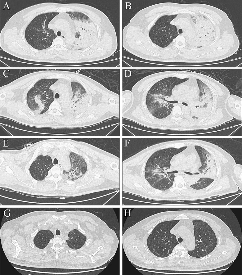
Fig. 2.

Serial chest computed tomography (CT) scans of a 43-year-old male farmer with severe psittacosis pneumonia. The initial CT scan (7 days after the onset) shows air-space consolidation with inflammatory exudation only appears in the superior lobe of left lung (a, b). The follow-up CT scan (16 days after the onset) shows exacerbation of the consolidated area in left lung and also in the middle and inferior lobes of right lung (c, d). On the follow-up CT scan (23 days after the onset), the area of consolidation has decreased (e, f) and 50 days after onset it has disappeared (g, h)
