Abstract
Background:
Pleomorphic adenoma (PA), mucoepidermoid carcinoma (MEC), and adenoid cystic carcinoma (AdCC) are the most common benign and malignant salivary gland tumors. Cyclooxygenase-2 (COX-2) is a key regulatory enzyme that its overexpression in various tumors is correlated with progression, metastasis, and apoptosis inhibition. Vascular endothelial growth factor (VEGF) is a potent angiogenic mediator that has an important role in neoplastic angiogenesis. The aim of this study was to immunohistochemically analyze the expression of COX-2 and VEGF and to compare the expression of benign and two malignant salivary gland tumors with varied structures.
Materials and Methods:
In this cross-sectional study, 90 specimens including 30 cases of each tumor were retrieved. Immunohistochemical staining of COX-2 and VEGF was performed for all the samples. The percentage of positive tumor cells and staining intensity was evaluated by two pathologists blindly. Data were analyzed by Chi-square and Gamma test and P < 0.05.
Results:
A statistically significant difference was noted between the expression and intensity of COX-2 and VEGF in PA, MEC, and AdCC (P < 0.05). A significant correlation was observed between COX-2 and VEGF expression in MEC and AdCC (P < 0.05). However, no significant correlation was found between the expression and intensity of COX-2 and VEGF with histologic grade and lymph node metastasis in MEC and AdCC (P < 0.05).
Conclusion:
High expression of VEGF and COX-2 in malignant tumors compared to PA suggested the role of both markers in malignant transformation. The significant correlation of VEGF expression with COX-2 may represent the role of COX-2 in tumor angiogenesis by modulating VEGF production.
Key Words: Adenoid cystic carcinoma, cyclooxygenase-2, mucoepidermoid carcinoma, pleomorphic adenoma, vascular endothelial growth factor
INTRODUCTION
Pleomorphic adenoma (PA) is the most common benign neoplasm with remarkable degree of morphological diversity. It usually occurs in the age range of 30–50 years. It presents with a minor preference in women.[1]
Mucoepidermoid carcinoma (MEC) is one of the most common salivary gland malignancies, mainly affecting parotid. The tumor occurs in the second to seventh decades of life and is also the most common malignant salivary gland tumor noted in children. MEC exhibits varied clinical presentations, that is, from a slow-growing mass to a destructive rapidly growing mass. The prognosis of MEC is usually related to clinical stage and histologic grade.[1]
Adenoid cystic carcinoma (AdCC) is one of the best-recognized salivary malignancies that can occur in any salivary gland site, but approximately 40%–45% develop within the minor salivary glands. AdCC is a persistent tumor that is prone to local recurrences and eventual distant metastasis.[1]
Many immunohistochemical studies in differential diagnosis of salivary gland tumors and identifying the prognosis of malignant salivary gland tumors have been published. However, few studies have focused on the expression of cyclooxygenase-2 (COX-2) and vascular endothelial growth factor (VEGF) and their significance. For example, CD44 expression in PA and carcinoma ex-PA and their adjacent normal salivary glands was evaluated.[2] Moreover, P63 expression was assessed in papillary cystadenoma and MEC of minor salivary glands.[3] These studies show the importance of finding immunohistochemistry markers in evaluating the prognosis of these head-and-neck tumors.
COX-2 is a key regulatory enzyme in the synthesis of prostaglandins in most tissues. The presence of COX-2 is usually associated with cellular activation including inflammation. Its overexpression has also been demonstrated in gastrointestinal tract, breast, lung, esophagus, pancreas, urinary bladder, prostate, and skin. COX-2 enzyme is not present in healthy tissues.[4] It seems that several significant processes for cancer development such as apoptosis and angiogenesis are influenced by COX-2.[5] On the other hand, the correlation of COX-2 overexpression and VEGF expression in head-and-neck cancer and oral squamous cell carcinoma has been demonstrated, but the correlation in salivary gland tumors is still elusive.[4,6]
Sakurai et al. showed that the expression of COX-2 in various histologic types of salivary gland adenoma and carcinoma was higher than normal salivary glands.[5]
VEGF is known as a powerful cytokine and a regulator of vasculogenesis and tumor angiogenesis in a number of malignancies. It is also related to vascular permeability and vasoactive molecule production.[7]
Lequerica-Fernández et al. and Fonseca et al. demonstrated that overexpression of VEGF in malignant salivary gland tumors might be associated with pathogenesis, progression, aggressiveness, and lymph node metastasis.[7,8]
The main aim of this study was to evaluate the combined immunohistochemical analysis of COX-2 with VEGF expression in PA, MEC, and AdCC of salivary glands.
MATERIALS AND METHODS
Specimen selection
The samples of this cross-sectional study were collected from 90 formalin-fixed, paraffin-embedded tissue blocks of PA, AdCC, and MEC obtained from the archives of the Pathology Department, Amir Alam Hospital, Tehran University of Medical Sciences, Tehran, Iran.
Thirty cases were diagnosed as PA, thirty cases as MEC, and thirty cases as AdCC. Hematoxylin and eosin-stained sections were used to confirm the diagnosis.
Clinicopathologic information on each case including age, sex, tumor location, and histologic grade was obtained from patient records and confirmed by reviewing the case slides. Cases without complete data, sufficient paraffin-embedded tumor material, appropriate fixation, incisional biopsy, and recurrent cases were excluded from the study.
Immunohistochemistry
4-μm sections were cut from all paraffin-embedded specimen blocks and mounted on silane-coated slides. The sections were deparaffinized with 100% xylene and rehydrated in graded ethanol series; they were immersed in Tris-buffered saline (TBS) of PH 6.0 and were heated in a microwave oven at 750 watts for antigen retrieval. After cooling into room temperature, the sections were incubated with primary antibodies: COX-2 (Monoclonal Mouse Anti-Human clone: SC-376861, Santa Cruz, USA) and VEGF (Polyclonal Rabbit Anti-Human clone: KLT9, Leica, USA) at 1:2000 for an hour through EnVision method. After washing in TBS, the sections were treated with a secondary antibody. DAB chromogen was applied to visualize the antibody and then counterstained with Mayer's hematoxylin. Ulcerative colitis and pyogenic granuloma were used as a positive control for COX-2 and VEGF, respectively.
Evaluation of immunohistochemistry
The COX-2 and VEGF immunoreactions in tumor cells were determined in 10 randomly selected fields by counting all positive cells (cytoplasmic staining) in each field according to the median index of positive cells obtained from 10 high-power fields and scored as follows:[9]0 (negative), 1%–25% (score 1), 26%–50% (score 2), 51%–75% (score 3), and 76%–100% (score 4). The intensity of staining was evaluated as follows: 0 = no positive cells, + = mild, ++ = moderate, and + 3 = strong.[4,6]
Histopathologic grade of AdCC samples was classified into tubular (Grade 1), cribriform (Grade 2), and solid (Grade 3) based on the histologic type, and the grade was identified.[10] MEC was categorized into low, intermediate, and high grade according to Auclair et al.[11] All the slides were evaluated by two pathologists, blindly and concurrently.
Statistical analysis
Statistical analysis was performed on the tabulated data using SPSS 18.0 software (SPSS Inc., Chicago, IL, USA). Chi-square test and Gamma test were used for data analysis. The significant level of all tests was set at P < 0.05.
RESULTS
The general characteristics of all patients included in this study are shown in Table 1.
Table 1.
Characteristics of all patients
| Variables | PA | MEC | AdCC |
|---|---|---|---|
| Sex | |||
| Male | 14 | 16 | 6 |
| Female | 16 | 14 | 24 |
| Age (mean) | 38.13±16.35 | 40.63±20.90 | 43.73±15.43 |
| Site of tumor | |||
| Palate | 18 | 1 | 2 |
| Parotid | 5 | 22 | 19 |
| Alveolar mucosa | 3 | 0 | 6 |
| Sublingual | 0 | 1 | 0 |
| submandibular | 1 | 1 | 1 |
| Tongue | 0 | 2 | 1 |
| Flour of the mouth | 0 | 1 | 0 |
| Cheek | 1 | 2 | 1 |
| Upper lip | 2 | 0 | 0 |
| Histopathological grade | |||
| High grade | 4 | ||
| Moderate grade | 7 | ||
| Low grade | 19 | ||
| Solid | 5 | ||
| Tubular | 6 | ||
| Cribriform | 19 | ||
| Size (cm) | |||
| Range | 1-4.5 | 1-7.5 | 0.7-7.5 |
| Mean | 2.02 | 5.19 | 3.01 |
| Lymph node metastasis | 3 | 2 |
PA: Pleomorphic adenoma; MEC: Mucoepidermoid carcinoma; AdCC: Adenoid cystic carcinoma
COX-2 was expressed in all cases of PA, MEC, and AdCC. In three groups, most cases were score 4 of expression. With respect to COX-2 intensity, 17 (56.7%) cases of PA, 27 (90%) cases of MEC, and 29 (96.7%) cases of AdCC showed strong intensity [Tables 2 and 3].
Table 2.
Cyclooxygenase-2 expression scores in present tumors
| Marker expression Tumor type | COX-2 expression (%) | Total | |||
|---|---|---|---|---|---|
| Score 1 (1-25) | Score 2 (26-50) | Score 3 (51-75) | Score 4 (76-100) | ||
| Tumor | |||||
| PA | |||||
| Count | 1 | 2 | 8 | 19 | 30 |
| Percentage | 3.3 | 6.7 | 26.7 | 63.3 | 100.0 |
| MEC | |||||
| Count | 0 | 0 | 0 | 30 | 30 |
| Percentage | 0.0 | 0.0 | 0.0 | 100.0 | 100.0 |
| AdCC | |||||
| Count | 1 | 2 | 4 | 23 | 30 |
| Percentage | 3.3 | 6.7 | 13.3 | 76.7 | 100.0 |
| Total | |||||
| Count | 2 | 4 | 12 | 72 | 90 |
| Percentage | 2.2 | 4.4 | 13.3 | 80.0 | 100.0 |
PA: Pleomorphic adenoma; MEC: Mucoepidermoid carcinoma; AdCC: Adenoid cystic carcinoma; COX-2: Cyclooxygenase-2
Table 3.
Cyclooxygenase-2 intensity in pleomorphic adenoma, mucoepidermoid carcinoma, and adenoid cystic carcinoma
| Marker intensity Tumor type | COX-2 intensity |
Total | ||
|---|---|---|---|---|
| Mild | Moderate | Strong | ||
| Tumor | ||||
| PA | ||||
| Count | 2 | 11 | 17 | 30 |
| Percentage | 6.7 | 36.7 | 56.7 | 100.0 |
| MEC | ||||
| Count | 0 | 3 | 27 | 30 |
| Percentage | 0.0 | 10.0 | 90.0 | 100.0 |
| AdCC | ||||
| Count | 0 | 1 | 29 | 30 |
| Percentage | 0.0 | 3.3 | 96.7 | 100.0 |
| Total | ||||
| Count | 2 | 15 | 73 | 90 |
| Percentage | 2.2 | 16.7 | 81.1 | 100.0 |
COX-2: Cyclooxygenase-2; PA: Pleomorphic adenoma; MEC: Mucoepidermoid carcinoma; AdCC: Adenoid cystic carcinoma
Chi-square test showed a significant difference between the intensity and expression of COX-2 in PA, MEC, and AdCC (P < 0.001).
Indeed, COX-2 expression showed a significant difference between MEC and AdCC (P = 0.011); however, there was no significant difference in COX-2 intensity between MEC and AdCC (P = 0.612).
VEGF expression was observed in all cases of PA, MEC, and AdCC. Twenty-three (76.7%) cases of PA, 29 (96.7%) cases of MEC, and 21 (70%) cases of AdCC exhibited score 4 of expression.
Considering VEGF intensity, 12 (40.0%) cases of PA, 27 (90%) cases of MEC, and 29 (96.7%) cases of AdCC showed strong intensity [Tables 4 and 5].
Table 4.
Vascular endothelial growth factor expression in pleomorphic adenoma, mucoepidermoid carcinoma, and adenoid cystic carcinoma
| Marker expression Tumor type | VEGF expression |
Total | |||
|---|---|---|---|---|---|
| 1-25 | 26-50 | 51-75 | 76-100 | ||
| Tumor | |||||
| PA | |||||
| Count | 3 | 1 | 3 | 23 | 30 |
| Percentage | 10.0 | 3.3 | 10.0 | 76.7 | 100.0 |
| MEC | |||||
| Count | 1 | 0 | 0 | 29 | 30 |
| Percentage | 3.3 | 0.0 | 0.0 | 96.7 | 100.0 |
| AdCC | |||||
| Count | 2 | 1 | 6 | 21 | 30 |
| Percentage | 6.7 | 3.3 | 20.0 | 70.0 | 100.0 |
| Total | |||||
| Count | 6 | 2 | 9 | 73 | 90 |
| Percentage | 6.7 | 2.2 | 10.0 | 81.1 | 100.0 |
VEGF: Vascular endothelial growth factor; PA: Pleomorphic adenoma; MEC: Mucoepidermoid carcinoma; AdCC: Adenoid cystic carcinoma
Table 5.
Vascular endothelial growth factor intensity in pleomorphic adenoma, mucoepidermoid carcinoma, and adenoid cystic carcinoma
| Marker intensity Tumor type | VEGF intensity |
Total | ||
|---|---|---|---|---|
| Mild | Moderate | Strong | ||
| Tumor | ||||
| PA | ||||
| Count | 2 | 16 | 12 | 30 |
| Percentage | 6.7 | 53.3 | 40.0 | 100.0 |
| MEC | ||||
| Count | 0 | 3 | 27 | 30 |
| Percentage | 0.0 | 10.0 | 90.0 | 100.0 |
| AdCC | ||||
| Count | 0 | 1 | 29 | 30 |
| Percentage | 0.0 | 3.3 | 96.7 | 100.0 |
| Total | ||||
| Count | 2 | 20 | 68 | 90 |
| Percentage | 2.2 | 22.2 | 75.6 | 100.0 |
VEGF: Vascular endothelial growth factor; PA: Pleomorphic adenoma; MEC: Mucoepidermoid carcinoma; AdCC: Adenoid cystic carcinoma
Data analysis showed a significant difference in VEGF intensity between three groups (P < 0.001).
Moreover, VEGF expression in MEC and AdCC showed a significant difference (P = 0.009). No significant relationship was observed between COX-2 and VEGF expression and intensity with histologic grade (P > 0.05) and lymph node metastasis (P > 0.05) in MEC and AdCC.
Gamma test showed a significant correlation between VEGF and COX-2 expression in PA (P = 0.03). Likewise, the correlation of VEGF and COX-2 intensity was seen in both PA (P = 0.016) and MEC (P = 0.001). No significant correlation was found between VEGF and COX-2 expression and intensity in AdCC (P > 0.05).
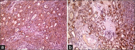
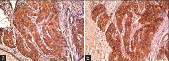
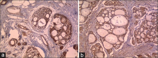
Figures 1-3 demonstrate the expression of VEGF and COX-2 in PA, MEC, and AdCC, respectively.
Figure 1.

Strong cytoplasmic expression of vascular endothelial growth factor (a) and cyclooxygenase-2 (b) in pleomorphic adenoma, ×200.
Figure 3.

Strong cytoplasmic expression of vascular endothelial growth factor (a) and cyclooxygenase-2 (b) in mucoepidermoid carcinoma, ×200.
Figure 2.

Strong cytoplasmic expression of vascular endothelial growth factor (a) and cyclooxygenase-2 (b) in adenoid cystic carcinoma, ×200.
DISCUSSION
Evaluation of COX-2 expression with invasive behavior in salivary gland malignancies has been reported. Moreover, high expression of VEGF correlated with lymph node metastasis in salivary gland carcinomas.[7] So far, comparison of COX-2 and VEGF expression in malignant and benign salivary tumors has not been studied. In this study, we studied the comparison of expression of COX-2 and VEGF in 30 samples of PA, 30 samples of MEC, and 30 samples of AdCC and their association with histopathological grade and lymph node metastasis.
The average age of patients with PA, MEC, and AdCC was 38.13, 40.63, and 43.7 years, respectively, which was similar to the studies of Fonseca et al.,[8] Aoki et al.,[12] Cho et al.,[13] and Merza[14] and inconsistent with Zyada et al.[15] and Stárek et al.[16]
In our study, the most common site of PA was palate which was consistent with the studies of Aoki et al.[12] and in contrast with the studies of Cho et al.,[13] Merza,[14] and Faur et al.[17] The most common location of MEC and AdCC was parotid which was consistent with the studies of Lequerica-Fernández et al.,[7] Fonseca et al.,[8] Cho et al.,[13] Zyada et al.,[15] Stárek et al.,[16] and Faur et al.[17] and was inconsistent with the study of Lim et al.[9]
In this study, immunohistochemical expression of COX-2 was seen in all samples of PA, MEC, and AdCC. Score 4 of COX-2 expression was 100% in MEC, 76.7% in AdCC, and 63.3% in PA. These results were consistent with the studies of Sakurai et al.,[5] Zyada et al.,[15] and Yi et al.[18]29 (96.7%) samples of AdCC, 27 (90%) samples of MEC, and 17 (56.7%) samples of PA exhibited strong COX-2 intensity.
In the present study, the expression of VEGF was observed in all samples of PA, MEC, and AdCC, which was in score 4 in most samples. In 27 (90%) of MEC, 29 (96.7%) of AdCC, and 12 (40%) of PA samples, VEGF intensity was strong. These results were consistent with the studies of Fonseca et al.,[8] Faur et al.,[17] Lim et al.,[9] Ou Yang et al.,[19] and Gupta et al.[20] Our results about COX-2 and VEGF expression were in contrast to Cho et al.,[13] Merza,[14] Rocha Tenorio et al.,[21] Lequerica-Fernández et al.,[7] and Li et al.[22] studies. This discrepancy may be attributed to different antibody manufactures and incubation time of primary antibody.
In our study, COX-2 expression in MEC and AdCC was higher than PA (P < 0.001). This result was consistent with the studies by Sakurai et al.,[5] Cho et al.,[13] Merza,[14] and Yi et al.[18] In this study, the intensity of VEGF and COX-2 in malignant tumors was higher than the benign tumor which was consistent with Sakurai et al.'s[5] study.
In this study, from 30 samples of MEC, 19, 7, and 4 cases were low, intermediate, and high grade, respectively. The expression of COX-2 was score 4 in all samples and for VEGF expression 29 samples were score 4. There was no significant relationship between histopathological grade of MEC and expression of VEGF and COX-2. This finding was consistent with the studies of Cho et al.[13], Zyada et al.[15] and Li et al.[22] and contrary to the study of Lim et al.[9] The difference may be due to different scoring methods.
In the present study, from 19 samples of AdCC with cribriform pattern, 13 samples were score 4 for COX-2 and VEGF expression. All samples of AdCC with tubular pattern and 4 samples of AdCC with solid pattern were score 4 for COX-2 expression, and 5 samples of AdCC with tubular pattern and 3 samples of AdCC with solid pattern were score 4 for VEGF expression. There was no significant relationship between the expression of COX-2 and VEGF with histologic grade in AdCC. This result was consistent with Lim et al.'s[9] study and in contrary to Li et al.[22] study.
In our study, three samples of MEC showed lymph node metastasis and all of them were score 3 for COX-2 and VEGF expression. Chi-square test showed no statistically significant difference between the expression of both markers and lymph node metastasis in MEC (P > 0.05). This finding was consistent with the study of Ou Yang et al.[19] and in contrary to the studies of Lequerica-Fernández et al.[7] and Zyada et al.[15] Furthermore, only two samples of AdCC showed lymph node metastasis and both of them were score 4 for VEGFexpression, and, in one sample, the expression of COX-2 was score 4. With Chi-square test, no significant difference was observed. This finding was in contrary to the studies of Lequerica-Fernández et al.[7] and Li et al.[22] As the number of samples with metastasis in our study was few, it seems that there could not reach a proper conclusion.
In this study, the coexpression of VEGF and COX-2 in salivary gland tumors was examined, in which there was a significant difference between the expression and intensity of both markers in PA and intensity of them in MEC, but no significant correlation was observed in AdCC. According to this study and Baghban et al.'s study,[4] COX-2 has probably a synergistic effect with VEGF in angiogenesis and progression of malignancies.
CONCLUSION
The high expression of VEGF and COX-2 in malignant tumors comparing PA suggests the role of both markers in malignant transformation. The significant correlation of VEGF expression with COX-2 may represent the role of COX-2 in tumor angiogenesis by modulating VEGF production.
Financial support and sponsorship
Nil.
Conflicts of interest
The authors of this manuscript declare that they have no conflicts of interest, real or perceived, financial or non-financial in this article.
REFERENCES
- 1.Neville BW, Damm DD, Allen CM, Bouguot JE. Oral & Maxillofacial Pathology. (4th ed) 2016:444–8. 454–7, 462–4. [Google Scholar]
- 2.Shamloo N, Taghavi N, Yazdani F, Shalpoush S, Ahmadi S. CD44 expression in pleomorphic adenoma, carcinoma ex pleomorphic adenoma and their adjacent normal salivary glands. Dent Res J (Isfahan) 2018;15:361–6. [PMC free article] [PubMed] [Google Scholar]
- 3.Fonseca FP, de Andrade BA, Lopes MA, Pontes HA, Vargas PA, de Almeida OP, et al. P63 expression in papillary cystadenoma and mucoepidermoid carcinoma of minor salivary glands. Oral Surg Oral Med Oral Pathol Oral Radiol. 2013;115:79–86. doi: 10.1016/j.oooo.2012.09.005. [DOI] [PubMed] [Google Scholar]
- 4.Akbarzadeh Baghban A, Taghavi N, Shahla M. Combined analysis of vascular endothelial growth factor expression with cyclooxygenase-2 and mast cell density in oral squamous cell carcinoma. Pathobiology. 2017;84:80–6. doi: 10.1159/000447778. [DOI] [PubMed] [Google Scholar]
- 5.Sakurai K, Urade M, Noguchi K, Kishimoto H, Ishibashi M, Yasoshima H, et al. Increased expression of cyclooxygenase-2 in human salivary gland tumors. Pathol Int. 2001;51:762–9. doi: 10.1046/j.1440-1827.2001.01280.x. [DOI] [PubMed] [Google Scholar]
- 6.Gallo O, Franchi A, Magnelli L, Sardi I, Vannacci A, Boddi V, et al. Cyclooxygenase-2 pathway correlates with VEGF expression in head and neck cancer. Implications for tumor angiogenesis and metastasis. Neoplasia. 2001;3:53–61. doi: 10.1038/sj.neo.7900127. [DOI] [PMC free article] [PubMed] [Google Scholar]
- 7.Lequerica-Fernández P, Astudillo A, de Vicente JC. Expression of vascular endothelial growth factor in salivary gland carcinomas correlates with lymph node metastasis. Anticancer Res. 2007;27:3661–6. [PubMed] [Google Scholar]
- 8.Fonseca FP, Basso MP, Mariano FV, Kowalski LP, Lopes MA, Martins MD, et al. Vascular endothelial growth factor immunoexpression is increased in malignant salivary gland tumors. Ann Diagn Pathol. 2015;19:169–74. doi: 10.1016/j.anndiagpath.2015.03.010. [DOI] [PubMed] [Google Scholar]
- 9.Lim JJ, Kang S, Lee MR, Pai HK, Yoon HJ, Lee JI, et al. Expression of vascular endothelial growth factor in salivary gland carcinomas and its relation to p53, Ki-67 and prognosis. J Oral Pathol Med. 2003;32:552–61. [PubMed] [Google Scholar]
- 10.Gnepp DR. Diagnostic Surgical Pathology of the Head and Neck. Philadelphia: Saunders; 2009. [Google Scholar]
- 11.Auclair PL, Goode RK, Ellis GL. Mucoepidermoid carcinoma of intraoral salivary glands, Cancer 69:2021–2030. 1992 doi: 10.1002/1097-0142(19920415)69:8<2021::aid-cncr2820690803>3.0.co;2-7. [DOI] [PubMed] [Google Scholar]
- 12.Aoki T, Tsukinoki K, Kurabayashi H, Sasaki M, Yasuda M, Ota Y, et al. Hepatocyte growth factor expression correlates with cyclooxygenase-2 pathway in human salivary gland tumors. Oral Oncol. 2006;42:51–6. doi: 10.1016/j.oraloncology.2005.06.012. [DOI] [PubMed] [Google Scholar]
- 13.Cho NP, Han HS, Soh Y, Son HJ. Overexpression of cyclooxygenase-2 correlates with cytoplasmic HuR expression in salivary mucoepidermoid carcinoma but not in pleomorphic adenoma. J Oral Pathol Med. 2007;36:297–303. doi: 10.1111/j.1600-0714.2007.00526.x. [DOI] [PubMed] [Google Scholar]
- 14.Merza MS. Comparative imuunohistochemical analysis of K-RAS oncogene and cyclooxygenase 2 enzyme expression in pleomorphic adenoma and adenoid cystic carcinoma of salivary glands. Eur Sci J. 2013;9:1857–7881. [Google Scholar]
- 15.Zyada MM, Grawish ME, Elsabaa HM. Predictive value of cyclooxygenase 2 and Bcl-2 for cervical lymph node metastasis in mucoepidermoid carcinoma. Ann Diagn Pathol. 2009;13:313–21. doi: 10.1016/j.anndiagpath.2009.06.003. [DOI] [PubMed] [Google Scholar]
- 16.Stárek I, Salzman R, Kučerová L, Skálová A, Hauer L. Expression of VEGF-C/-D and lymphangiogenesis in salivary adenoid cystic carcinoma. Pathol Res Pract. 2015;211:759–65. doi: 10.1016/j.prp.2015.07.001. [DOI] [PubMed] [Google Scholar]
- 17.Faur AC, Lazar E, Cornianu M. Vascular endothelial growth factor (VEGF) expression and microvascular density in salivary gland tumours. APMIS. 2014;122:418–26. doi: 10.1111/apm.12160. [DOI] [PubMed] [Google Scholar]
- 18.Yi Z, Zhong-Cheng G, Xiong-Hong N, Hong, Jian-Hua Z, Zhao-Quan L. Expression of COX-2 and EZH2 in mucoepidermoid carcinoma of salivary glands. J Logist Univ CAPF (Med Sci) 2012;21(7):506–508. 512, 582. [Google Scholar]
- 19.Ou Yang KX, Liang J, Huang ZQ. Association of clinicopathologic parameters with the expression of inducible nitric oxide synthase and vascular endothelial growth factor in mucoepidermoid carcinoma. Oral Dis. 2011;17:590–6. doi: 10.1111/j.1601-0825.2011.01813.x. [DOI] [PubMed] [Google Scholar]
- 20.Gupta B, Chandra S, Raj V, Gupta V. Immunohistochemical expression of vascular endothelial growth factor in orofacial lesions – A review. J Oral Biol Craniofac Res. 2016;6:231–6. doi: 10.1016/j.jobcr.2016.01.006. [DOI] [PMC free article] [PubMed] [Google Scholar]
- 21.Rocha Tenorio JD, Da Silva LP, Ferreira Do Nascimento GJ, Medeiros De Souza GF, Veras Sobral AP. Immunohistochemical study of cyclin D1, COX-2, and PCNA in pleomorphic adenomas. Oral Surg Oral Med Oral Pathol Oral Radiol. 2014;117:212. [Google Scholar]
- 22.Li Z, Tang P, Xu Z. Clinico-pathological significance of microvascular density and vascular endothelial growth factor expression in adenoid cyctic carcinoma of salivary glands. Chin J Stomatol. 2001;36:212–4. [PubMed] [Google Scholar]


