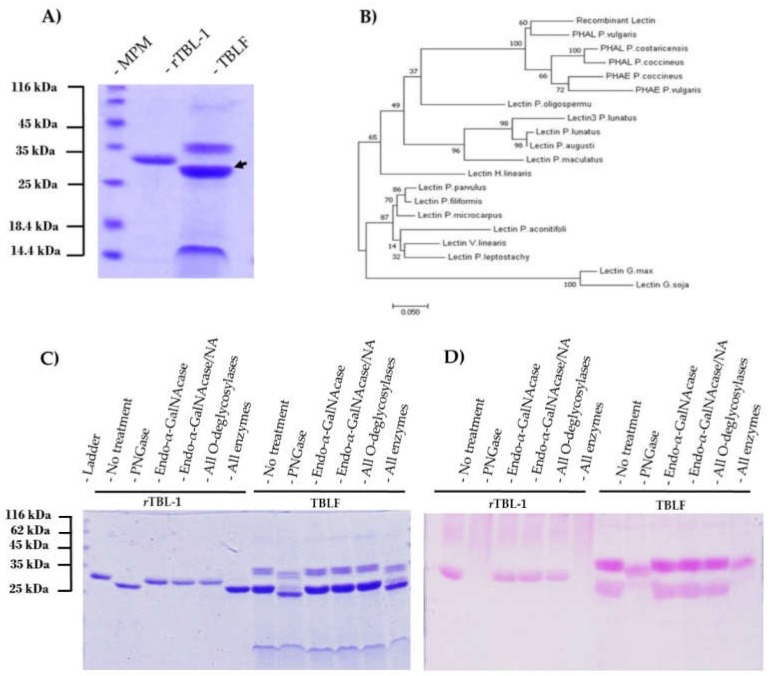
Figure 3.
Molecular comparison of recombinant and native TBL-1: (A) SDS-PAGE electrophoresis of rTBL-1 and Tepary Bean Lectin Fraction (TBLF) (gels stained for total protein). The black arrow shows native TBL-1. (B) Comparative phylogenetic sequence analysis of several legume lectins. (C) SDS-PAGE of rTBL-1 and Tepary bean lectin fraction (TBLF) +/− deglycosylation treatments. Lane 1, rTBL-1 no enzyme control; lanes 2–5, rTBL-1 treated with PNGaseF, endo-α-GalNAcase, endo-α-GalNAcase, and neuraminidase (NA), respectively; lane 6, rTBL-1 treated with all O-deglycosylases (β1,4-galactosidase, endo-α-GalNAcase, NA, and β-N-acetylglucosaminidase); lane 7, rTBL-1 treated with all deglycosylases (N-glycosidase F, β1,4-galactosidase, endo-α-GalNAcase, NA, and β-N-acetylglucosaminidase); lane 8, TBLF without treatment; lanes 9–11 TBLF treated with PNGaseF, endo-α-GalNAcase, endo-α-GalNAcase, and NA, respectively; lane 12, TBLF treated with all O-desglycosylases (β1,4-galactosidase, endo-α-GlcNAcase, NA, and β-N-acetylglucosaminidase); lane 13, TBLF treated with all deglycosylases (N-glycosidase-F, β1,4-galactosidase, endo-α-GalNAcase, NA, and β-N-Acetylglucosaminidase). (D) SDS-PAGE of rTBL-1 and TBLF +/– deglycosylation treatments stained with Schiff-PAS reagent for the detection of glycoproteins. Sample arrangement is as described for SDS-PAGE gel.

