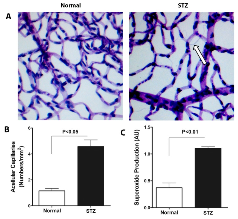
Figure 4.
Evaluation of acellular capillary formation and superoxide anion generation in T1D mice. Representative images of trypsin-digested retinal vascular preparations from 4-month old control mice and 4-month old mice with 2-month duration of T1D (A) The arrow indicates an acellular capillary. Quantitative measurement of acellular capillaries was significantly increased in diabetic eyes (n = 6). (B) STZ-induced diabetic retina showed >1.5 fold increase in superoxide anion levels, p < 0.01 (C) The errors bars represent SEM.

