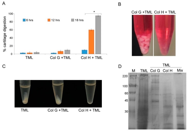
Figure 1.
Performance of ColG and ColH in tissue disgregation. (A) Cartilage dissociation assay using ColG and ColH in the presence of TML at different times (6, 12, and 18 h). (B) Cartilage digested with Col H or G plus TML after 18 h of treatment. (C) Samples containing powered cartilage treated 24 h with only TML, ColG plus TML, or ColH plus TML. (D) SDS PAGE of protein extracted from cartilage digested with thermolysin (TLM) alone or added of ColG, ColH, or both ColG and H (Mix). M = High molecular weight marker. The data represent the mean ± SD of three independent experiments.

