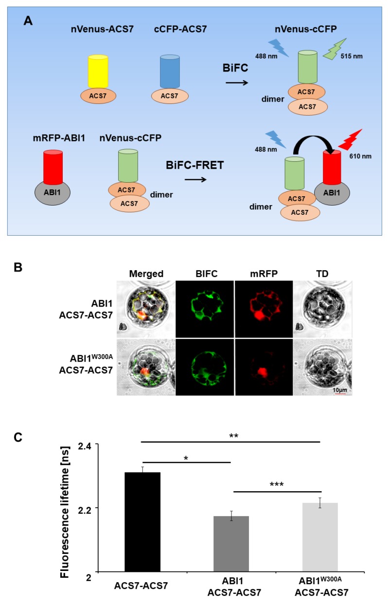
Figure 5.
Multicolor BiFC–FRET–FLIM analysis. (A) Summary of mcBiFC–FRET–FLIM experiment. Interaction of nVenus–ACS7 and cCFP–ACS7 results in ACS7 dimer reconstruction (nVenus–cCFP) and green light emission (515 nm). Reconstructed ACS7 dimer (nVenus–cCFP donor) excited by 488 nm laser light transfers energy to ABI1–RFP; (B) multicolor BiFC–FRET–FLIM analyses of protein interactions between the ACS7 homodimer and WT ABI1 and ABI1 W300A mutant protein in A. thaliana protoplasts. (C) Co-expression of nVenus–ACS7 and cCFP–ACS7 in protoplasts leads to reconstruction of fluorescent nVenus–cGFP protein by BiFC due to the formation of the ACS7 homodimer. This reconstructed nVenus-cCFP acts as donor. The acceptor mRFP is fused to ABI1 or ABI1 W300A. Fluorescence lifetime of the donor molecule was measured in picoseconds (ps). Error bars indicate the SD (standard deviation, n > 10), and the asterisk indicates a significant difference between the sample in the presence and absence of an acceptor (*p = 0.004; ** p = 0.014; *** p < 0.00001). Mean value of reconstructed nVenus-cCFP lifetime is Tamp: 2.28 ns. χ2 ~1 was considered a perfect fit; scale bar, 10 µm.

