Abstract
Objectives:
To determine the secondary metabolites from Verbascum mucronatum Lam. and evaluate their antimicrobial activity.
Materials and Methods:
Antimicrobial activities of the isolated metabolites were determined using broth microdilutions against the bacteria (Escherichia coli ATCC 25922, Enterococcus faecalis ATCC 29212, Pseudomonas aeruginosa ATCC 27853, Staphylococcus aureus ATCC 29213) and fungi (Candida albicans ATCC 90028, Candida krusei ATCC 6258, Candida parapsilosis ATCC 90018).
Results:
Four iridoid glycosides; ajugol (1), aucubin (2), lasianthoside I (3), catalpol (4), two triterpenic saponins; ilwensisaponin C (5), ilwensisaponin A (=mimengoside A) (6), and one phenylethanoid glycoside; verbascoside (=acteoside) (7) were isolated from the water soluble parts of the methanolic extract gained flowery parts of V. mucronatum Lam.
Conclusion:
Within the obtained compounds, ajugol and ilwensisaponin A showed moderate antimicrobial activity, especially against fungi.
Keywords: Scrophulariaceae, Verbascum mucronatum Lam., secondary metabolites, antimicrobial activity
INTRODUCTION
Verbascum is a widespread genus of the family Scrophulariaceae, which comprises more than 300 species of the world’s flora.1 This genus is represented by 233 species, 196 of which are endemic in Turkish flora.2,3,4 Infusions prepared with the leaves and flowers of Verbascum species have been used as an expectorant and mucolytic5 wound healer6 for the treatment of hemorrhoids and rheumatism7 in folk medicine. Turker and Camper8 showed that Klebsiella pneumoniae and Staphylococcus aureus showed sensitivity to Mullein (Verbascum thapsus), which may explain why Mullein is used in folk medicine to treat respiratory disorders (caused by K. pneumoniae and S. aureus) and urinary tract infections (caused by K. pneumoniae). Antibacterial and antifungal activities of Verbascum L. species have been previously reviewed and the activity of the genus against several bacteria and fungi has been revealed.9 The antimicrobial activity of Verbascum mucronatum has also been determined using disc diffusion tests by our research group.10 In addition, V. mucronatum Lam. has been used as a Hemostatic in Turkish traditional medicine.11
Previous investigations on Turkish Verbascum L. species by our research group led to the isolation and characterization of a number of secondary metabolites such as iridoids, monoterpene glucosides, saponins, phenylethanoids, neolignans, and flavonoid glycosides.12,13,14,15,16 As a part of our ongoing studies on the secondary metabolites of Verbascum L. species, we have now investigated the methanolic extract of the flowery parts of V. mucronatum, and isolated four iridoids; ajugol (1), aucubin (2), lasianthoside I (3), catalpol (4), two saponins; ilwensisaponin C (5) and ilwensisaponin A (6), along with a phenylethanoid glycoside, verbascoside (=acteoside) (7) by means of various chromatographic techniques (Figure 1). The current paper deals with the isolation and structure elucidation of the compounds (1-7) from the title plant and the evaluation of their antimicrobial activities.
Figure 1.
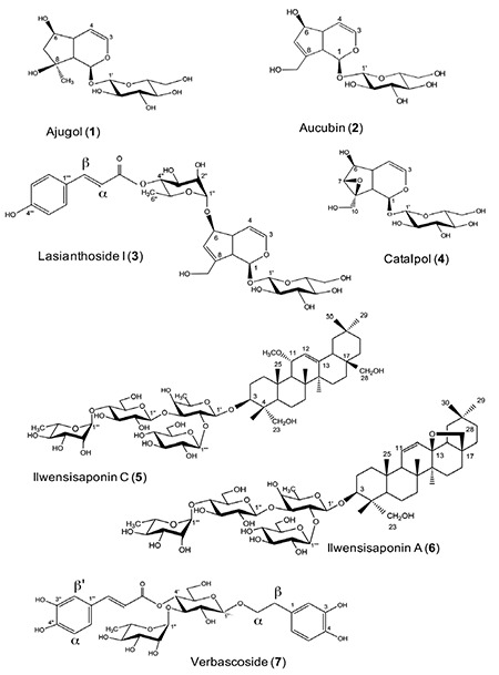
Isolated secondary metabolites from Verbascum mucronatum Lam.
MATERIALS AND METHODS
General experimental procedures
The ultraviolet (UV) spectra (λmax) were recorded on a Agilent 8453 spectrophotometer. The infrared (IR) spectra (νmax) were determined on a Perkin Elmer 2000 fourier transform (FT)-IR spectrophotometer. The 1D and 2D nuclear magnetic resonance (NMR) spectra were obtained on a Bruker Avance DRX 500 and 400 FT spectrometer operating at 500 and 400 MHz for 1H NMR, and 125 and 100 MHz for 13C NMR. For the 13C NMR spectra, multiplicities were determined using distortionless enhancement with a polarization transfer (DEPT) experiment. LC-ESIMS data were obtained using a Bruker BioApex FT-mass spectrometry instrument in the ESI mode. Reversed-phase material (C-18, LiChroprep 25-40 µm) and polyamide were used for vacuum liquid chromatography (VLC), reversed-phase material (C-18, LiChroprep 25-40 µm) was used for middle pressure liquid chromatography (MPLC), and Si gel (230-400 mesh) (Merck) was used for column chromatography (CC). Pre-coated silica gel 60 F254 aluminum sheets (Merck) were used for thin-layer chromatography (TLC); developing systems, CHCl3-MeOH-H2O (61:32:7 and 80:20:2). Plates were examined using UV fluorescence and sprayed with 1% vanillin in concentrated H2SO4, followed by heating at 105°C for 1-2 min.
Plant material
V. mucronatum Lam. was collected from Aksaray, 17 km from Aksaray to Ulukışla, in July 2007. A voucher specimen has been deposited in the Herbarium of the Faculty of Science, Gazi University, Ankara, Turkey (GAZI 10097). The flowery parts of the plant, which were air dried in the shade, were used in the phytochemical studies.
Extraction and isolation
Air-dried and powdered flowery parts of the plant (586.2 g) were extracted with MeOH (3x2.5 L). The MeOH extract was evaporated to dryness in vacuo to yield 70.4 g of crude extract, then MeOH extract was dissolved with 100 mL distilled water and partitioned in CHCl3 (2x100 mL). H2O and CHCl3 phases were evaporated to dryness in vacuo to yield 65.8 g H2O and 3.6 g CHCl3 extracts. The H2O phase was fractionated using CC on polyamide (150 g) using H2O-MeOH (100:0→0:100) (each 500 mL), respectively, to yield 6 fractions (Frs. A-F). Fraction D (4.9 g), eluted with 75% methanol, was subjected to VLC using reversed-phase material (C-18, LiChroprep 25-40 µm, 150 g), using MeOH-H2O mixtures (0-100%) to give catalpol (4) (62.1 mg), aucubin (2) (139.3 mg), ajugol (1) (48.6 mg), Fr. D3 (1.19 g) and Fr. D4 (625.3 mg). Frs. D3 and D4 were rechromatographed. Fr. D3 was applied to MPLC using reversed-phase material (C-18, LiChroprep 25-40 µm) using MeOH-H2O mixtures (100:0→30-70) to yield ilwensisaponin C (5) (14.7 mg), ilwensisaponin A (6) (51.5 mg), and lasianthoside I (3) (6.7 mg). Fr. D4 was rechromatographed on a silica gel column (55 mg) and eluted CHCl3-MeOH (70:30→60:40) mixtures to give verbascoside (=acteoside) (7) (14.8 mg).
Antimicrobial activity-broth microdilution method
Antibacterial and antifungal activities were determined using the broth microdilution test as recommended by Clinical and Laboratory Standards Institute.17,18 Plant extracts were tested against four bacteria including two Gram-positive (S. aureus ATCC 29213, Enterococcus faecalis ATCC 29212) and two Gram-negative microorganisms (Escherichia coli ATCC 25922, Pseudomonas aeruginosa ATCC 27853), as well as for antifungal activities against three yeasts (Candida albicans ATCC 90028, Candida krusei ATCC 6258, Candida parapsilosis ATCC 90018). The antibacterial activity test was performed in Mueller-Hinton broth (MHB, Difco Laboratories, Detroit, MI, USA); for antifungal test, RPMI-1640 medium with L-glutamine (ICN-Flow, Aurora, OH, USA), buffered with MOPS buffer (ICN-Flow, Aurora, OH, USA) was used. The inoculum densities were approximately 5×10⁵ CFU/mL and 0.5-2.5×10³ CFU/mL for bacteria and fungi, respectively.
Each plant extract was dissolved in 2.44 mL DMSO. Finally, two-fold concentrations were prepared in the wells of the microtiter plates, between 1024-1 µg/mL. Ampicillin and fluconazole were used as reference antibiotics for bacteria and fungi, respectively (64-0.0625 µg/mL). Microtiter plates were incubated at 35°C for 18-24 h for bacteria and 48 h for fungi. After the incubation period, minimum inhibitory concentration (MIC) values were defined as the lowest concentration of the extracts that inhibits the visible growth of the microorganisms.
RESULTS
Ajugol (1): UV λmax (MeOH) 220 nm, IR (KBr) νmax 3410 (OH), 1660 (C=C) cm-1, Positive ion LC-ESIMS m/z 371 [M+Na]+ (calc. for C15H24O9), 1H NMR (400 MHz, DMSO-d6) of 1: δH 6.10 (1H, dd, J=6/1.6 Hz, H-3), 5.29 (1H, d, J=2 Hz, H-1), 4.78 (1H, dd, J=6/2.8 Hz, H-4), 4.43 (1H, d, J=7.6 Hz, H-1’), 3.71 (1H, d, J=2.8 Hz, H-6), 3.71-3.65 (2H, *, H-6’), 3.05-2.93 (1H, *, H-2’, H-3’, H-4’, H-5’), 2.47 (1H, m, H-5), 2.32 (1H, t, J=10 Hz, H-9), 1.84 (1H, dd, J=12.8/6.0 Hz, H-7b), 1.63 (1H, dd, J=13.2/6.0 Hz, H-7a), 1.13 (3H, s, H-10), and 13C NMR (100 MHz, DMSO-d6) (see Table 1).
Table 1. 13C NMR (DMSO-d6) data of compounds of 1, 2, 3 and 4.
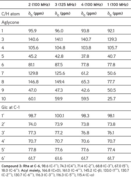
Aucubin (2): UV λmax (MeOH) 205 nm, (KBr) νmax 3275 (OH), 1650 (C=C) cm-1, Positive ion LC-ESIMS m/z 369 [M+Na]+ (calc. for C15H22O9), 1H NMR (400 MHz, DMSO-d6) of 2: δH 6.30 (1H, dd, J=4.8/1.6 Hz, H-3), 5.65 (1H, bs, H-7) 5.01 (1H, d, J=4.8 Hz, H-4), 4.95 (1H, d, J=5.6 Hz, H-1), 4.85 (1H, d, J=7.7 Hz, H-1’), 4.40, (1H, d, J=6.4 Hz, H-6), 4.14 (1H, dd, J=12.4/4.0 Hz, H-10b), 3.96 (1H, dd, J=12.4/4.0 Hz, H-10a), 3.66 (1H, dd, J=12.8/4.8 Hz, H-6’a), 3.42 (1H, dd, J=12.0/4.8 Hz, H-6’b), 3.16 (1H, m, H-3’), 3.11 (1H, m, H-4’), 3.04 (1H, m, H-5’), 3.00 (1H, m, H-2’), 2.72 (1H, t, J=7.2 Hz, H-9), 2.50 (1H, m, H-5), and 13C NMR (100 MHz, DMSO-d6) (see Table 1).
Lasianthoside I (3): UV λmax (MeOH) 216, 277 nm, IR (KBr) νmax 3405 (OH), 1704 (C=O), 1655 (C=C), 1508, 1451 (aromatic ring) cm-1, Positive ion LC-ESIMS m/z 611 [M+Na]+ (calc. for C30H38O15), 1H NMR (400 MHz, DMSO-d6) of 3: δH 6.37 (1H, dd, J=4.8/1.2 Hz, H-3), 5.26 (1H, d, J=4.4 Hz, H-4), 5.10 (1H, d, J=4.0 Hz, H-1), 4.91 (1H, d, J=7.6 Hz, H-1’), 4.18 (1H, d, J=6.0 Hz, H-10b), 3.86 (1H, d, J=4 Hz, H-6’b), 3.78 (1H, t, J=6.8 Hz, H-6), 3.66 (1H, *, H-10a), 3.64 (1H, dd, J=10.8/6.4 Hz, H-6’a), 3.35 (1H, s, H-7), 2.31 (1H, t, J=7.6 Hz, H-9), 3.13-3.19 (1H, *, H-3’, H-4’, H-5’), 3.02 (1H, dd, J=10/6.4 Hz, H-2’), 2.12 (1H, m, H-5), and 13C NMR (125 MHz, DMSO-d6) (see Table 1).
Catalpol (4): UV λmax (MeOH) nm 208 nm, IR (KBr) νmax 3450 (OH), 1670 (C=C) cm-1, Positive ion LC-ESIMS m/z 385 [M+Na]+ (calc. for C15H22O10), 1H NMR (400 MHz, DMSO-d6) of 4: δH 6.37 (1H, dd, J= 4.8/1.2 Hz, H-3), 5.26 (1H, d, J=4.4 Hz, H-4), 5.10 (1H, d, J=4.0 Hz, H-1), 4.91, (1H, d, J=7.6 Hz, H-1’), 4.18 (1H, d, J=6.0 Hz, H-10b), 3.86 (1H, d, J=4 Hz H-6’b), 3.78 (1H, t, J=6.8 Hz, H-6), 3.66 (1H, *, 10a), 3.64 (1H, dd, J=10.8/6.4 Hz, H-6’a), 3.35 (1H, s, H-7), 3.13-3.19 (*, H-3’, H-4’, H-5’), 3.02 (1H, dd, J=10/6.4 Hz, H-2’), 2.31 (1H, t, J=7.6 Hz, H-9), 2.12 (1H, m, H-5), and 13C NMR (100 MHz, DMSO-d6) (see Table 1).
Ilwensisaponin C (5): UV λmax (MeOH) 205 nm, IR (KBr) νmax 3400 (OH), 1665 (C=C) cm-1, Positive ion LC-ESIMS m/z 1127 [M+Na]+ (calc. for C55H92O22), 1H NMR (400 MHz, pyridine) of 5: δH 5.78 (1H, bs, H-1’’’), 5.54 (1H, d, J=7.0 Hz, H-1’’’’), 5.46 (1H, bs, H-12), 5.21 (1H, d, J=7.0 Hz, H-1’’), 4.91 (1H, d, J=6.6 Hz, H-1’), 4.35 (1H, *, H-2’), 4.33 (1H, *, H-23b), 4.10 (1H, *, H-2’’’’), 4.10 (1H, *, H-3), 3.89 (1H, *, H-2’’), 3.82 (1H, *, H-11), 3.81 (1H, d, J=11.7 Hz, H-28b), 3.69 (1H, d, J=8.3 Hz, H-23a), 3.57 (1H, d, J=10.2 Hz, H-28a), 1.68 (3H, d, J=5.5 Hz, H-6’’’), 1.35 (3H, d, J=4.8 Hz, H-6’), 1.30 (3H, s, H-27), 1.08 (3H, s, H-24), 1.07 (3H, s, H-25), 0.96 (3H, s, H-26), 0.95 (3H, s, H-30), 0.88 (3H, s, H-29), CH3O: 3.21 (3H, s), and 13C NMR (125 MHz, pyridine) (see Table 2).
Table 2. 13C NMR (125 MHz, pyridine-d5/5, CD3OD/6) data of compounds 5 and 6.
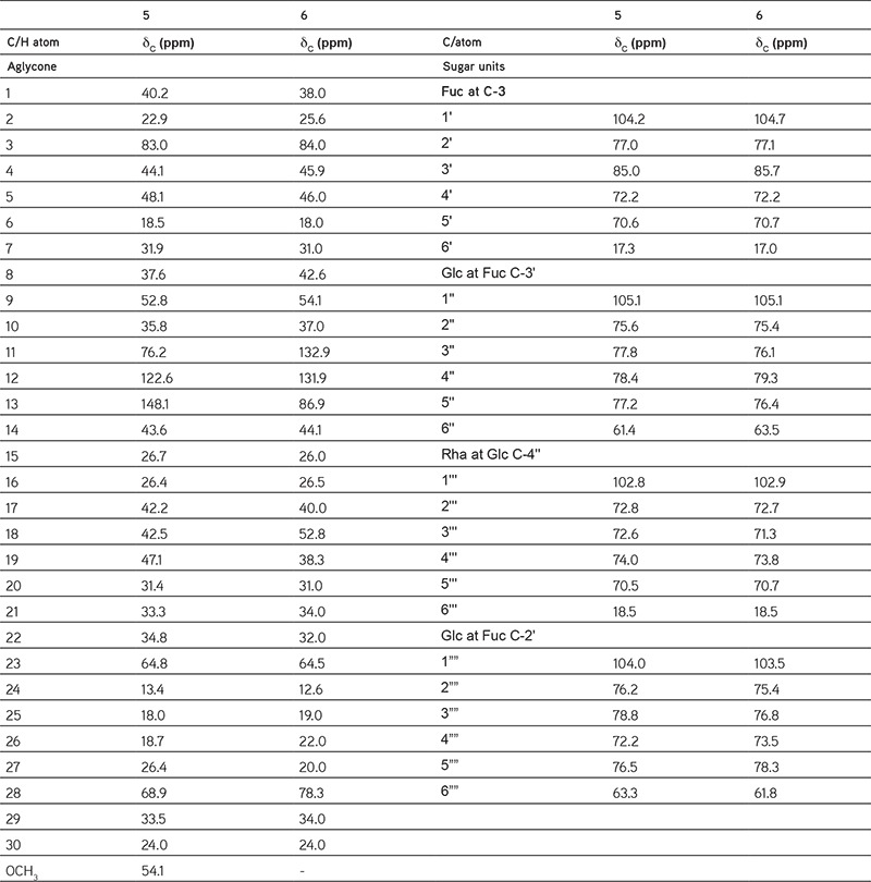
Ilwensisaponin A (6): UV λmax (MeOH) 206 nm, IR (KBr) νmax 3434 (OH), 1645 (C=C) cm-1, Positive ion LC-ESIMS m/z 1095 [M+Na]+ (calc. for C54H88O21), 1H NMR (500 MHz, pyridine) of 6: δH 5.94 (1H, d, J=10.4 Hz, H-11), 5.77 (1H, d, J=1.5 Hz, H-1’’’), 5.53 (1H, *, H-12), 5.20 (1H, d, J=7.6 Hz, H-1’’), 5.53 (1H, d, J=7.9 Hz, H-1’’’’), 4.91 (1H, d, J=7.7 Hz, H-1’), 4.58 (1H, *, H-2’’’), 4.34 (1H, *, H-23b), 4.25 (1H, *, H-2’), 4.11 (1H, *, H-3), 4.05 (1H, *, H-2’’’’), 3.90 (1H, *, H-2’’), 3.72 (1H, *, H-28b), 3.70 (1H, *, H-23a), 3.33 (1H, d, J=6.2 Hz, H-28a), 1.68 (1H, d, J=6.1 Hz, H-6’’’), 1.38 (3H, bs, H-6’), 1.31 (3H, s, H-26), 1.04 (3H, s, H-24), 0.98 (3H, s, H-27), 0.96 (3H, s, H-25), 0.87 (3H, s, H-29), 0.82 (3H, s, H-30), and 13C NMR (125 MHz, CD3OD) (see Table 2).
Verbascoside (Acteoside) (7): UV λmax (MeOH) 220, 332 nm, IR (KBr) νmax 3392 (OH), 1699 (C=O), 1631 (C=C), 1604, 1525 (aromatic ring) cm-1, Positive ion LC-ESIMS m/z 647 [M+Na]+ (calc. for C29H36O15), 1H NMR (500 MHz, DMSO-d6) of 7: δH 7.48 (1H, d, J=15.8 Hz, H-β’), 7.04 (1H, s, H-2’’’), 6.97 (1H, d, J=7.5 Hz, H-6’’’), 6.79 (1H, d, J=7.7 Hz, H-5’’’), 6.67 (1H, bs, H-2), 6.67 (1H, bs, H-5), 6.52 (1H, d, J=7.5 Hz, H-6), 6.20 (1H, d, J=15.8 Hz, H-α’), 5.07 (1H, bs, H-1’’), 4.75 (1H, t, J=9.4 Hz, H-4’), 4.37 (1H, d, J=7.7 Hz, H-1’), 3.72 (1H, *, H-2’’), 3.91, (1H, m, H-αb), 3.67, (1H, m, H-αa), 2.73 (2H, s, H-β), 3.68 (1H, *, H-3’), 3.45-3.70 (2H, *, H-6’), 3.45 (1H, *, H-5’), 3.36 (1H, *, H-5’’), 3.35 (1H, *, H-3’’), 3.26 (1H, t, J=8.3 Hz, H-2’), 3.15 (1H, *, H-4’’), 1.00 (3H, d, J=5.8 Hz, H-6’’), and 13C NMR (125 MHz, CDCl3) (see Table 3).
Table 3. 13C NMR (125 MHz, CDCl3) data of compound 7.
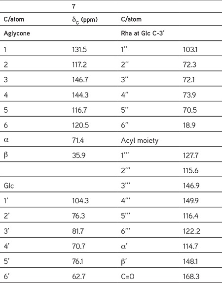
*(overlapped)
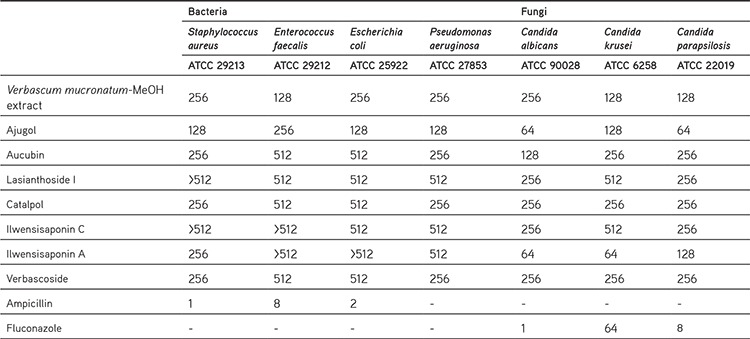
The methanolic extract of the flowery part of V. mucronatum and isolated compounds possessed moderate antimicrobial activity, especially against fungi. Iridoid glycoside ajugol was found to be the most active compound against C. albicans and C. parapsilosis with an MIC value of 64 µg/mL, as well as ilwensisaponin A inhibited C. albicans and C. krusei with the same MIC value as ajugol. These active compounds were found to be much more effective against fungi than the V. mucronatum extract (Table 4).
Table 4. Minimum inhibitory concentrations (μg/mL) of the methanolic extract and the secondary metabolites.
DISCUSSION
Compound 1 was isolated as a white amorphous powder with the molecular formula C15H24O9 (LC-ESIMS m/z 371 [M+Na]+). An iridoid enolether system (220 nm) in UV spectrum; hydroxyl group (3410 cm-1) and double-bond (1660 cm-1) absorption bands in IR spectra were observed. Compound 1 was identified as ajugol when comparing 1H and 13C NMR spectra with those of ajugol.19
Compound 2 (see Figure 1) was isolated as white amorphous powder with the molecular formula C15H22O9 (LC-ESIMS m/z 369 [M+Na]+). An iridoid enolether system (205 nm) in UV spectrum; hydroxyl group (3275 cm-1) and double-bond (1650 cm-1) absorption bands in IR spectra were observed. Compound 2 was identified as aucubin when comparing 1H and 13C NMR spectra with those of aucubin.20,21
Compound 3 (see Figure 1) was isolated as a white amorphous powder with the molecular formula C30H38O15 (LC-ESIMS m/z 661 [M+Na]+). The presence of an iridoid enolether system (216 nm) and an aromatic acid (277 nm) moiety in UV spectrum and absorption bands for a hydroxyl group (3405 cm-1), a conjugated ester carbonyl (1704 cm-1), a double-bond (1655 cm-1) and an aromatic ring (1451 cm-1, 1508 cm-1) in IR spectra were observed. The 1H and 13C NMR spectra of 3 were similar to those of lasianthoside I. Based on this evidence, compound 3 was identified as lasianthoside I.22
Compound 4 (Figure 1) was isolated as a white amorphous powder with the molecular formula C15H22O10 (LC-ESIMS m/z 385 [M+Na]+). Its UV spectrum supported the presence of an iridoid enolether system (208 nm) and absorption bands were for a hydroxyl group (3450 cm-1), and a double-bond (1670 cm-1) in the IR spectra were observed. The 1H and 13C NMR spectra of compound 4 were similar to those of catalpol. Thus, compound 4 was identified as catalpol.23
Compounds 5 and 6 (Figure 1) were obtained as amorphous compounds with molecular weights of 1104 {LC-ESIMS: m/z 1127 ([M+Na]+)}, and 1072 {LC-ESIMS: m/z 1095 ([M+Na]+)}, as calculated for C55H92O22 and C54H88O21, respectively.
In their IR spectra, the observed absorbances were consistent with the presence of olefinic double bonds. The 1H and 13C NMR data of compounds 5 and 6 suggested that they had similar structures, possessing the same sugar moieties but differing in their aglycones.
In the 1H NMR spectrum of compound 5, characteristic resonances for anomeric protons were observed at δH 4.91 (d, J= 6.6 Hz), 5.21 (d, J= 7.0 Hz), 5.54 (d, J= 7.0 Hz), 5.78 (bs), and, in the 13C NMR spectrum, anomeric carbons at δc 104.2 (β-D-fucopyranose), 105.1 (β-D-glucopyranose-inner), 104.0 (β-D-glucopyranose-terminal) and 102.8 (α-L-rhamnopyranose), as well as 2 proton signals at δH 1.35 (d, J= 4.8 Hz) and 1.68 (d, J= 5.5 Hz), arising from the secondary methyl groups in the sugar moieties. By means of HMBC correlations, the sequence of the saccharidic chain was determined as [α-L-rhamnopyranosyl-(1→4)-β-D-glucopyranosyl-(1→3)]-[β-D-glucopyranosyl-(1→2)]-β-D fucopyranoside.
The 1H NMR of compound 5 showed 6 tertiary methyl signals at δH 0.88, 0.95, 0.96, 1.07, 1.08 and 1.30. The proton signal at δH 3.21 (3H) was attributed to methoxy protons, and δH 5.46 (br s) to the olefinic proton of the aglycone. It was determined that the aglycone was an oleanane-Δ12 type confirmed by presence of δc 122.6 and 148.1 signals in the 13C NMR spectrum. The assignment of the remaining NMR signals was achieved by means of 1H-1H COSY, HMQC, and HMBC experiments.
The location of the methoxy group was determined using HMBC correlations between methoxy protons and C-11, whereas a chemical shift of C-11 (δc 76.2) was also evident. From the chemical shift of C-11 (δc 76.2) in compound 5, it can be concluded that the methoxyl group had an α-configuration as reported for saikosaponin-b4.24 The H-3 methine proton, H-23 and H-28 methylene protons showed downfield shifts due to hydroxy substitutions.
Consequently, the structure was elucidated to be 3-O-{[α-L-rhamnosyl-(1→4)-(β-D-glucopyranosyl-(1→3)]-β-D-glucopyranosyl]-(1→2)-β-D-fucopyranosyl-11-methoxy-olean-12-ene-3β,23,28-triol (=ilwensisaponin C).25
Compound 6 was distinguished from compound 5 by differences in the aglycone parts in 1H and 13C NMR spectra.
The 1H NMR of compound 6 showed 6 tertiary methyl signals at δH 0.82, 0.87, 0.96, 0.98, 1.04 and 1.31. The olefinic protons H-11 and H-12 were determined at 5.94 (br d, J= 10.4 Hz), δc 132.9 and δH 5.53 (*), δc 131.9, respectively. Thus, aglycone was identified as an oleanane-Δ11 type and no signals of a methoxy group in 1H and 13C NMR spectra of compound 6 were observed compared with those of compound 5.
Due to presence of an oxo-bridge between C-28 and C-13, a chemical shift of C-28 methylene protons (δH 3.33-3.72) appeared in the higher field in comparison with those of C-23 hydroxylated methylene protons (δH 3.70-4.34). Based on this evidence, the aglycone of compound 6 was determined as 13β, 28-epoxyolean-11-ene-3β,23-diol.26
As a result, the structure of compound 6 was determined as 3-O-{[α-L-rhamnosyl-(1→4)-(β-D-glucopyranosyl-(1→3)]- β-D-glucopyranosyl]-(1→2)-β-D-fucopyranosyl}-13β,28-epoxyolean-11-ene-3β,23-diol (=ilwensisaponin A25=mimen- goside A).27
Compound 7 (Figure 1) was obtained as an amorphous powder. Its structure was identified as verbascoside by comparing its 1H and DEPT-13C NMR data with previously published data and by direct comparison with the authentic sample on a TLC plate.
It has been reported that Verbascum L. species contained diverse iridoid glycosides such as ajugol5,13, aucubin28, lasianthoside I22 and catalpol23; saponins such as ilwensisaponin C13 and ilwensisaponin A13; and phenylethanoid glycosides such as verbascoside.13 Ilwensisaponin A has previously been found to be active against Aspergillus fumigatus;29 it showed moderate antifungal activity in the current study.
CONCLUSIONS
This paper is the first to report the presence of these compounds from V. mucronatum Lam. Our continuing studies will be of assistance in clarifying the chemotaxonomic classification of the genus Verbascum L. On the other hand, when the antimicrobial activity results were evaluated, the higher activities of ajugol and ilwensisaponin A than the V. mucronatum extract suggest that more active compounds may be found in further phytochemical studies.
Acknowledgments
The authors would like to thank Prof. Dr. Hayri Duman, Gazi University, Faculty of Science, Department of Botany, Etiler, Ankara, Turkey, for the authentication of the plant specimen.
Footnotes
Conflict of Interest: No conflict of interest was declared by the authors.
References
- 1.Tutin TG. Flora Europaea Vol 3. Cambridge; University Press. 1972. [Google Scholar]
- 2.Davis PH, Mill RR, Tan K. Flora of Turkey and the East Aegean Islands. Edinburg; University Press. 1988. [Google Scholar]
- 3.Ekim T, Verbascum L. In: Güner A, Özhatay N, Ekim T, Başer KHC, eds. Flora of Turkey and East Aegean Islands. Edinburg University Press. 2000:193–194. [Google Scholar]
- 4.Huber-Morath A, Verbascum L. In: Davis P, ed. Flora of Turkey and the East Aegean Islands. Edinburgh University Press; 1978:461–463. [Google Scholar]
- 5.Baytop A. Therapy with Medicinal Plants in Turkey (Past and Present) Nobel Tip Kitabevleri Ltd. 1999. [Google Scholar]
- 6.Sezik E, Yeşilada E, Honda G, Takaishi Y, Takeda Y, Tanaka T. Traditional medicine in Turkey X. Folk medicine in Central Anatolia. J Ethnopharmacol. 2001;75:95–115. doi: 10.1016/s0378-8741(00)00399-8. [DOI] [PubMed] [Google Scholar]
- 7.Tuzlaci E, Alparslan DF. Turkish folk medicinal plants, part V: Babaeski (Kırklareli) J Pharm Istanbul University. 2007;39:11–23. [Google Scholar]
- 8.Turker AU, Camper ND. Biological activity of common mullein, a medicinal plant. J Ethnopharmacol. 2002;82:117–125. doi: 10.1016/s0378-8741(02)00186-1. [DOI] [PubMed] [Google Scholar]
- 9.Tatli I, Akdemir ZS. Traditional uses and biological activities of Verbascum species. FABAD J Pharm Sci. 2006;31:85–96. [Google Scholar]
- 10.Kahraman C, Ekizoglu M, Kart D, Akdemir ZS, Tatli I. Antimicrobial activity of some Verbascum species growing in Turkey. FABAD J Pharm Sci. 2011;36:11–15. [Google Scholar]
- 11.Cubukcu B, Atay M, Sarıyar G, Ozhatay N. Folk medicines in Aydın. Journal of Traditional and Folcloric Drugs. 1994;1:1–58. [Google Scholar]
- 12.Akdemir ZS, Tatli I, Bedir E, Khan IA. Two new iridoid glucosides from Verbascum salviifolium Boiss. Z Naturforsch B. 2005;60:113–117. [Google Scholar]
- 13.Tatli I, Akdemir ZŞ, Bedir E, Khan IA. Saponin, iridoid, phenylethanoid and monoterpene glycosides from Verbascum pterocalycinum var. mutense. Turk J Chem. 2004;28:111–122. [Google Scholar]
- 14.Akdemir ZŞ, Tatli I, Bedir E, Khan IA. Neolignan and phenylethanoid glycosides from Verbascum salviifolium boiss. Turk J Chem. 2004;28:621–628. [Google Scholar]
- 15.Akdemir ZS, Tatli I, Bedir E, Khan IA. Iridoid and phenylethanoid glycosides from Verbascum lasianthum. Turk J Chem. 2004;28:227–234. [Google Scholar]
- 16.Akdemir ZS, Tatli II, Bedir E, Khan IA. Antioxidant flavonoids from Verbascum salviifolium boiss. FABAD J Pharm Sci. 2004;28:71–75. [Google Scholar]
- 17.Wayne P. Reference method for broth dilution antifungal susceptibility testing of yeasts: Approved standard. 3rd ed. M 27-A3 ed. Clinical and Laboratory Standards Institute. 2008. [Google Scholar]
- 18.Wayne P. Methods for dilution antimicrobial susceptibility tests for bacteria that grow aerobically: Approved standard. 8th ed. M 07-A8 ed. Clinical and Laboratory Standards Institute. 2008. [Google Scholar]
- 19.Pardo F, Perich F, Torres R, Delle Monache F. Phytotoxic iridoid glucosides from the roots of Verbascum thapsus. J Chem Ecol. 1998;24:645–653. [Google Scholar]
- 20.Bianco A, Passacantilli P, Polidori G. 1H and 13CNMR data of C-6 epimeric iridoids. Org Magn Resonance. 1983;21:460–461. [Google Scholar]
- 21.Chaudhuri RK, Sticher O. New iridoid glucosides and a lignan diglucoside from Globularia alypum L. Helv Chim Acta. 1981;64:3–15. [Google Scholar]
- 22.Tatli I, Khan IA, Akdemir ZS. Acylated iridoid glycosides from the flowers of Verbascum lasianthum Boiss. ex Bentham. Z Naturforsch B. 2006;61:1183–1187. [Google Scholar]
- 23.Tatli I, Akdemir ZS, Bedir E, Khan IA. 6-O-alpha-L-rhamnopyranosylcatalpol derivative iridoids from Verbascum cilicicum. Turk J Chem. 2003;27:765–772. [Google Scholar]
- 24.Ishii H, Seo S, Tori K, Tozyo T, Yoshimura Y. The Structures of Saikosaponin-E and Acetylsaikosaponins, minor components isolated from Bupleurum falcatum L. determined by C-13 Nmr-spectroscopy. Tetrahedron Lett. 1977:1227–1230. [Google Scholar]
- 25.Calis I, Zor M, Basaran AA, Wright AD, Sticher O. Ilwensisaponin A, B, C and triterpene saponins from Scrophularia ilwensis. Helv Chim Acta. 1993;76:1352–1360. [Google Scholar]
- 26.Tori K, Yoshimura Y, Seo S, Sakurawi K, Tomita Y, Ishii H. Carbon-13 NMR spektra of saikogenins. Stereochemical dependence in hydroxilation effects upon carbon-13 chemical shifts of oleanene-type triterpenoids. Tetrahedron Lett. 1976;17:4163–4166. [Google Scholar]
- 27.Ding N, Yahara S, Nohara T. Structure of mimengosides A and B, new triterpenoid glycosides from Buddleja flos produced in China. Chem Pharm Bull. 1992;40:780–782. [Google Scholar]
- 28.Akdemir ZS, Tatli I, Bedir E, Khan IA. Acylated iridoid glycosides from Verbascum lasianthum. Turk J Chem. 2004;28:101–109. [Google Scholar]
- 29.Tatlı I, Akdemir Z. Antimicrobial and antimalarial activities of secondary metabolites from some Turkish Verbascum species. FABAD J Pharm Sci. 2005;30:84–92. [Google Scholar]


