Abstract
Objectives:
The objective of the present study was to formulate Desloratadine-Eudragit® RS100 nanoparticles and investigate the characteristics of the prepared nanoparticles.
Materials and Methods:
The nanoparticles were prepared by spray drying method and the quantification of desloratadine (DL) was carried out with a high performance liquid chromatography (HPLC) method.
Results:
DL was successfully loaded to polymer and the developed HPLC method was found to be linear, reproducible, precise, accurate, specific and selective. Characterization of the nanoparticles including entrapment efficiency, particle size, zeta potential, morphology, polidispersity index, solid state characterizations and drug release was performed. In vitro release studies of DL loaded nanoparticles were also examined in the simulated intestinal fluid (pH 7.4). In vitro release of DL from nanoparticle formulations followed Korsemeyer-Peppas model.
Conclusion:
A validated HPLC method was developed for the determination of DL. Proposed spray drying method can be successfully applicable to the nanoparticle preparation containing DL. In addition, the release studies of all nanoparticles and active substance have been studied comparatively. Hence, it could be concluded that DL loaded nanoparticles seem to be a promising drug delivery system for the active agent.
Keywords: Desloratadine, Eudragit® RS100, HPLC, nanoparticle
INTRODUCTION
Perennial allergic rhinitis affects up to 21% of the general population in some countries.1 With the prevalence of the disease increasing, even greater numbers of the population will be affected in the future. Convenient treatment is so important to ease the signs and symptoms of allergic rhinitis like sneezing, rhinorrhea, nasal congestion and to improve patients quality of life. Desloratadine (DL) is a tricyclic antihistamine. It has an orally active nonsedating, peripheral histamine H1-receptor antagonist. DL inhibited histamine release from human mast cells in vitro. It has been used to treat allergy symptoms.2 Old histamine H1 receptor antagonists have the potential for adverse central nervous system effects and are characterized by poor receptor specificity.3DL is slightly soluble in water and it has a reduced drug dissolution rate in gastrointestinal fluid following oral administration, and consequently, reduced bioavailability.4
In the recent decades nanoparticles containing drugs have gained increasing interest in pharmaceutic fields owing to their potential for site-specific drug delivery. The drugs are incorporated into a polymeric matrix, covalently bind to the polymer backbone or form electrolyte complexes of oppositely charged polymer-drug systems.5 Drug dissolution rate could be controlled with different approaches such as formulation of nanoparticles.6
Eudragit® RS100 is a co-polymer of poly (ethylacrylate, methyl-methacrylate and chlorotrimethyl-ammonioethyl methacrylate), contains 4.5-6.8% of quaternary ammonium groups.7 Biodegradable drug delivery systems consist of non-toxic, biodegradable polymers and are well tolerated by the human body. They are preferred for increasing the absorption of active substance, for increasing bioavailability and for targeting therapeutic agents to specific organs.8 Eudragit RS-100 is a widely used polymer for the preparation of controlled release oral pharmaceutical dosage forms.5,6,9As a result, Eudragit® RS100 is a promising polymer for the transport of the active substance to the targeted region.
In this study, we investigate the possibility of the preparation of stable DL loaded nanoparticles by using spray-drying method without any cross-linked agent. The physicochemical properties of the obtained nanoparticles were characterized. In addition, the performed formulation and characterization studies results will be helpful to the future planned in vivo studies and in order to formulate DL nanoparticles to alternative dosage forms such as tablets or capsules for the usage in antihistaminic treatment.
MATERIALS AND METHOD
Materials
DL was a kind gift from Morepen Labs. (New Jesey, USA), Eudragit® RS100 was purchased from Evonic India Pvt. Ltd. Methanol and acetonitrile were obtained from Merck Chemical Co. (Germany). All the other chemicals were of analytical grade.
Preparation of nanoparticles
In this study, nanoparticles of DL with the different ratios of DL/Eudragit® RS100 were prepared using Buchi B-190 mini spray dryer (Büchi, Switzerland).10,11 The spray dryer was connected to the Inert Loop B-295 (Büchi Labortechnik AG, Flawil, Switzerland) because of the organic solvent. Carbondioxide gas was used at a flow rate of 120 L/min. The inlet temperature was selected as 120°C for having maximum dried nanoparticles.12,13 The residual oxygen level in the system was controlled below 4%. The fully weighed Eudragit polymer was dissolved in methanol by mixing at magnetic stirrer for 2 hours at 250 rpm. After the clear solution was obtained, DL was added and stirred for 10 more min at 250 rpm stirring speed. The transparent solution was run using 30 min of methanol for conditioning the device to the desired level in terms of parameters such as spraying, pump level, inlet temperature, outlet temperature, gas flow and ambient temperature. After the required conditions had been met, the clear solution was transferred to a dry nano-spray dryer, peristaltic pump with an inlet temperature of 120°C and an outlet temperature of 64°C and delivered to a drying zone with a 4 µm nozzle diameter. After the drying nanoparticles were collected in the collection chamber.
Characterization studies
The particle size distribution and zeta potential of the nanoparticles were measured at 25°C by dynamic light scattering (DLS) (Malvern Zetasizer-Nano ZS, Malvern Instruments Limited, UK). The morphology of the nanoparticles were carried out by with scanning electron microscope (SEM) and also fourier transform infrared spectroscopy (FTIR) analysis were done, respectively. The stability of spheres was tested using differential scanning calorimeter (DSC) analysis and nuclear magnetic resonance analysis (1H-NMR) analysis. In order to determine release properties in vitro drug release mechanism was examined in the simulated biological fluid at pH 7.4. Encapsulation efficiency was also analyzed for the determination of the drug amount in the nanoparticle formulations.
Measurement of particle size and zeta potential
The particle size, size distribution and zeta potential of the nanoparticles were measured at 25°C by DLS (Malvern Zetasizer-Nano ZS, Malvern Instruments Limited, Worcestershire, UK). Samples of all nanoparticles were dispersed in double-distilled water (adjusted to a constant conductivity of 50 µS•cm-1 using 0.9% NaCl) just prior to analyses. All analyses were repeated in triplicate.
Scanning electron microscopy analysis
The particle shape and surface properties of the freshly prepared nanoparticle formulation and DL were investigated by SEM (Zeiss Ultra Plus Fesem, Germany) after spreading the formulation and DL onto the double-sided carbon tape pre-affixed on a specimen stub and were then allowed to dry at room-temperature. Samples were coated with a thin layer of gold (100 Å) by a sputter coater under 50 mA for 2 min before observed under SEM. Images were taken at high vacuum mode with varying magnifications at an accelerating voltage of 3.0 kV.
Differential scanning calorimetry
Thermal analysis of the DL formulation was carried out in a pressure-assisted aluminum sample vessel using DSC (Schimadzu DSC-60, Japan) apparatus at a flow rate of nitrogen of 50 mL/dk-1 and at a temperature 25°C to a final temperature of 180°C at 10°C min-1 against aluminum reference. DSC experiments with pure DL were previously carried out to identify the melting point peak. As a control, physical mixtures of DL and polymer were also analyzed.14
Fourier transform infrared spectroscopy
The FTIR spectrum of the DL formulations were determined at a wavelength of 4000-500 cm-1 using FTIR (Schimadzu IR Prestige-21, Japan) instrument.
Nuclear magnetic resonance
1H-NMR of the DL formulation prepared was carried out by dissolving in deutero chloroform (CDCl3), NMR (Bruker 500 MHz UltraShield NMR, Germany).
Entrapment efficiency
In order to determine the amount of DL in drug loaded nanoparticles, drug entrapment efficiency study was studied by validated high performance liquid chromatography (HPLC) method with the ultraviolet (UV) detection set in 262 nm. A reversed phase Lichrospher C18 column (150x2.3 mm i.d., pore size 5 µm) was used. The mobile phase consisted of a mixture of acetonitrile:water (60:40 v/v) and the flow rate was set at 0.8 mL/min. In the first step, in order to find free DL, the spray-dried nanoparticles (5 mg) were dissolved in 2 mL of distilled water in Eppendorf tube. After being held in the ultrasonic bath for 20 min, the upper transparent part was collected by centrifuging for 5 min at 13.000 rpm and the sample was analyzed through the necessary dilutions (1:1) with mobile phase. In the second step, 2 mL of the mobile phase, in which DL and Eudragit® RS100 are soluble, is added to the remaining residue formulation in the Eppendorf tube. Five min ultrasonic bath is used to obtain a clear solution and the sample was also analyzed through the necessary dilutions (1:1) with mobile phase in order to determine the amount of active substance entrapped. The experiments were repeated 3 times for each formulation. Before the injections, all solutions were previously filtered through a membrane filter (pore size 0.22 µm, Millipore). Entrapment efficiency (EE) was calculated using equation given below.15
EE (%)= Total DL - Free DL/Total DL x 100
In vitro drug release studies
Studies were performed in simulated intestinal fluid (SIF: pH 7.4), for a period of 24 h.16,17,18 A dialysis (cellulose membrane) method was used to identify release behavior of nanoparticles. 5 mg nanoparticle formulation was weighed into the dialysis bag with 1 mL of dissolution medium. The dialysis bag which was sealed with clamps was placed in 50 mL of the SIF at 37±0.5°C under sink conditions and stirred at 100 rpm using magnetic stirrer. In order to determine the amount of DL released from the dialysis bag at different time intervals (5 min, 10 min, 15 min, 30 min, 45 min, 1 h, 2 h, 3 h, 4 h, 5 h 6 h, 24 h), 1 mL of the samples were picked up and then replaced with the 1 mL of fresh SIF. The concentration of drug released to the medium was determined by measuring the absorbance at 262 nm using HPLC Shimadzu liquid chromatography equipped with a model LC-10ATVP binary pump and model SPD-M10AVP PDA detector using the stationary phase 150x4.6 mm LiChrospher® 100 RP-18 octadecyl silane column (5 µm particle size) (Merck, Darmstadt, Germany) with integration by LC Solution Version 1.23 SP1 Software (Shimadzu Corporation, Kyoto, Japan). The mobile phase consisted of acetonitrile:water (60:40). The mobile phase was prepared daily and degassed by sonication under reduced pressure and filtered through 0.45 µm membrane filter. The flow rate was set at 0.8 mL/min resulting in a run time of 7 min per sample. The injection volume was 20 µL. Detection was performed at 262 nm and samples were analyzed at room temperature. In order to study the mechanism of drug release from nanoparticles, the release data were fitted to different equations with DDSolver programme.
RESULTS AND DISCUSSION
In the current study, a simple spray drying methods was used for the preperation of formulations. Differrent amounts of drug/polymer was formulated as given in Table 1.
Table 1. Composition of DL loaded eudragit RS®100 nanoparticles.
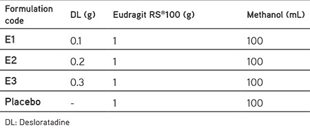
Solubility characteristic of a drug is one of the main factor to decide about the method of encapsulation.5 Spray drying methods was selected for the formulations.10,11,12Spray-drying parameters are given in Table 2.
Table 2. Spray-drying parameters.

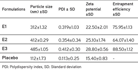
All the prepared formulations were in nanometer size and the size distributions were relatively monodisperse in all the formulations with the polydispersity index (PDI) values between 0.113±0.25-0.412±0.30 (Table 3). Most researchers recognize PDI values ≤0.3 as optimum values; however, values ≤0.5 are also acceptable. According to our Results given in Table 3 formulations were in homogenous size distributions even after DL was loaded to polymer.14,15,19
Table 3. Drug loading and particle size distribution of nanoparticle formulations.
Particle size is one of the important physical properties of colloidal systems. Particle size distribution of the formulation is especially significant in the physical stability (Figure 1) and activity of colloidal systems was also found that the size of nanoparticles plays a key role in their adhesion to and interaction with biological cells.20
Figure 1.
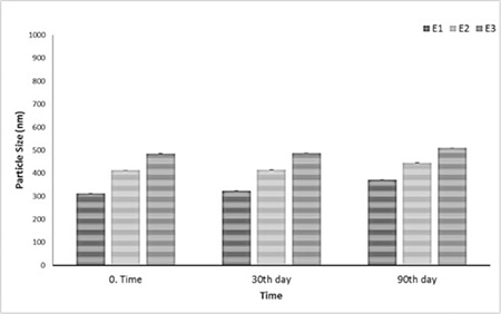
Particle size distribution of formulations during 3 months storage at 25°C
The mean particle size of DL loaded nanoparticles of all formulations ranged from 312±1.32 nm to 485±1.05 nm with a relative monodisperse distribution (Table 3). Smaller particles have higher surface area/volume ratio, which makes it easier for the encapsulated drug to be released from the nanoparticles by diffusion and surface erosion and also have advantages for the drug loaded nanoparticles to penetrate into, and permeate through the physiological drug barriers.21 The acceptable value for PDI is 0.05-0.7; values greater than 0.7 indicate very broad size distribution and probably no suitability for DLS technique.19,20Smaller size helps in targeting and increased penetration of drug through biological membranes.22 Since it was reported that, change in the organic phase ratio may result the change in the viscosity of the system, which in turn can change the characteristics of the nanoparticles (e.g., particle size, encapsulation efficiency, surface morphology, etc.), therefore, different ratios of formulations were evaluated. It was noticed that the mean particle size increase with increases of drug concentration. Increase in particle size with the increase in drug concentration might have occurred due to the fact that it produces a significant increase in the viscosity and thus leading to an increase of the nanoparticle size, polymer concentration had profound effect on average particle size. This effect can be attributed to the effect of polymer concentration on the viscosity of the polymeric solution and need more energy to disperse the system.23,24
The measurement of the zeta potential allows predictions of storage stability of colloidal dispersions.20,21 Zeta potential of nanoparticles is commonly used to characterize the surface property of nanoparticles. The mean zeta potential of all formulations ranged from 15.40±0.83 mV to 28.80±0.56 mV, which may be attributed to the positive charges on polymer matrices indicating good physical stability as shown in Table 3. Cationic property of nanoparticles was determined due to the effect of positively charged quaternary ammonium groups in Eudragit® RS100 interact with the negatively charged mucus and open up the tight junctions of epithelial cells to allow the paracellular transport pathway resulting in an increase in bioavailability.25,26As a result of the stability studies of the formulations over 3 months, no statistically significant change was observed (p>0.05). This indicates the stability of the formulations.
From the SEM, it was observed that nanoparticles were usually spherical (Figure 3). The surface of the drug-loaded Eudragit RS100 nanoparticles manifested the presence of drug particles.27
Figure 3.
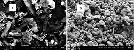
a) Scanning electron microscope photography of drug, b) scanning electron microscope photography of drug-loaded Eudragit RS100 nanoparticles (E1)
DSC has shown to be an important tool to quickly obtain information about possible interactions between the active and the excipients, according to the appearance, shift, or disappearance of endothermic or exothermic peaks.28 DSC gives an insight into melting and recrystallization behaviors of polymeric nanoparticles. No loss in typical peaks or no appearances of new peaks were recorded upon DSC analyses. This indicates that there was no physical or chemical interaction or incompatibility between the formulations prepared.29 Accordingly, DSC measurements were performed on each of the components, both in their pure forms and the corresponding drug loaded forms (Figure 4).
Figure 4.
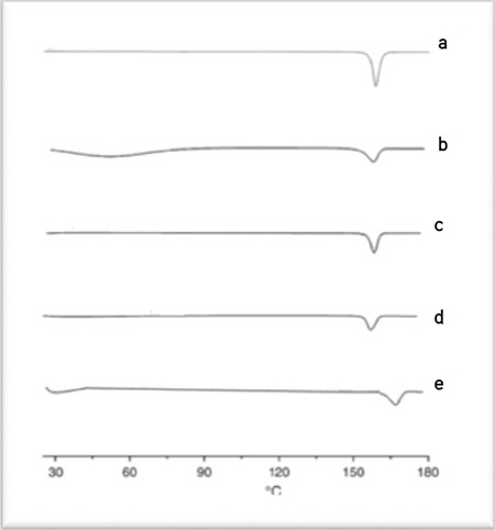
Differential scanning calorimeter curves of desloratadine (A), E1 (B), E2 (C), EC (D) and Eudragit® RS100 (E)
FTIR spectroscopy is used as an important technique to investigate possible chemical interactions between drug and active substance. The formation of new absorption bands and the expansion of the absorption bands give the main signals of the interaction between the drug and the active substance.28 The FTIR spectra show that the characteristic bands of DL and polymer individually were not altered in binary mixtures, which indicates no interactions between DL and the selected polymer when contact is established in spray dryer. The spectra of DL show prominent bands at 1.141 and 1.435 cm-1, which is attributed to the C-C stretching of aromatic rings, while the bands at 1.172 and 778 cm-1 correspond to C-N amines and C-Cl stretching, respectively and is given in Figure 5.
Figure 5.
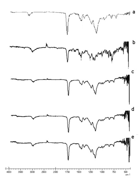
FTIR bands of Eudragit® RS100 (A), desloratadine (B), E1 (C), E2 (D) and E3 (E) formulations
These results are in agreement with other published datas.27,29 The spectra of DL loaded polymer formulations show the same absorption bands, indicating that the DL did not decompose in the range of applied temperatures and successfully loaded to polymers. Therefore, DL is sufficiently thermally stable to allow analysis by the DSC method.
1H-NMR investigations were used to obtain information about the mobility and structure of both the nanoparticle system and the incorporated drug. Comparing the drug-loaded nanoparticle formulations with each other no essential difference could be observed. On the other hand, the signals of pure drug are also seen in the same as the drug loaded formulations. This indicates that the active substance has been successfully loaded into the formulations. Peaks in 7-8 ppm interval belong to aromatic C-H peaks, peaks in 3-4 ppm belongs to -CH3 in the spectrum of DL.30,31 Similar spectra were also obtained for the spectra of drug loaded nanoparticle formulations indicating the successful incorporation of drug into the nanoparticles. Physicochemical characterization was undertaken both by DSC and 1H-NMR measurements. The combination of both analytical methods proved very suitable to obtain information about the structure of the DL loaded polymers.31,32
A simple and validated HPLC method was adopted and developed for the determination of DL. The method was validated for accuracy, precision and linearity with reference to the International Council for Harmonisation guidelines.33 The separation and resolution of the peaks of the blank reagent and the formulation could be achieved using a mixture of acetonitrile:water (60:40 v/v) as the mobile phase with UV detection at 262 nm.34 The method was found to be linear and reproducible. For the developed method the linear regression anaysis of the datas gave the following equation A= 4096.6x+13453 with a correlation coefficient of, r=0.9999. Calibration curve of DL was constructed by plotting absorbance versus concentration which showed linearity over the concentration ranges of 100-700 µg/mL (Figure 7). The limit of quantification was determined as 0.290 µg/mL-1, and the limit of detection was calculated as 0.628 µg/mL-1.35Meanwhile the developed method was spesific for the determination of DL. The developed method is found to be precise, accurate, specific and selective. The method was also found to be linear and reproducible. According to the conditions described, the retention time was about 1.7 min for DL.
Figure 7.
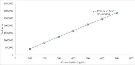
Calibration curve of desloratadine
The accuracy of the proposed method was assessed by recovery studies at different concentrations of 100 µg/mL-1, 400 µg/mL-1, 700 µg/mL-1. The recovery studies were carried out by adding the known amount of standard solution of the pure drug. The solutions were analysed by proposed method, the results were given in Table 4.
Table 4. Accuracy results of desloratadine.
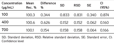
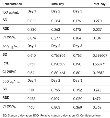
The precision of the method was assessed by carrying out six replicate determination of three different concentrations of DL both inter-day and intra-day. The results are shown in Table 5. The method was found to be precise as indicated by the results showing % relative standard deviation less than 2.
Table 5. The results of intra-day and Inter-day precision of desloratadine.
The encapsulation efficiency explains that it is the percentage of the amount of the drug that loaded to drug carrier agent. In this study, the % EE of formulations E1, E2 and E3 were found as 75.95±1.13, 64.07±1.40 and 88.50±1.12 respectively. Drug-polymer composition is most likely the main factor for release rate, however it seems that a complex phenomena between drug and polymer molecules may occur, including entrapment of drug within polymer molecules and the adsorption of drug molecules on the surface of polymeric matrix as a result of electrostatic adhesions.5,21
Eudragit® RS100 as a water-insoluble carrier was identified to be able to control drug release in both nanoparticles and solid dispersions.9,35 Figure 8 demonstrates the DL release profiles from Eudragit® RS100 polymers. There were no noteworthy differences between formulations as stated by the release profiles. All the nanoparticles revealed slower drug release rate in comparison with the intact drug. The release rate was not affected by increasing the Eudragit® RS100 relative amount in the nanoparticles. The released profiles of DL loaded formulations exhibited a burst release in one hour time which was attributed to the drug loaded the surface of nanoparticles.5,20 When nanoparticles of microcrystalline drug particles, are exposed to aqueous media, the carrier dissolves and the drug is released as a fine colloidal dispersion, thus resulting in higher surface area and consequently, enhanced dissolution rate and bioavailability of poorly water soluble drugs.36,37
Figure 8.
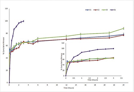
In vitro release of formulations and pure desloratadine (n=7)
In order to study the mechanism of drug release from nanoparticles, the release data were fitted to different equations with DDSolver programme (Table 6).38 Furthermore a kinetic parameter can be used to study the influence of formulation factors on the drug release for optimization as well as control of release.38 The kinetic models used were zero-order, first-order, Higuchi, Korsemeyer-Peppas models. According to highest k and r2 values and according to lowest AIC values, in vitro release of all DL loaded polymer formulations followed Korsemeyer-Peppas model.39 This model indicates a diffusion controlled drug release mechanism from polymer matrix.40
Table 6. Mathematical modelling of desloratadine loaded nanoparticles.

CONCLUSION
As indicated with this study, formulations of DL-Eudragit® RS100 nanoparticles applying single spray drying procedure is able to improve the physicochemical characteristics of the drug. The HPLC method that was validated for DL was also found to be a simple and rapid method. The DSC and NMR studies confirmed the decrease of drug crystallinity in the nanoparticles. The intermolecular interaction between DL and Eudragit® RS100 was identified in the FTIR spectrum of the nanoparticles. DL was successfully formulated as a model drug in this study. It was shown that all nanoparticles displayed a slowed release pattern with a burst release in comparison with the pure drug powder. The advances in the formulation technology of nanoparticle delivery systems has been widely accepted approach as compared to conventional immediate release formulations of the same drug. Hence it could be concluded that DL loaded nanoparticles seem to be a promising delivery system for the drug.
Additionally, formulation studies that we have undertaken in this study will be helpful to the future planned in vivo studies to convert DL nanoparticles into alternative dosage forms such as tablets or capsules for use in antihistaminic treatment.
Figure 2.
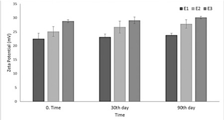
Zeta potential cahange of formulations during 3 months storage at 25°C
Figure 6.
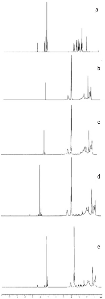
1H-NMR-spectrum of Eudragit® RS100 (A), DL (B), E1 (C), E2 (D) and E3 (E) formulations
Acknowledgments
The author would like to thank to BIBAM (Anadolu University) management for spray dryer equipments and the author would like to thank to Exp. Serkan Levent from Anadolu University, Faculty of Pharmacy, Department of Pharmaceutical Chemistry for NMR and FTIR analyses.
Footnotes
Conflict of Interest: No conflict of interest was declared by the author.
References
- 1.Simons FE, Prenner BM, Finn A Jr; Desloratadine Study Group. Efficacy and safety of desloratadine in the treatment of perennial allergic rhinitis. J Allergy Clin Immunol. 2003;111:617–622. doi: 10.1067/mai.2003.168. [DOI] [PubMed] [Google Scholar]
- 2.Zheng J, Rustum AM. Rapid separation of desloratadine and related compounds in solid pharmaceutical formulation using gradient ion-pair chromatography. J Pharm Biomed Anal. 2010;51:146–152. doi: 10.1016/j.jpba.2009.08.024. [DOI] [PubMed] [Google Scholar]
- 3.Veronez IP, Daniel JSP, Junior CEC, Garcia JS, Trevisan MG. Development, characterization, and stability studies of ethinyl estradiol solid dispersion. J Therm Anal Calorim. 2014;120:573–581. [Google Scholar]
- 4.Kolašinac N, Kachrimanis K, Homšek I, Grujić B, Đurić Z, Ibrić S. Solubility enhancement of desloratadine by solid dispersion in poloxamers. Int J Pharm. 2012;436:161–170. doi: 10.1016/j.ijpharm.2012.06.060. [DOI] [PubMed] [Google Scholar]
- 5.Adibkia K, Javadzadeh Y, Dastmalchi S, Mohammadi G, Niri FK, Alaei- Beirami M. Naproxen-eudragit RS100 nanoparticles: preparation and physicochemical characterization. Colloids Surf B Biointerfaces. 2011;83:155–159. doi: 10.1016/j.colsurfb.2010.11.014. [DOI] [PubMed] [Google Scholar]
- 6.Adibkia K, Mohajjel Nayebi A, Barzegar-Jalali M, Hosseinzadeh S, Ghanbarzadeh S, Shiva A. Comparison of the Analgesic Effect of Diclofenac Sodium-Eudragit® RS100 Solid Dispersion and Nanoparticles Using Formalin Test in the Rats. Adv Pharm Bull. 2015;5:77–81. doi: 10.5681/apb.2015.010. [DOI] [PMC free article] [PubMed] [Google Scholar]
- 7.Kiliçarslan M, Baykara T. The effect of the drug/polymer ratio on the properties of the verapamil HCl loaded microspheres. Int J Pharm. 2003;252:99–109. doi: 10.1016/s0378-5173(02)00630-0. [DOI] [PubMed] [Google Scholar]
- 8.Bodmeier R, Chen H. Preparation and characterization of microspheres containing the anti-inflammatory agents, indomethacin, ibuprofen, and ketoprofen. J Control Release. 1989;10:167–175. [Google Scholar]
- 9.Barzegar-Jalali M, Alaei-Beirami M, Javadzadeh Y, Mohammadi G, Hamidi A, Andalib S, Adibkia K. Comparison of physicochemical characteristics and drug release of diclofenac Sodium-Eudragit® RS100 nanoparticles and solid dispersions. Powder Technology. 2012;219:211–216. [Google Scholar]
- 10.Lee SH, Heng D, Ng WK, Chan HK, Tan RB. Nano spray drying: a novel method for preparing protein nanoparticles for protein therapy. Int J Pharm. 2011;403:192–200. doi: 10.1016/j.ijpharm.2010.10.012. [DOI] [PubMed] [Google Scholar]
- 11.Li X, Anton N, Arpagaus C, Belleteix F, Vandamme TF. Nanoparticles by spray drying using innovative new technology: the Büchi nano spray dryer B-90. J Control Release. 2010;147:304–310. doi: 10.1016/j.jconrel.2010.07.113. [DOI] [PubMed] [Google Scholar]
- 12.Pradhan R, Kim SY, Yong CS, Kim JO. Preparation and characterization of spray-dried valsartan-loaded Eudragit® E PO solid dispersion microparticles. Asian J Pharm Sci. 2016;11:744–750. [Google Scholar]
- 13.Paudel A, Loyson Y, Van den Mooter G. An investigation into the effect of spray drying temperature and atomizing conditions on miscibility, physical stability, and performance of naproxen-PVP K 25 solid dispersions. J Pharm Sci. 2013;102:1249–1267. doi: 10.1002/jps.23459. [DOI] [PubMed] [Google Scholar]
- 14.Gill P, Moghadam TT, Ranjbar B. Differential Scanning Calorimetry Techniques: Applications in Biology and Nanoscience. J Biomol Tech. 2010;21:167–193. [PMC free article] [PubMed] [Google Scholar]
- 15.Karabey-Akyurek Y, Nemutlu E, Bilensoy E, Oner L. An Improved and Validated HPLC Method for the Determination of Methylprednisolone Sodium Succinate and its Degradation Products in Nanoparticles. Curr Pharm Anal. 2017;13:162–168. [Google Scholar]
- 16.Borges O, Cordeiro-da-Silva A, Romeijn SG, Amidi M, de Sousa A, Borchard G, Junginger HE. Uptake studies in rat Peyer’s patches, cytotoxicity and release studies of alginate coated chitosan nanoparticles for mucosal vaccination. J Control Release. 2006;114:348–358. doi: 10.1016/j.jconrel.2006.06.011. [DOI] [PubMed] [Google Scholar]
- 17.Kalaria DR, Sharma G, Beniwal V, Ravi Kumar MN. Design of biodegradable nanoparticles for oral delivery of doxorubicin: in vivo pharmacokinetics and toxicity studies in rats. Pharm Res. 2009;26:492–501. doi: 10.1007/s11095-008-9763-4. [DOI] [PubMed] [Google Scholar]
- 18.Kawadkar J, Chauhan Meenakshi K, Ram A. Evaluation of potential of Zn-pectinate gel (ZPG) microparticles containing mesalazine for colonic drug delivery. DARU. 2010;18:211–220. [PMC free article] [PubMed] [Google Scholar]
- 19.Büyükköroğlu G, Kaytaz Şenel B, Karabacak RB. Preparation and In Vitro Evaluation of DNA-Bonded Polymeric Nanoparticles as New Approach for Transcutaneous Vaccination. Lat Am J Pharm. 2017;36:730–739. [Google Scholar]
- 20.Kırımlığlu-Yurtdaş G, Yazan Y. Formulation and In Vitro Characterization of Polymeric Nanoparticles Designed for Oral Delivery of Levofloxacin Hemihydrate. Eur Int J Scie Technol. 2016;5:148–157. [Google Scholar]
- 21.Thagele R, Mishra A, Pathak AK. Formulation and characterization of clarithromycin based nanoparticulate drug delivery system. Int J Pharm Life Sci. 2011;2:510–515. [Google Scholar]
- 22.Vasir JK, Reddy MK, Labhasetwar VK. Nanosystems in Drug Targeting: Opportunities and Challenges. Curr Nanosci. 2005;1:47–64. [Google Scholar]
- 23.Sharma N, Madan P, Lin S. Effect of process and formulation variables on the preparation of parenteral paclitaxel-loaded biodegradable polymeric nanoparticles: A co-surfactant study. Asian J Pharm Sci. 2016;11:404–416. [Google Scholar]
- 24.Yeo Y, Baek N, Park K. Microencapsulation methods for delivery of protein drugs. Biotechnology and Bioprocess Engineering. 2001;6:213–230. [Google Scholar]
- 25.Trivedi P, Verma A, Garud N. Preparation and characterization of aceclofenac microspheres. Asian J Pharm. 2008;2:110–115. [Google Scholar]
- 26.Ubrich N, Schmidt C, Bodmeier R, Hoffman M, Maincent P. Oral evaluation in rabbits of cyclosporin-loaded Eudragit RS or RL nanoparticles. Int J Pharm. 2005;288:169–175. doi: 10.1016/j.ijpharm.2004.09.019. [DOI] [PubMed] [Google Scholar]
- 27.Joshi AS, Patil CC, Shiralashetti SS, Kalyane NV. Design, characterization and evaluation of Eudragit microspheres containing glipizide. Drug Invention Today. 2013;5:229–234. [Google Scholar]
- 28.Daniel JSP, Veronez IP, Rodrigues LL, Trevisan MG, Garcia JS. Risperidone-solid-state characterization and pharmaceutical compatibility using thermal and non-thermal techniques. Thermochimica Acta. 2013;568:148–155. [Google Scholar]
- 29.Büyükköroğlu G, Yazan EY, Öner AF. Preparation and physicochemical characterizations of solid lipid nanoparticles containing DOTAP® for DNA delivery. Turk J Chem. 2015;39:1012–1024. [Google Scholar]
- 30.Devane MA, Shaikh SR. Formulation and evaluation of Desloratadine orodispersible tablets by using ß-cyclodextrin and superdisintegrants. J Pharm Res. 2011;4:3327–3330. [Google Scholar]
- 31.Mu S, Liu Y, Gong M, Liu DK, Liu CX. Synthesis and biological evaluation of substituted desloratadines as potent arginine vasopressin V2 receptor antagonists. Molecules. 2014;19:2694–2706. doi: 10.3390/molecules19022694. [DOI] [PMC free article] [PubMed] [Google Scholar]
- 32.Ali SM, Upadhyay SK, Maheshwari A. NMR spectroscopic study of the inclusion complex of desloratadine with β-cyclodextrin in solution. J Incl Phenom Macrocycl Chem. 2007;59:351–355. [Google Scholar]
- 33.ICH-Q2 (R1) International Conference on Harmonisation of Technical Requirements for Registration of Pharmaceuticals for Humen Use 2005; [Accessed: 20.02.2015]. [Internet] http//www.ich.org . [DOI] [PMC free article] [PubMed]
- 34.El-Enany N, El-Sherbiny D, Belal F. Spectrophotometric, spectrofluorometric and HPLC determination of desloratadine in dosage forms and human plasma. Chem Pharm Bull (Tokyo). 2007;55:1662–1670. doi: 10.1248/cpb.55.1662. [DOI] [PubMed] [Google Scholar]
- 35.“Guidance for Industry Bioanalytical Method Validation, US Department of Health and Human Services, Food and Drug Administration,” Center for Drug Evaluation and Research, Rockville, MD, May, 2001. (accessed September 1, 2004). [Internet] http://www. fda.gov/eder/guidance/4252fnl.pdf .
- 36.Lee VH. Nanotechnology: challenging the limit of creativity in targeted drug delivery. Adv Drug Deliv Rev. 2004;56:1527–1528. [Google Scholar]
- 37.Solid dispersion of poorly water-soluble drugs: Early promises, subsequent problems, and recent breakthroughs. J Pharm Sci. 1999;88:1058–1066. doi: 10.1021/js980403l. [DOI] [PubMed] [Google Scholar]
- 38.Zhang Y, Huo M, Zhou J, Zou A, Li W, Yao C, Xie S. DDSolver: an add-in program for modeling and comparison of drug dissolution profiles. AAPS J. 2010;12:263–271. doi: 10.1208/s12248-010-9185-1. [DOI] [PMC free article] [PubMed] [Google Scholar]
- 39.Barzegar-Jalali M, Adibkia K, Valizadeh H, Shadbad MR, Nokhodchi A, Omidi Y, Mohammadi G, Nezhadi SH, Hasan M. Kinetic analysis of drug release from nanoparticles. J Pharm Pharm Sci. 2008;11:167–177. doi: 10.18433/j3d59t. [DOI] [PubMed] [Google Scholar]
- 40.Singhvi G, Singh M. Review: in vitro drug release characterization models. Int J Pharm Stud Res. 2011;2:77–84. [Google Scholar]


