Abstract
Objectives:
Extracellular matrix components, including vitronectin (VN), soluble epithelial-cadherin (sE-cadherin) and transforming growth factor-beta 1 (TGF-β1), play a key role in the invasion and metastasis of cancer. The objective of the study was to determine the clinical significance of serum levels of these molecules in patients with endometrial and ovarian cancers.
Materials and Methods:
Serum levels of VN, sE-cadherin and TGF-β1 in patients with endometrial (n=28) and ovarian cancers (n=40) and healthy controls (n=41) were measured by ELISA using commercial kits.
Results:
A significant difference was found in VN, sE-cadherin and TGF-β1 levels between patients and healthy controls (p<0.01, p<0.01 and p<0.05, respectively). Serum VN and sE-cadherin levels were decreased significantly in both endometrial and ovarian cancer patients compared to controls (p<0.01, p<0.01, respectively). Conversely, TGF-β1 levels were increased significantly in patients with ovarian cancer as compared to controls (p<0.01). There was no significant difference between healthy controls and endometrial cancer patients.
Conclusion:
In conclusion, our study reveals that serum VN, sE-cadherin and TGF-β1 levels can be candidate targets for providing new diagnostic procedures in endometrial and ovarian cancers.
Keywords: Vitronectin, sE-cadherin, TGF-ß1, endometrial cancer, ovarian cancer
INTRODUCTION
Currently, cancer ranks the second most common cause of death following cardiovascular diseases.1 Among the gynecological malignancies; endometrial and ovarian cancers have the highest mortality rate.2 In Turkey the most frequently detected gynecological cancer is endometrium cancer.3 According to The Association of Public Health Professionals report in Turkey the ovarian cancer is the seventh most common cause of cancer related mortality with a frequency of 5%.4
The adherence of cells to the extracellular matriks (ECM) underlies maintenance of tissue integrity, cellular movement, and extracellular recognition processes.5 It is well known that cancer cell formation and dispersion is closely associated with loss of cell-cell adhesion and tissue integrity, due to excessive proteolytic degradation of the ECM, and altered cell-ECM adhesion.6 Thus, in recent years, some relatively small molecules have generated great interest as a new research target in tumor pathogenesis. Vitronectin (VN) is a major plasma glycoprotein that execute multiple functions in the regulation of cell differentiation, proliferation, and morphogenesis as a cell adhesion molecule.7 Recent studies have been shown that VN has a role in pathophysiological processes and its biosynthesis may be regulated in disease states.8,9 Plenty of in vitro studies suggest that tumor invasion is enhanced with modulated VN activities in some cancer types.10,11
Epithelial-cadherin (E-cadherin) is an epithelial adhesion molecule, the intact function of which is crucial for the establishment and maintenance of epithelial tissue polarity, structural integrity, cell proliferation and recognition.12 E-cadherin consists of an extracellular domain, a transmembrane segment, and a cytoplasmic domain.13 It plays a pivotal role in the suppression of tumor invasion and metastasis.14 Decreased E-cadherin expressions were observed in epithelial tumor cells, and its expression levels were found to be closely linked to loss of cell to cell adhesion and carcinogenesis.15 But, there are conflicting results about soluble E-cadherin (sE-cadherin) levels in cancer. Some suggest that serum sE-cadherin levels were higher,16 while others were revealed that sE-cadherin levels were significantly lower17 in patients compared to controls.
Transforming growth factor-beta (TGF-β) is a member of a family of multifunctional polypeptides and has a significant role in regulating cell growth and differentiation, apoptosis, cell motility, ECM production, angiogenesis, and cellular immunity.18 TGF-β are widely expressed in all tissues and has a dual role in cancer, acting both as a tumor suppressor in the early stages of tumorigenesis by arresting cell cycle progression in late G1 phase in epithelial cells and as a promoter of an epithelial to mesenchymal transition that has been associated with increased tumor growth, cell motility, invasion and metastasis.19 Some studies have found that TGF-β1 is quickly activated and released into the blood and serum and tissue levels of it are significantly enhanced in cancer patients.20,21 Increased TGF-β1 levels were reported in various cancers.22,23
There are inadequate published data to verify the importance of serum VN, sE-cadherin and TGF-β1 concentrations in endometrial and ovarian cancers, up to now. Therefore, the investigation of changes in VN, sE-cadherin and TGF-β1 expressions and compare patients with healthy individuals was aimed for both diagnostic and protective purposes in these cancers.
MATERIALS AND METHODS
Patients
Newly diagnosed sixty eight gynecological cancer patients (mean age 58.62±1.70 years) and forty one age-matched healthy volunteers (mean age 56.56±1.78 years) were collected from Ankara Oncology Training and Research Hospital, Ankara, Turkey. Control group consists of 41 healthy individual with no systemic or benign/malign diseases. Patients with gynecological cancer were divided into two groups; forty of the patients (n=40) were ovarian cancer and the rest of them were endometrial cancer (n=28). None of the patients had any additional disease or had previously undergone any treatment and none of the control groups had a history of gynecological cancer. The study was approved by Gazi University Oncology Training and Research Hospital Medical Ethics Committee, and written informed consent was obtained from the patients or their relatives. The Declaration of Helsinki was adhered to in this study. Gynecological cancer was diagnosed by pathology reports, definitely.
Methods
Sample collection and analysis
Peripheral venous blood samples were collected from the patients and healthy controls and immediately placed into sterile test tubes. The blood samples were centrifugated at 1000Xg for 15 min at 4°C to obtain serum and kept in eppendorf tubes at -80°C until analysis. In the present study, the serum levels of VN, sE-cadherin and TGF-β1 from the patients with endometrial and ovarian cancers and healthy controls were analyzed and compared by ELISA.
Measurement of serum vitronectin levels
The VN levels in serum were measured spectrophotometrically by using a commercial assay kit, according to the manufacturer’s instructions (GenWay, Nancy Ridge Drive San Diego, CA, USA). To carry out the immunological reaction, 100 µL of antibody-peroxidase conjugate solution was pipetted into each well of antibody coated microtiterplate, and subsequently 50 µL of standard or diluted sample (500-1000 fold) solution was transferred to the plate. Then the plate was sealed with a foil and incubated for 2 hours at room temperature by gentle mixing. Each well was aspirated and washed four times with 400 µL of washing buffer. 100 µL substrate solution was added to each well, then incubated at room temperature for 15 min for substrate incubation. After incubation period, 100 µL stop solution was added into each well in the same order as for substrate and the plate was tapped gently to mix. After stopping the reaction, optical density at 450 nm of each well was measured by Standard microplate reader, and a blank well was set as zero. According to the standard concentrations and corresponding optical density values, the standard curve was obtained. The concentration of the VN in each sample was determined by interpolating from the VN concentration (X axis) to the absorbance value (Y axis). The range of standard curve is 5-320 ng/mL. The intra-assay CV is <4.4% and the inter-assay precision is <5.6%.
Measurement of serum sE-cadherin levels
The sE-cadherin levels in serum were measured spectrophotometrically by using a commercial assay kit, according to the manufacturer’s instructions (Aviscera Bioscience, Suite C Santa Clara, CA, USA). 100 µL of Dilution Buffer was pipetted to Blank Wells and the same voume of standard, diluted sample (20 fold) or positive control was added per well. Then plate was covered with plate sealer and incubated for 2 hours on micro-plate shaker at room temperature. Subsequently, each well was aspirated and washed four times with 300 µL 1X Washing Buffer. 100 µL detection antibody working solution was added to each well, then incubated at room temperature for 2 hours on micro-plate shaker. Later on, aspiration and washing step were repeated for four times. After the washing step, 100 µL of Streptavidin-Horseradish Peroxidase Conjugate was transferred to each well and the plate was incubated again on micro-plate shaker, at room temperature for 45 min. Next, aspiration and washing steps were repeated for four times. 100 µL substrate solution was pipetted to each well and incubated one more time, on micro-plate shaker, at room temperature for 10 min by protecting the plate from light. And finally, the reaction was stopped by adding 100 µL of stop solution into the wells. Optical density of each well was determined within 15 min, using a micro-plate reader at 450 nm. By plotting the concentrations of standards against optical values, standard curve was obtained. The corresponding sample concentrations were then determined by comparing the optical density values of samples to the standard curve. The range of standard curve is 187.5-12000 pg/mL and the sensitivity is 93 pg/mL. The intra-assay precision is <4.8% and the inter-assay precision is <6.6%.
Measurement of serum TGF-β1 levels
The TGF-β1 levels in serum were measured spectrophotometrically by using a commercial assay kit, according to the manufacturer’s instructions (BOSTER Biological Tech., Fremont, Pleasanton, USA). 100 µL of prepared with different concentration human TGF-β1 standard solutions were pipetted into the precoated 96-well plate. For control well, 100 µL sample diluent buffer was used. And 100 µL of properly diluted sample (7 fold) of serum was transferred to each empty well. After sealing the plate with a cover, it was incubated at 37°C for 90 min. Plate content was discarded. Following, 100 µL biotinylated anti-human TGF-β1 antibody working solution was poured into each well and the plate was incubated again at 37°C for 1 hour. Then, the plate in question was washed three times with 0.01 M tris-buffered saline (TBS). After the third washing, 100 µL prepared ABC working solution was added into each well and the plate was incubated again at 37°C for 30 min. Plate washed five times with the enough volume of 0.01 M TBS. 90 µL prepared TMB color developing agent was trasferred into each well and the plate incubate at 37°C in dark for 25 min. Then, 100 µL TMB stop solution was added and the optical density of each well was read at 450 nm in a micro-plate reader within 30 min after adding the stop solution. According to the standard concentrations and corresponding optical density values, the standard curve was drawn. The corresponding sample concentrations were then determined by comparing the optical density values of samples to the standard curve. The range of standard curve is 15.6-1000 pg/mL and the sensitivity is <1 pg/mL. The intra-assay precision is <4.1% and the inter-assay precision is <6.0%.
Measurement of other blood parameters
Serum levels of total cholesterol, high-density lipoprotein (HDL)-cholesterol and triglycerides were measured spectrophotometrically on the Roche-Hytachi P-800 autoanalyser by using a commercial kit (Roche, Mannheim, Germany) in all subjects recruited in the study. Concentration of serum low-density lipoprotein (LDL)-cholesterol was calculated by the standard Friedewald formula.24
Statistical analysis
SPSS packed programme (version 17 software, SPSS Inc. Chicago, Illinois, USA) was used for statistical analyses. The results were presented as mean ± standard deviation. Student’s t-test was used to compare the results between groups. Mann-Whitney U test was used to evaluate the results between subgroups. Pearson correlation coefficients were calculated for relationship between measured parameters. The value of p<0.05 was considered as statistically significant, in all statistical analyses.
RESULTS
General characteristics and some biochemical parameters of patients and control groups were presented in Table 1. No significant difference was determined between groups in terms of age and quetelet index (p>0.05).
Table 1. General characteristics and biochemical parameters from gynecological cancer and control.
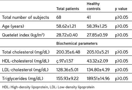
Similarly, no significant differences were determined in the comparison of total cholesterol, HDL-cholesterol LDL-cholesterol and triglyceride levels between total patients and healthy controls (p>0.05).
Serum VN (112.72±4.34 µg/mL) and sE-cadherin (5.15±0.40 ng/mL) levels of patients were found to be significantly lower than healthy controls (134.87±4.59 µg/mL and 23.48±2.12 ng/mL recpectively) (p<0.01). However, in patients, serum TGF-β1 levels (173.74±12.31 pg/mL) were found to be higher than healthy controls (133.50±14.37 pg/mL) (p<0.05, Figure 1).
Figure 1.
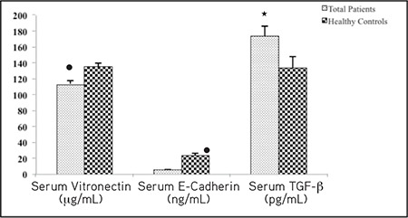
The comparison of serum concentrations of VN, sE-cadherin and TGF-β1 between total patient group and control group
●: A significant difference from control group (p<0.01), ★: A significant difference from control group (p<0.05), VN: Vitronectin, TGF- β1: Transforming growth factor-beta 1, sE-cadherin: Soluble epithelial-cadherin
In patients with endometrial and ovarian cancer, serum VN levels (117.07±5.59 µg/mL and 109.68±6.27 µg/mL, respectively) were found to be lower than healthy controls (134.87±4.59 µg/mL) (p<0.01) (Figure 2). The lower VN levels were determined in ovarian cancer group compared to endometrial cancer group. Similarly serum sE-cadherin levels in endometrial and ovarian cancer patient groups (4.76±0.63 ng/mL and 5.42±0.53 ng/mL, respectively) were found to be lower than healthy controls (23.48±2.12 ng/mL) (p<0.01), but the sE-cadherin levels were found to be lower in patients with endometrial cancer in comparison to ovarian cancer patient group. Serum TGF-β1 levels were found to be increased significantly in ovarian cancer patiens when compared to control group (180.31±11.61 pg/mL and 133.50±14.37 pg/mL, recpectively) (p<0.05). However, no significant difference was found between endometrial cancer patients and healthy controls (164.36±25.10 pg/mL and 133.50±14.37 pg/mL, recpectively) (p>0.05) (Figure 2).
Figure 2.
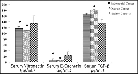
The comparison of serum concentrations of VN, sE-cadherin and TGF-β1 between endometrial and ovarian cancers and control group
●: A significant difference from control group (p<0.01), VN: Vitronectin, TGF- β1: Transforming growth factor-beta 1, sE-cadherin: Soluble epithelialcadherin
Using bivariate correlation analysis of the measured parameters, positive correlations were found between serum VN and sE-cadherin levels (r=0.775, p<0.01) (Figure 3a) and negative correlations were found between both VN and TGF-β1 (r=-0.379, p<0.05) (Figure 3b) and sE-cadherin and TGF-β1 (r=-0.373, p<0.05) (Figure 3c) in control group.
Figure 3a.
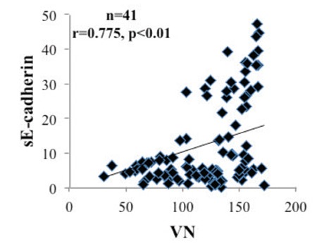
Correlation between serum VN and sE-cadherin in control group, The solid line represents the calculated regression line with a correlation coefficient (r) of 0.775
VN: Vitronectin, sE-cadherin: Soluble epithelial-cadherin
Figure 3b.
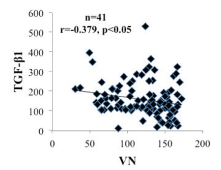
Correlation between serum VN and TGF-β1 in control group, The solid line represents the calculated regression line with a correlation coefficient (r) of -0.379
VN: Vitronectin, TGF- β1: Transforming growth factor-beta 1,
Figure 3c.
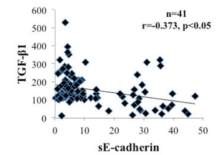
Correlation between serum sE-cadherin and TGF-β1 in control group, The solid line represents the calculated regression line with a correlation coefficient (r) of -0.373
TGF- β1: Transforming growth factor-beta 1, sE-cadherin: Soluble epithelialcadherin
DISCUSSION
Endometrial and ovarian cancers are the most common of the gynecologic malignancies in women. Regulation of cellular adhesion is provided by signaling pathways between tumor cells and the ECM. Cellular adhesion is controlled by the cell surface receptor family and integrins. VN, sE-cadherin and TGF-β1 are important components of the ECM proteins. Thus, we mainly tried to demonstrate the clinical significance of serum levels of these molecules in terms of diagnostic purpose in endometrial and ovarian cancers.
There are inconsistent results in the literature concerning serum VN levels in various cancer types. In one of the studies, Kadowaki et al.25 showed that serum VN levels were elevated in breast cancer patients. They concluded that serum VN level is very valuable for evaluating clinical assessment of breast cancer. Contrary to the aforementioned study, Hao et al.26 found that the mean value of serum VN level in patients with breast cancer at early and late stages were lower than normal individuals. Yamada et al.27 studied the plasma concentration of VN in control subjects and hepatocellular carcinoma (HCC). They obtain similar lower plasma VN levels in patients with HCC as compared to control group. No significant difference in VN levels was reported by Tugcu et al.28 between control and patient groups in various types of cancers apart from gynecological cancers. In this study concerning the VN levels when compared to control group a significant difference was found both for endometrial and for ovarian cancer patient groups. Decrease in serum VN levels may be attributed to matrix metalloproteinase-2 secreted by tumor cells in ECM.
Cell to cell adhesion is basically mediated by cadherins. There are conflicting results in the literature about sE-cadherin in cancer. Elevated sE-cadherin levels were reported in gastric cancer by Katayama et al.29, later on it was confirmed by Gofuku et al.30 In newly diagnosed bladder cancer patients, serum sE-cadherin levels were found to be significantly higher than normal controls and they suggested that high levels of sE-cadherin correlate with higher grade tumors.31 Similarly, Liang et al.16 were revealed that the serum sE-cadherin in breast cancer patients were significantly higher than controls. Shirahama et al.32 indicated that the levels of serum sE-cadherin did not vary significantly from controls in basal cell carcinoma. In colorectal patients, Velikova et al.33 determined that there was no statistically significant difference between sE-cadherin levels between controls and patients. However, Bonaldi et al.17 suggested that sE-cadherin was lower in patients with prostate cancer compared to controls. Several studies concluded that decreased expression of E-cadherin facilitated tumor invasion and metastasis in various tumors such as ovarian, endometrial and cervical cancers.34,35 In our study we determined that sE-cadherin levels were decreased in both cancer groups. We think that low serum sE-cadherin levels may be result of decreased E-cadherin expression in these gynecological cancers.
For many cancer, circulating levels of TGF-β1 have been measured up to now. Elevated serum TGF-β1 levels have been observed in various cancer types and have been linked to cancers in previous studies. Shim et al.20 evaluated the serum levels of TGF-β1 in patients with colorectal carcinoma versus healthy controls and their results showed that TGF-β1 serum levels in patients were significantly higher than controls. TGF-β1 has been reported to be enhanced in serum in invasive breast cancer patients.36 In a similar manner, it was demonstrated that serum concentrations of TGF-β1 in gastric cancer patients were significantly higher than controls.37 Han et al.23 were revealed that patients with cholangiocarcinomas, HCCs and gastric carcinomas presented increased serum TGF-b1 levels than non-cancer counterparts. It has been shown that the blockage of TGF-β causes upregulation of E-cadherin resulting in the reduction of migration and invasion of carcinoma cells38 and then TGF-β1 expression negatively correlated with E-cadherin expression.39 Our results are consistent with the literature in terms of elevated serum TGF-β1 levels and reduced sE-cadherin levels. It was assumed that increased TGF-β1 levels may be associated with decreased E-Cadherin expression in endometrial and ovarian cancers.40
CONCLUSION
As a conclusion, our study revealed that when compared to control group serum levels of VN and sE-cadherin are significantly decreased whereas TGF-β1 level is elevated in patients with gynecological cancer. In ovarian cancer, we have found marked increase of TGF-β1 and decrease of VN. A remarkable decrease in sE-cadherin concentration has been determined in patients with endometrial cancer. Although large-scale studies are required, it was suggested that alterations in serum levels of mentioned relatively small molecules in ovarian and endometrial cancer may be associated with disease progression. Therefore these molecules may be promising targets for both diagnosis and therapy.
Footnotes
Conflict of Interest: No conflict of interest was declared by the authors.
References
- 1.Jemal A, Siegel R, Ward E, Murray T, Xu J, Thun MJ. Cancer statistics, 2007. CA Cancer J Clin. 2007;57:43–66. doi: 10.3322/canjclin.57.1.43. [DOI] [PubMed] [Google Scholar]
- 2.Nguyen L, Cardenas-Goicoechea SJ, Gordon P, Curtin C, Momeni M, Chuang L, Fishman D. Biomarkers for early detection of ovarian cancer. Womens Health (Lond). 2013;9:171–187. doi: 10.2217/whe.13.2. [DOI] [PubMed] [Google Scholar]
- 3.Turgut A, Ozler A, Sak ME, Evsen MS, Soydinç HE, Alabalık U, Gül T. Retrospective analysis of the patients with gynecological cancer: 11- Year Experience. J Clin Exp Invest. 2012;3:209–213. [Google Scholar]
- 4.Ergör G. Non Contagious Diseases in Turkey. Cancer Mortality. In: Unal B, ed. Turkish Public Health Report. 2012:286–287. [Google Scholar]
- 5.Preissner KT. Structure and Biological Role of Vitronectin. Annu Rev Cell Biol. 1991;7:275–310. doi: 10.1146/annurev.cb.07.110191.001423. [DOI] [PubMed] [Google Scholar]
- 6.De Wever O, Mareel M. Role of tissue stroma in cancer cell invasion. J Pathol. 2003;200:429–447. doi: 10.1002/path.1398. [DOI] [PubMed] [Google Scholar]
- 7.Tomasini BR, Mosher DF. Vitronectin. Prog Hemost Thromb. 1990;10:269–305. [PubMed] [Google Scholar]
- 8.Juliano RL, Varner JA. Adhesion molecules in cancer: the role of integrins. Curr Opin Cell Biol. 1993;5:812–818. doi: 10.1016/0955-0674(93)90030-t. [DOI] [PubMed] [Google Scholar]
- 9.Madsen CD, Sidenius N. The interaction between urokinase receptor and vitronectin in cell adhesion and signalling. Eur J Cell Biol. 2008;87:617–629. doi: 10.1016/j.ejcb.2008.02.003. [DOI] [PubMed] [Google Scholar]
- 10.Kashyap AS, Hollier BG, Manton KJ, Satyamoorthy K, Leavesley DI, Upton Z. Insulin-like growth factor-I: vitronectin complex-induced changes in gene expression effect breast cell survival and migration. Endocrinology. 2011;152:1388–1401. doi: 10.1210/en.2010-0897. [DOI] [PubMed] [Google Scholar]
- 11.Pola C, Formenti SC, Schneider RJ. Vitronectin-alphavbeta3 integrin engagement directs hypoxia-resistant mTOR activity and sustained protein synthesis linked to invasion by breast cancer cells. Cancer Res. 2013;73:4571–4578. doi: 10.1158/0008-5472.CAN-13-0218. [DOI] [PubMed] [Google Scholar]
- 12.Mărgineanu E, Cotrutz CE, Cotrutz C. Correlation between E-cadherin abnormal expressions in different types of cancer and the process of metastasis. Rev Med Chir Soc Med Nat Iasi. 2008;112:432–436. [PubMed] [Google Scholar]
- 13.Chetty R, Serra S. Nuclear E-cadherin immunoexpression: from biology to potential application in diagnostic pathology. Adv Anat Pathol. 2008;15:234–240. doi: 10.1097/PAP.0b013e31817bf566. [DOI] [PubMed] [Google Scholar]
- 14.Ho CM, Cheng WF, Lin MC, Chen TC, Huang SH, Liu FS, Chien CC, Yu MH, Wang TY, Hsieh CY. Prognostic and predictive values of E-cadherin for patients of ovarian clear cell adenocarcinoma. Int J Gynecol Cancer. 2010;20:1490–1497. doi: 10.1111/IGC.0b013e3181e68a4d. [DOI] [PubMed] [Google Scholar]
- 15.Holcomb K, Delatorre R, Pedemonte B, McLeod C, Anderson L, Chambers J. E-cadherin expression in endometrioid, papillary serous, and clear cell carcinoma of the endometrium. Obstet Gynecol. 2002;100:1290–1295. doi: 10.1016/s0029-7844(02)02391-8. [DOI] [PubMed] [Google Scholar]
- 16.Liang Z, Sun XY, Xu LC, Fu RZ. Abnormal expression of serum soluble E-cadherin is correlated with clinicopathological features and prognosis of breast cancer. Med Sci Monit. 2014;20:2776–2782. doi: 10.12659/MSM.892049. [DOI] [PMC free article] [PubMed] [Google Scholar]
- 17.Bonaldi C, Azzalis LA, Junqueira VB, de Oliveira CG, Vilas Boas VA, Gáscon TM, Gehrke FS, Kuniyoshi RK, Alves BC, Fonseca FL. Plasma levels of E-cadherin and MMP-13 in prostate cancer patients: correlation with PSA, testosterone and pathological parameters. Tumori. 2015;101:185–188. doi: 10.5301/tj.5000237. [DOI] [PubMed] [Google Scholar]
- 18.Massague J. TGF-β signal transduction. Annu Rev Biochem. 1998;67:753–791. doi: 10.1146/annurev.biochem.67.1.753. [DOI] [PubMed] [Google Scholar]
- 19.Akhurst RJ, Derynck R. TGF-β signaling in cancer-a double-edged sword. Trends Cell Biol. 2001;11:44–51. doi: 10.1016/s0962-8924(01)02130-4. [DOI] [PubMed] [Google Scholar]
- 20.Shim KS, Kim KH, Han WS, Park EB. Elevated serum levels of transforming growth factor-beta1 in patients with colorectal carcinoma: its association with tumor progression and its significant decrease after curative surgical resection. Cancer. 1999;85:554–561. doi: 10.1002/(sici)1097-0142(19990201)85:3<554::aid-cncr6>3.0.co;2-x. [DOI] [PubMed] [Google Scholar]
- 21.Zhao J, Liang Y, Yin Q, Liu S, Wang Q, Tang Y, Cao C. Clinical and prognostic significance of serum transforming growth factor-beta1 levels in patients with pancreatic ductal adenocarcinoma. Braz J Med Biol Res. 2016;25:49. doi: 10.1590/1414-431X20165485. [DOI] [PMC free article] [PubMed] [Google Scholar]
- 22.Loh JK, Lieu AS, Su YF, Cheng CY, Tsai TH, Lin CL, Lee KS, Hwang SL, Kwan AL, Wang CJ, Hong YR, Howng SL, Chio CC. The alteration of plasma TGF-β1 levels in patients with brain tumors after tumor removal. Kaohsiung J Med Sci. 2012;28:316–321. doi: 10.1016/j.kjms.2011.11.012. [DOI] [PubMed] [Google Scholar]
- 23.Han B, Cai H, Chen Y, Hu B, Luo H, Wu Y, Wu J. The role of TGFBI (βig-H3) in gastrointestinal tract tumorigenesis. Mol Cancer. 2015;14:64. doi: 10.1186/s12943-015-0335-z. [DOI] [PMC free article] [PubMed] [Google Scholar]
- 24.Friedewald WT, Levy RI, Fredrıckson DS. Estimation of the concentration of low-density lipoprotein cholesterol in plasma, without use of the preparative ultracentrifuge. Clin Chem. 1972;18:499–502. [PubMed] [Google Scholar]
- 25.Kadowaki M, Sangai T, Nagashima T, Sakakibara M, Yoshitomi H, Takano S, Sogawa K, Umemura H, Fushimi K, Nakatani Y, Nomura F, Miyazaki M. Identification of vitronectin as a novel serum marker for early breast cancer detection using a new proteomic approach. J Cancer Res Clin Oncol. 2011;137:1105–1115. doi: 10.1007/s00432-010-0974-9. [DOI] [PubMed] [Google Scholar]
- 26.Hao W, Zhang X, Xiu B, Yang X, Hu S, Liu Z, Duan C, Jin S, Ying X, Zhao Y, Han X, Hao X, Fan Y, Johnson H, Meng D, Persson JL, Zhang H, Feng X, Huang Y. Vitronectin: a promising breast cancer serum biomarker for early diagnosis of breast cancer in patients. Tumour Biol. 2016;7:8909–8916. doi: 10.1007/s13277-015-4750-y. [DOI] [PubMed] [Google Scholar]
- 27.Yamada S, Kobayashi J, Murawaki Y, Suou T, Kawasaki H. Collagenbinding activity of plasma vitronectin in chronic liver disease. Clin Chim Acta. 1996;252:95–103. doi: 10.1016/0009-8981(96)06320-6. [DOI] [PubMed] [Google Scholar]
- 28.Tugcu D, Devecioglu O, Unuvar A, Ekmekci H, Ekmekci OB, Anak S, Ozturk G, Akcay A, Aydogan G. Plasma Levels of Plasminogen Activator Inhibitor Type 1 and Vitronectin in Children With Cancer. Clin Appl Thromb Hemost. 2016;22:28–33. doi: 10.1177/1076029614531450. [DOI] [PubMed] [Google Scholar]
- 29.Katayama M, Hirai S, Kamihagi K, Nakagawa K, Yasumoto M, Kato I. Soluble E-cadherin fragments increased in circulation of cancer patients. Br J Cancer. 1994;69:580–585. doi: 10.1038/bjc.1994.106. [DOI] [PMC free article] [PubMed] [Google Scholar]
- 30.Gofuku J, Shiozaki H, Doki Y, Inoue M, Hirao M, Fukuchi N, Monden M. Characterization of soluble E-cadherin as a disease marker in gastric cancer patients. Br J Cancer. 1998;78:1095–1101. doi: 10.1038/bjc.1998.634. [DOI] [PMC free article] [PubMed] [Google Scholar]
- 31.Griffiths TR, Brotherick I, Bishop RI, White MD, McKenna DM, Horne CH, Shenton BK, Neal DE, Mellon JK. Cell adhesion molecules in bladder cancer: soluble serum E-cadherin correlates with predictors of recurrence. Br J Cancer. 1996;74:579–584. doi: 10.1038/bjc.1996.404. [DOI] [PMC free article] [PubMed] [Google Scholar]
- 32.Shirahama S, Furukawa F, Wakita H, Takigawa M. E- and P-cadherin expression in tumor tissues and soluble E-cadherin levels in sera of patients with skin cancer. J Dermatol Sci. 1996;13:30–36. doi: 10.1016/0923-1811(95)00493-9. [DOI] [PubMed] [Google Scholar]
- 33.Velikova G, Banks RE, Gearing A, Hemingway I, Forbes MA, Preston SR, Hall NR, Jones M, Wyatt J, Miller K, Ward U, Al-Maskatti J, Singh SM, Finan PJ, Ambrose NS, Primrose JN, Selby PJ. Serum concentrations of soluble adhesion molecules in patients with colorectal cancer. Br J Cancer. 1996;77:1857–1863. doi: 10.1038/bjc.1998.309. [DOI] [PMC free article] [PubMed] [Google Scholar]
- 34.Varras M, Skafida E, Vasilakaki T, Anastasiadis A, Akrivis C, Vrachnis N, Nikolopoulos G. Expression of E-cadherin in primary endometrial carcinomas: clinicopathological and immunohistochemical analysis of 30 cases. Eur J Gynaecol Oncol. 2013;34:31–35. [PubMed] [Google Scholar]
- 35.Zhou XM, Zhang H, Han X. Role of epithelial to mesenchymal transition proteins in gynecological cancers: pathological and therapeutic perspectives. Tumour Biol. 2014;35:9523–9530. doi: 10.1007/s13277-014-2537-1. [DOI] [PubMed] [Google Scholar]
- 36.Sheen-Chen SM, Chen HS, Sheen CW, Eng HL, Chen WJ. Serum levels of transforming growth factor beta1 in patients with breast cancer. Arch Surg. 2001;136:937–940. doi: 10.1001/archsurg.136.8.937. [DOI] [PubMed] [Google Scholar]
- 37.Li X, Yue ZC, Zhang YY, Bai J, Meng XN, Geng JS, Fu SB. Elevated serum level and gene polymorphisms of TGF-beta1 in gastric cancer. J Clin Lab Anal. 2008;22:164–171. doi: 10.1002/jcla.20236. [DOI] [PMC free article] [PubMed] [Google Scholar]
- 38.Fransvea E, Angelotti U, Antonaci S, Giannelli G. Blocking transforming growth factor-beta up-regulates E-cadherin and reduces migration and invasion of hepatocellular carcinoma cells. Hepatology. 2008;47:1557–1566. doi: 10.1002/hep.22201. [DOI] [PubMed] [Google Scholar]
- 39.Liu GL, Yang HJ, Liu T, Lin YZ. Expression and significance of E-cadherin, N-cadherin, transforming growth factor-β1 and Twist in prostate cancer. Asian Pac J Trop Med. 2014;7:76–82. doi: 10.1016/S1995-7645(13)60196-0. [DOI] [PubMed] [Google Scholar]
- 40.Cho IJ, Kim YW, Han CY, Kim EH, Anderson RA, Lee YS, Lee CH, Hwang SJ, Kim SG. E-cadherin antagonizes transforming growth factor β1 gene induction in hepatic stellate cells by inhibiting RhoA-dependent Smad3 phosphorylation. Hepatology. 2010;52:2053–2064. doi: 10.1002/hep.23931. [DOI] [PMC free article] [PubMed] [Google Scholar]


