Abstract
Objectives:
Chronic myelogenous leukemia (CML) is a type of blood cancer that is initially treated with imatinib (first Abl kinase inhibitor). However, some patients with CML develop imatinib resistance. Several new generation drugs have been developed, but do not overcome this problem. Glycyrrhetic acid (GA) is a plant-derived pentacyclic triterpenoid that exhibits multiple pharmacological properties for the treatment of cancers. The current study aimed to investigate the effects of GA on the K562 cell line (Bcr-Abl positive leukemia).
Materials and Methods:
The MTT cell proliferation assay was employed to evaluate the cytotoxic effect of GA compared with imatinib (positive control) against leukemia and normal blood cells. For detection of cell death, an apoptotic/necrotic/healthy assay was performed against the K562 cell line. To investigate the kinase inhibitory activity of GA, the Abl1 kinase profiling assay and a molecular docking study were performed.
Results:
GA showed Abl kinase inhibitory activity with an IC50 value of 29.2 μM and induced apoptosis in the K562 cell line after 6 h of treatment.
Conclusion:
The current findings indicate that this class of plant extract could be a potential candidate for treatment of CML.
Keywords: Pentacyclic triterpenoid, glycyrrhetic acid, Abl kinase, apoptosis, K562 cells
INTRODUCTION
One of the major health problems all over the world is cancer. In 2015, over 8.7 million patients died from cancer globally, with approximately 17.5 million new cases.1,2 Despite extensive efforts over the last several decades, cancer is still one of the most common causes of death in many developing nations. The death rate from cancer is predicted to be 17 million with 26 million new cases each year by 2030.3,4 Due to this global increase in the cancer burden, a massive research effort has been made for the discovery of effective and less toxic chemotherapeutic agents.5,6,7,8 Over the past few years tyrosine kinases have attracted growing interest as drug targets in anticancer drug discovery that play an important role in signal transduction, mitogenesis, and other cellular activation processes.9,10,11 More than 25 kinase inhibitors are available for cancer therapy and there are currently many more promising candidates in clinical development12,13 after the approval of imatinib as the first Bcr-Abl tyrosine kinase inhibitor in 2002.14 Although imatinib continues to be the initial choice in chronic myelogenous leukemia (CML) treatment, some patients with CML develop drug resistance to imatinib.14,15 This major limitation is becoming a significant concern for patients with imatinib-resistant chronic-phase CML.16 Several new generation drugs have been developed, but there are still no alternative drugs available to overcome this problem.17,18Pentacyclic triterpenoids have emerged as a unique class of natural compounds and have been studied extensively for more than a century due to their effective therapeutic applications for the treatment of a wide spectrum of diseases and their high safety profile.19,20 Recent studies have indicated that two different types of pentacyclic triterpenoids, celastrol and gypsogenin, exert anti-Abelson kinase 1 (Abl1) and anti-CML leukemia effects.21,22
Glycyrrhetic acid (GA) is an olenane-type natural pentacyclic triterpenoid extracted from liquorice that exhibits promising anticancer activity on many cancer cells including human ovarian cancer, breast cancer, hepatocellular carcinoma, pituitary adenoma, human bladder cancer, lung cancer, and leukemia.19,23,24,25,26,27 However, there is no reported research yet in the literature that displays Bcr-Abl inhibitory activity of GA. In the present study, we explored the biological activities of GA against leukemia cell lines (Jurkat, MT-2, and K562) and normal cells of human blood peripheral blood mononuclear cells (PBMCs) and then evaluated its antityrosine kinase activity. Furthermore, apoptotic/necrotic analysis against the K562 cell line and molecular docking with the Abl kinase domain were carried out using GA.
MATERIALS AND METHODS
Cell culture conditions and drug treatment
The K562, Jurkat, and MT-2 cell lines were cultured in RPMI 1640 (Wako Pure Chemical Industries, Osaka, Japan) medium with 10% fetal bovine serum (Equitech-Bio, Kerrville, TX, USA) and 89 μM/mL streptomycin (Meiji Seika Pharma, Tokyo, Japan) in a humid atmosphere at 37°C and 5% CO2. PBMCs (Precision Bioservices, Frederic, MD, USA) were incubated in RPMI 1640 medium with 10% human serum AB (Gemini, Woodland, CA, USA) and 89 μM/mL streptomycin at 37°C (humid atmosphere, 5% CO2). In the experiments, the leukemia and PBMCs were incubated in 24-well culture plates at 105 and 106 cells/mL concentration respectively for 48 h. The stock solutions of GA (Tokyo Chemical Industry Co. Ltd., Tokyo, Japan) and imatinib (Wako Pure Chemical Industries, Osaka, Japan) in concentrations of 2.5 mM, 5 mM, 10 mM, 20 mM, and 30 mM were prepared in DMSO (Wako Pure Chemical Industries, Osaka, Japan). The concentration of DMSO in the final culture medium was 1%.
MTT assay for cytotoxicity
The MTT test was performed routinely as described in the literature.28,29 GA and imatinib were cultured with cells in different concentrations (3-300 μM). After 48 h of treatment, cells were incubated with MTT (Dojindo Molecular Technologies, Kumamoto, Japan) solution in medium for 4 h. At the end of incubation, the solution was taken out and 100 µL of DMSO was added to each well. The absorbance of the solution was measured in a microplate reader, Infinitive M1000 (Tecan, Groding, Austria), at a wavelength of 550 nm with background subtraction at 630 nm. All experiments were run in triplicate and cell viability was calculated as the percentage of viable control cells. IC50 values were estimated from the results of the MTT test described as the drug concentrations that reduced absorbance to 50% of control values.
Detection of cell death
After treatment of K562 cells with GA or imatinib at IC50 concentrations for 6 h, a apoptotic/necrotic/healthy detection kit (PromoKine, Heidelberg, Germany) was used according to PromoKine’s instructions with modifications.30 After the cells were harvested and washed with PBS, the cells were suspended with binding buffer (1X). After that, 50 µL of binding buffer, 4 µL of FITC-annexin V solution, 4 µL of ethidium homodimer III solution, and 4 µL of Hoechst 33342 solution were added to the cells for 30 min at room temperature in the dark. Then the cells were analyzed by a fluorescence microscope, Biorevo Fluorescence BZ-9000 (Keyence, Osaka, Japan). The number of apoptotic cells (annexin V), late apoptotic or necrotic cells (annexin V and ethidium homodimer III), and necrotic cells (ethidium homodimer III) were counted as previously described.31
Abl1 tyrosine kinase profiling system
The Abl1 kinase profiling assay (Promega Corporation, Madison, WI, USA) was performed as previously described with modifications.21 In this system, Abl1 kinase strip and its substrate were diluted with 95 µL of 2.5X kinase reaction buffer and 15 µL of 100 µM ATP. Then 2 µL of kinase working stock and 2 µL of ATP/substrate working stock were dispensed in the 384-well plate wells along with 1 µL of compound solution at varying concentrations (10-300 µM) in a buffer. The kinase reaction was incubated for 1 h at room temperature and then the activity of Abl1 kinase was detected using the ADP-Glo kinase assay (Promega Corporation). Abl1 inhibition profiling of GA in dose-response mode was measured by a luminescence microplate reader, Infinitive M1000 (Tecan). IC50 values of GA and imatinib required to reduce kinase activity by 50% were calculated using the software Image J.
Molecular modeling
To investigate the binding modes of GA with Abl1 kinase, a molecular docking study was performed using Molecular Operating Environment 2015.10 (Chemical Computing Group, Montreal, Canada). The co-crystal structure of Abl tyrosine kinase with imatinib was obtained as the docking template from the Protein Data Bank (PDB code: 1IEP).32 Then the Abl kinase and GA were prepared for molecular docking analysis including the addition of hydrogen atoms, the assignment of bond order, assessment of the correct protonation state, and other default parameters. All molecular docking calculations were performed as previously described.33,34
RESULTS
In the present study, we first performed the MTT assay to investigate the antiproliferative effects of GA and imatinib against multiple human leukemia cells (K562 CML, Jurkat, and MT-2) at various concentrations (10-300 μM). Imatinib was selected as a model drug, considering its wide use in the treatment of CML. GA (Figure 1) and imatinib were dissolved in DMSO, diluted with culture medium, and then treated with cultured cells for 48 h. The IC50 values of these compounds on three cancer cell lines are shown in Table 1. GA exhibited a concentration-dependent inhibitory effect with IC50 values that were less than 75 μM against all three cancer cell lines. It possessed the most potent cytotoxic activity against imatinib-sensitive K562 cells with an IC50 value of 51.6 μM (Figure 2A), while the IC50 values of GA on Jurkat and MT-2 cells were 55.1 μM and 70.2 μM, respectively, weaker than those of the positive control. Next the activity of the target compound was examined on normal PBMCs and compared with imatinib (Figure 2B). GA did not show considerable cytotoxicity against PBMCs, with an IC50 value of 117.5 μmM, and exhibited ~3.5 times lower cytotoxicity than imatinib (Figure 2B). These results indicate that GA can act as an anti-CML agent and exhibits good selectivity for K562 cell lines over normal cells.
Figure 1.
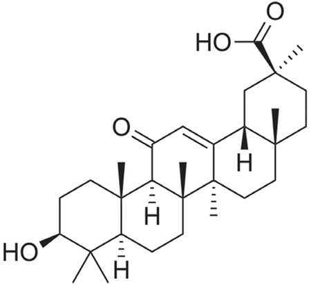
Structure of glycyrrhetic acid
Table 1. IC50 values of GA and imatinib for K562, Jurkat, MT-2 and peripheral blood mononuclear cells after 48 h treatment.
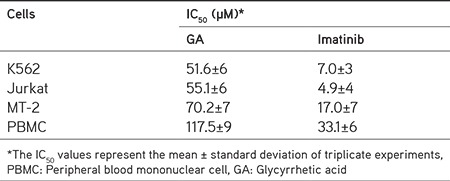
Figure 2.
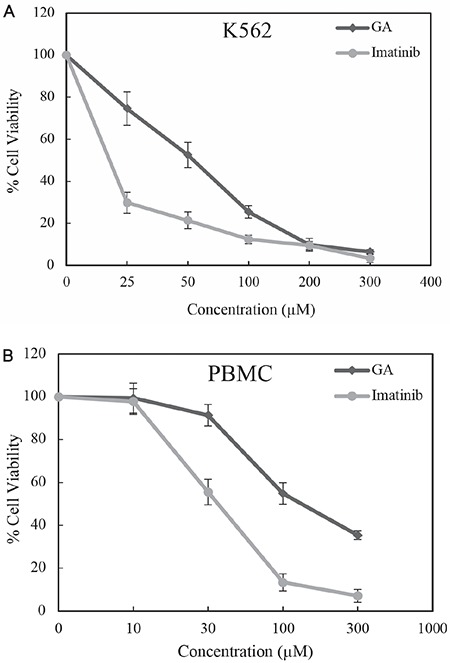
Cell proliferation inhibition of GA and imatinib on (A) K562 and (B) PBMC cells after 48 h incubation
PBMC: Peripherol blood mononuclear cell, GA: Glycyrrhetic acid
In order to investigate the process of apoptosis and necrosis, K562 cells treated with GA or imatinib at IC50 concentrations were subjected to the annexin V/ethidium homodimer III and Hoechst 33342 staining method and then observed by a fluorescence microscope (Figure 3). In the control experiment (1% DMSO), all cells were healthy (blue staining) at 6 h (Figure 3A). On the other hand, the cells treated with GA and imatinib were stained mostly with healthy cells (blue) and then apoptotic cells (green), and only a few necrotic cells (red) and late apoptotic or necrotic cells (both green and red) were detected at 6 h (Figure 3A), suggesting that the main cell death pathway of GA and imatinib was apoptosis at an earlier time. The results showed that GA has 71% apoptotic, 9% necrotic, and 20% late apoptotic/necrotic activities (Figure 3B). In contrast, the response of K562 cells upon 6 h imatinib treatment was 62% apoptosis, 11% necrosis, and 27% late apoptosis/necrosis (Figure 3B). Surprisingly, the results demonstrated that GA is able to induce more cell apoptosis than imatinib in Bcr-Abl positive cells.
Figure 3.
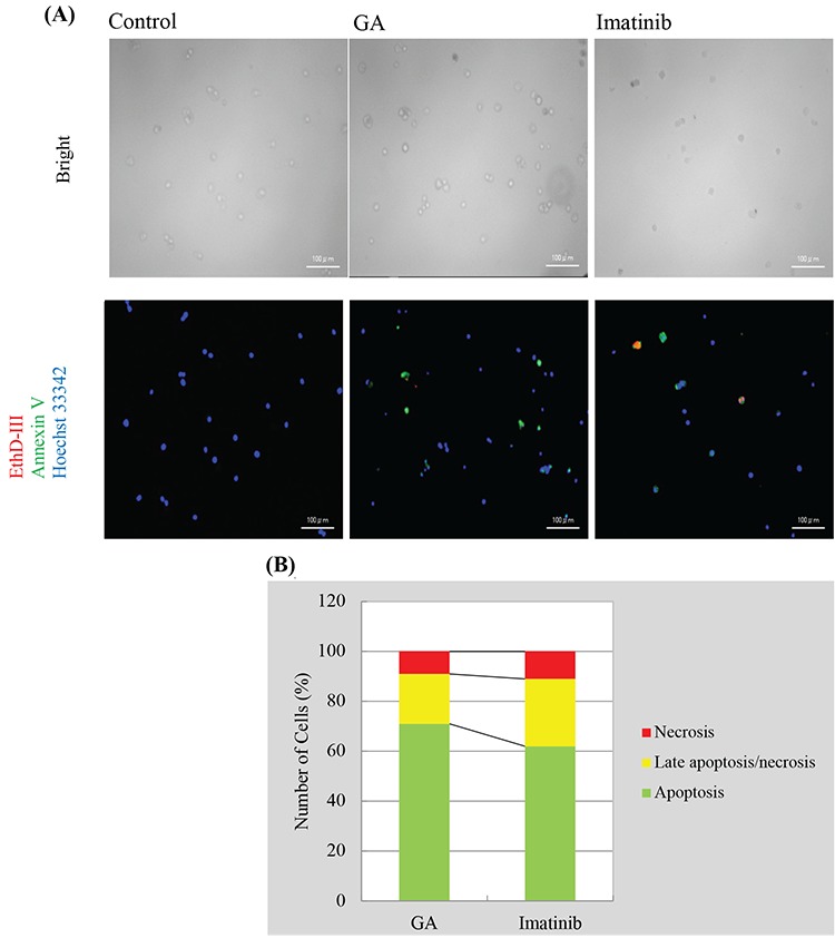
Alteration in K562 cells at IC50 concentrations of control, GA, and imatinib (A) for 6 h. (B) A total of approximate 100 stained cells were selected randomly in each experiment of (A) and were classified into 3 types: “apoptosis” (green), “necrosis or late apoptosis” (both green and red), and “necrosis” (red)
GA: Glycyrrhetic acid
To explore the inhibition profile of GA on Bcr-Abl, we used the Abl1 tyrosine activity-based kinase assay. In this system, GA was screened at multiple concentrations (10-300 μM) to determine its inhibitory profile on the target kinase (Abl1 tyrosine kinase). GA displayed potential Bcr-Abl inhibitory activity with an IC50 value of 29.2 μM as shown in Figure 4. Imatinib was included for comparison and showed a stronger inhibitory effect than GA on Abl1. In order to understand the Bcr-Abl kinase inhibitory activity of GA, we next examined molecular modeling based on the co-crystal structure of Abl with imatinib as the docking model (PDB ID code: 1IEP). GA fitted into the pocket, forming five noncovalent interactions with four amino acid residues, namely His361, Arg362, Asp381, and Ala380 (Figure 5). It is clear that GA carboxylate plays a pivotal role in activity by forming two H bonds with the basic amino acid Arg362. Binding energy values of GA and imatinib into the pocket are -7.2 and -11.1 kcal/mol, respectively, in agreement with the experimental results.
Figure 4.
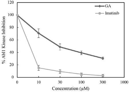
Effect of GA and imatinib on Abl1 tyrosine kinase at varying concentrations (10-300 μM), GA: Glycyrrhetic acid
Figure 5.
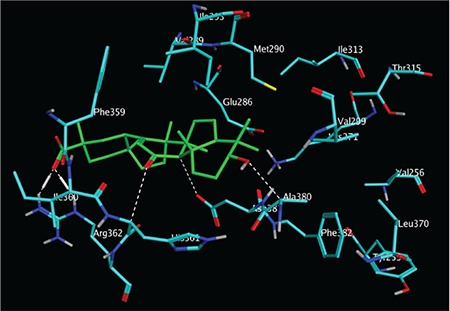
The binding poses of GA in Bcr-Abl tyrosine kinase (PDB ID: 1IEP). GA is shown as green sticks, key amino acid residues are visualized in cyan sticks, and settled interactions are indicated as dotted white lines)
GA: Glycyrrhetic acid
DISCUSSION
CML is a cancer of white blood cells mainly caused by Bcr-Abl. Bcr-Abl tyrosine kinase inhibitors including imatinib have demonstrated significant therapeutic effects in many CML patients. However, resistance and toxicity of these inhibitors have been frequently reported in recent years. Therefore, novel Bcr-Abl inhibitors with high efficacy and low toxicity for treatment of CML are still sought. The accumulated evidence shows that GA has antitumor activities in breast cancer, ovarian cancer, and acute promyelocytic leukemia, but its activity against CML is yet to be investigated.
In the present study, we explored the cytotoxic activity of GA against different leukemic cell lines (K562, Jurkat, and MT-2) and found that GA possesses a remarkable antiproliferative effect on the K562 Bcr-Abl positive cell line. Moreover, GA induced programmed cell death in CML cells more efficiently than imatinib at 6 h of treatment and showed significant tumor selectivity on blood cells (PBMC and K562). To get more insights into GA’s molecular mechanism, we assessed its effect on Abl1 kinase, which is amplified in K562 cells. As anticipated, GA inhibited Abl1 kinase with an IC50 of 29.2 μM. Molecular modeling simulation provided mechanistic information on the possible binding mode of GA into the ATP binding site of Abl1 kinase.
CONCLUSION
Recently, we have also revealed the activity of gypsogenin, which is another pentacyclic triterpenoid, and its derivatives against the K562 cell line.21 Considering previous and current data together, our findings suggest that PTs have promising anticancer roles and deserve particular attention in the treatment of CML. We think that derivatization of GA will enhance its binding affinity into Bcr-Abl kinase, which in turn will enhance its anticancer activity. Further derivatizations and biological investigations for improvement of GA activity are ongoing.
Acknowledgments
The author thanks Prof. Masami Otsuka, Dr. Mikako Fujita, Dr. Mohamad O. Radwan, and Dr. Nizar Turker for their helpful discussions on molecular modeling and English editing. The work was supported by Project P16111 from Japan Society for the Promotion of Science (JSPS).
Footnotes
Conflict of Interest: No conflict of interest was declared by the authors.
References
- 1.Fitzmaurice C, Allen C, Barber RM, Barregard L, Bhutta ZA, Brenner H, Dicker DJ, Chimed- Orchir O, Dandona R, Dandona L, Fleming T, Forouzanfar MH, Hancock J, Hay RJ, Hunter-Merrill R, Huynh C, Hosgood HD, Johnson CO, Jonas JB, Khubchandani J, Kumar GA, Kutz M, Lan Q, Larson HJ, Liang X, Lim SS, Lopez AD, MacIntyre MF, Marczak L, Marquez N, Mokdad AH, Pinho C, Pourmalek F, Salomon JA, Sanabria JR, Sandar L, Sartorius B, Schwartz SM, Shackelford KA, Shibuya K, Stanaway J, Steiner C, Sun J, Takahashi K, Vollset SE, Vos T, Wagner JA, Wang H, Westerman R, Zeeb H, Zoeckler L, Abd-Allah F, Ahmed MB, Alabed S, Alam NK, Aldhahri SF, Alem G, Alemayohu MA, Ali R, Al-Raddadi R, Amare A, Amoako Y, Artaman A, Asayesh H, Atnafu N, Awasthi A, Saleem HB, Barac A, Bedi N, Bensenor I, Berhane A, Bernabé E, Betsu B, Binagwaho A, Boneya D, Campos-Nonato I, Castañeda-Orjuela C, Catalá-López F, Chiang P, Chibueze C, Chitheer A, Choi JY, Cowie B, Damtew S, das Neves J, Dey S, Dharmaratne S, Dhillon P, Ding E, Driscoll T, Ekwueme D, Endries AY, Farvid M, Farzadfar F, Fernandes J69, Fischer F, G/Hiwot TT, Gebru A, Gopalani S, Hailu A, Horino M, Horita N, Husseini A, Huybrechts I, Inoue M, Islami F, Jakovljevic M, James S, Javanbakht M, Jee SH, Kasaeian A, Kedir MS, Khader YS, Khang YH, Kim D, Leigh J, Linn S, Lunevicius R, El Razek HMA, Malekzadeh R, Malta DC, Marcenes W, Markos D, Melaku YA, Meles KG, Mendoza W, Mengiste DT, Meretoja TJ, Miller TR, Mohammad KA, Mohammadi A, Mohammed S, Moradi-Lakeh M, Nagel G, Nand D, Le Nguyen Q, Nolte S, Ogbo FA, Oladimeji KE, Oren E, Pa M, Park EK, Pereira DM, Plass D, Qorbani M, Radfar A, Rafay A, Rahman M, Rana SM, Søreide K, Satpathy M, Sawhney M, Sepanlou SG, Shaikh MA, She J, Shiue I, Shore HR, Shrime MG, So S, Soneji S, Stathopoulou V, Stroumpoulis K, Sufiyan MB, Sykes BL, Tabarés- Seisdedos R, Tadese F, Tedla BA, Tessema GA, Thakur JS, Tran BX, Ukwaja KN, Uzochukwu BSC, Vlassov VV, Weiderpass E, Wubshet Terefe M, Yebyo HG, Yimam HH, Yonemoto N, Younis MZ, Yu C, Zaidi Z, Zaki MES, Zenebe ZM, Murray CJL, Naghavi M; Global Burden of Disease Cancer Collaboration. Global, Regional, and national cancer incidence, mortality, years of life lost, years lived with disability, and disability-adjusted life-years for 32 cancer groups, 1990 to 2015: A Systematic Analysis for the Global Burden of Disease Study. JAMA Oncol. 2017;3:524–548. doi: 10.1001/jamaoncol.2016.5688. [DOI] [PMC free article] [PubMed] [Google Scholar]
- 2.Ferlay J, Soerjomataram I, Dikshit R, Eser S, Mathers C, Rebelo M, Parkin DM, Formann D, Bray F. Cancer incidence and mortality worldwide: sources, methods and major patterns in GLOBOCAN 2012. Int J Cancer. 2015;136:359–386. doi: 10.1002/ijc.29210. [DOI] [PubMed] [Google Scholar]
- 3.Thun MJ, DeLancey JO, Center MM, Jemal A, Ward EM. The global burden of cancer: Priorities for prevention. Carcinogenesis. 2010;31:100–110. doi: 10.1093/carcin/bgp263. [DOI] [PMC free article] [PubMed] [Google Scholar]
- 4.Torre LA, Bray F, Siegel RL, Ferlay J, Lortet-tieulent J, Jemal A. Global cancer statistics, 2012. CA Cancer J Clin. 2015;65:87–108. doi: 10.3322/caac.21262. [DOI] [PubMed] [Google Scholar]
- 5.Hoelder S, Clarke PA, Workman P. Discovery of small molecule cancer drugs: Successes, challenges and opportunities. Mol Oncol. 2012;6:155–176. doi: 10.1016/j.molonc.2012.02.004. [DOI] [PMC free article] [PubMed] [Google Scholar]
- 6.Cragg GM, Pezzuto JM. Natural products as a vital source for the discovery of cancer chemotherapeutic and chemopreventive agents. Med Princ Pract. 2016;25(Suppl 2):41–59. doi: 10.1159/000443404. [DOI] [PMC free article] [PubMed] [Google Scholar]
- 7.Ma X, Yu H. Global burden of cancer. Yale J Biol Med. 2006;79:85–94. [PMC free article] [PubMed] [Google Scholar]
- 8.Altıntop MD, Ciftci HI, Radwan MO, Sever B, Kaplancıklı ZA, Ali TFS, Koga R, Fujita M, Otsuka M, Özdemir A. Design, synthesis, and biological evaluation of novel 1,3,4-thiadiazole derivatives as potential antitumor agents against chronic myelogenous leukemia: Striking effect of nitrothiazole moiety. Molecules. 2017;23. doi: 10.3390/molecules23010059. [DOI] [PMC free article] [PubMed] [Google Scholar]
- 9.Schenk PW, Snaar-Jagalska BE. Signal perception and transduction: The role of protein kinases. Biochim Biophys Acta. 1999;1449:1–24. doi: 10.1016/s0167-4889(98)00178-5. [DOI] [PubMed] [Google Scholar]
- 10.Fabbro D, Fendrich G, Guez V, Meyer T, Furet P, Mestan J, Griffin JD, Manley PW, Cowan-Jacob SW. Targeted therapy with imatinib: An exception or a rule? Handbook of Experimental Pharmacology. 2005;167:361–389. [Google Scholar]
- 11.Wang Z, Cole PA. Catalytic mechanisms and regulation of protein kinases. Methods Enzymol. 2014;548:1–21. doi: 10.1016/B978-0-12-397918-6.00001-X. [DOI] [PMC free article] [PubMed] [Google Scholar]
- 12.Borriello A, Caldarelli I, Bencivenga D, Stampone E, Perrotta S, Oliva A, Della Ragione F. Tyrosine kinase inhibitors and mesenchymal stromal cells: Effects on self-renewal, commitment and functions. Oncotarget. 2017;8:5540–5565. doi: 10.18632/oncotarget.12649. [DOI] [PMC free article] [PubMed] [Google Scholar]
- 13.Gross S, Rahal R, Stransky N, Lengauer C, Hoeflich KP. Targeting cancer with kinase inhibitors. J Clin Invest. 2015;125:1780–1789. doi: 10.1172/JCI76094. [DOI] [PMC free article] [PubMed] [Google Scholar]
- 14.Mughal A, Aslam HM, Khan AM, Saleem S, Umah R, Saleem M. Bcr-Abl tyrosine kinase inhibitors- current status. Infec Agents Cancer. 2013;8:23. doi: 10.1186/1750-9378-8-23. [DOI] [PMC free article] [PubMed] [Google Scholar]
- 15.Soverini S, Hochhaus A, Nicolini FE, Gruber F, Lange T, Saglio G, Pane F, Müller MC, Ernst T, Rosti G, Porkka K, Baccarani M, Cross NC, Martinelli G. BCR-ABL kinase domain mutation analysis in chronic myeloid leukemia patients treated with tyrosine kinase inhibitors: Recommendations from an expert panel on behalf of European LeukemiaNet. Blood. 2011;118:1208–1215. doi: 10.1182/blood-2010-12-326405. [DOI] [PubMed] [Google Scholar]
- 16.Barouch-Bentov R, Sauer K. Mechanisms of drug-resistance in kinases. Expert Opin Investig Drugs. 2011;20:153–208. doi: 10.1517/13543784.2011.546344. [DOI] [PMC free article] [PubMed] [Google Scholar]
- 17.Weisberg E, Manley PW, Cowan-Jacob SW, Hochhaus A, Griffin JD. Second generation inhibitors of BCR-ABL for the treatment of imatinib-resistant chronic myeloid leukaemia. Nat Rev Cancer. 2017;7:345–356. doi: 10.1038/nrc2126. [DOI] [PubMed] [Google Scholar]
- 18.Giles FJ, O’Dwyer M, Swords R. Class effects of tyrosine kinase inhibitors in the treatment of chronic myeloid leukemia. Leukemia. 2009;23:1698–1707. doi: 10.1038/leu.2009.111. [DOI] [PubMed] [Google Scholar]
- 19.Salvador JAR, Leal AS, Valdeira AS, Gonçalves BMF, Alho DPS, Figueiredo SAC, Silvestre SM, Mendes VIS. Oleanane-, ursane-, and quinone methide friedelane-type triterpenoid derivatives: Recent advances in cancer treatment. Eur J Med Chem. 2017;142:130. doi: 10.1016/j.ejmech.2017.07.013. [DOI] [PubMed] [Google Scholar]
- 20.Radwan MO, Ismail MAH, El-Mekkawy S, Ismail NSM, Hanna AG. Synthesis and biological activity of new 18β-glycyrrhetinic acid derivatives. Arab J Chem. 2016;9:390–399. [Google Scholar]
- 21.Ciftci HI, Ozturk SE, Ali TFS, Radwan MO, Tateishi H, Koga R, Ocak Z, Can M, Otsuka M, Fujita M. The first pentacyclic triterpenoid gypsogenin derivative exhibiting anti-Abl1 kinase and anti-chronic myelogenous leukemia activities. Biol Pharm Bull. 2018;141:570–574. doi: 10.1248/bpb.b17-00902. [DOI] [PubMed] [Google Scholar]
- 22.Lu Z, Jin Y, Qiu L, Lai Y, Pan J. Celastrol, a novel HSP90 inhibitor, depletes Bcr-Abl and induces apoptosis in imatinib-resistant chronic myelogenous leukemia cells harboring T315I mutation. Cancer Lett. 2010;290:182–191. doi: 10.1016/j.canlet.2009.09.006. [DOI] [PubMed] [Google Scholar]
- 23.Sharma G, Kar S, Palit S, Das PK. 18β-glycyrrhetinic acid induces apoptosis through modulation of Akt/FOXO3a/Bim pathway in human breast cancer MCF-7 cells. J Cell Physiol. 2012;227:1923–1931. doi: 10.1002/jcp.22920. [DOI] [PubMed] [Google Scholar]
- 24.Zhu J, Chen M, Chen N, Ma A, Zhu C, Zhao R, Jiang M, Zhou J, Ye L, Fu H, Zhang X. Glycyrrhetinic acid induces G1-phase cell cycle arrest in human non-small cell lung cancer cells through endoplasmic reticulum stress pathway. Int J Oncol. 2015;46:981–988. doi: 10.3892/ijo.2015.2819. [DOI] [PMC free article] [PubMed] [Google Scholar]
- 25.Liu D, Song D, Guo G, Wang R, Lv J, Jing Y, Zhao L. The synthesis of 18β-glycyrrhetinic acid derivatives which have increased antiproliferative and apoptotic effects in leukemia cells. Bioorgan Med Chem. 2007;15:5432–5439. doi: 10.1016/j.bmc.2007.05.057. [DOI] [PubMed] [Google Scholar]
- 26.Pirzadeh S, Fakhari S, Jalili A, Mirzai S, Ghaderi B, Haghshenas V. Glycyrrhetinic Acid induces apoptosis in leukemic HL60 cells through upregulating of CD95 / CD178. Int J Mol Cell Med. 2014;3:272–278. [PMC free article] [PubMed] [Google Scholar]
- 27.Gao Z, Kang X, Hu J, Ju Y, Xu C. Induction of apoptosis with mitochondrial membrane depolarization by a glycyrrhetinic acid derivative in human leukemia K562 cells. Cytotechnology. 2012;64:421–428. doi: 10.1007/s10616-011-9419-9. [DOI] [PMC free article] [PubMed] [Google Scholar]
- 28.Ali TF, Iwamaru K, Ciftci HI, Koga R, Matsumoto M, Oba Y, Kurosaki H, Fujita M, Okamoto Y, Umezawa K, Nakao M, Hide T, Makino K, Kuratsu J, Abdel-Aziz M, Abuo-Rahma Gel-D, Beshr EA, Otsuka M. Novel metal chelating molecules with anticancer activity. Striking effect of the imidazole substitution of the histidine-pyridine-histidine system. Bioorg Med Chem. 2015;23:5476–5482. doi: 10.1016/j.bmc.2015.07.044. [DOI] [PubMed] [Google Scholar]
- 29.Bayrak N, Yıldırım H, Tuyun AF, Kara EM, Celik BO, Gupta GK, Ciftci HI, Fujita M, Otsuka M, Nasiri HR. Synthesis, computational study, and evaluation of in vitro antimicrobial, antibiofilm, and anticancer activities of new sulfanyl aminonaphthoquinone derivatives. Lett Drug Des Discov. 2017;14:647–661. [Google Scholar]
- 30.Karabacak M, Altıntop MD, İbrahim Çiftçi H, Koga R, Otsuka M, Fujita M, Özdemir A. Synthesis and evaluation of new pyrazoline derivatives as potential anticancer agents. Molecules. 2015;20:19066–19084. doi: 10.3390/molecules201019066. [DOI] [PMC free article] [PubMed] [Google Scholar]
- 31.Tateishi H, Monde K, Anraku K, Koga R, Hayashi Y, Ciftci HI, DeMirci H, Higashi T, Motoyama K, Arima H, Otsuka M, Fujita M. A clue to unprecedented strategy to HIV eradication: “Lock-in and apoptosis”. Sci Rep. 2017;7:8957. doi: 10.1038/s41598-017-09129-w. [DOI] [PMC free article] [PubMed] [Google Scholar]
- 32.Nagar B, Bornmann WG, Pellicena P, Schindler T, Veach DR, Miller WT, Clarkson B, Kuriyan J. Crystal structures of the kinase domain of c-Abl in complex with the small molecule inhibitors PD173955 and imatinib (STI-571) Cancer Res. 2002;62:4236–4243. [PubMed] [Google Scholar]
- 33.Radwan MO, Sonoda S, Ejima T, Tanaka A, Koga R, Okamoto Y, Fujita M, Otsuka M. Zinc-mediated binding of a low-molecular-weight stabilizer of the host anti-viral factor apolipoprotein B mRNA-editing enzyme, catalytic polypeptide-like 3G. Bioorgan Med Chem. 2016;24:4398–4405. doi: 10.1016/j.bmc.2016.07.030. [DOI] [PubMed] [Google Scholar]
- 34.Koga R, Radwan MO, Ejima T, Kanemaru Y, Tateishi H, Ali TFS, Ciftci HI, Shibata Y, Taguchi Y, Inoue J, Otsuka M, Fujita M. A Dithiol Compound Binds to the Zinc finger protein TRAF6 and suppresses its ubiquitination. ChemMedChem. 2017;12:1935–1941. doi: 10.1002/cmdc.201700399. [DOI] [PubMed] [Google Scholar]


