Abstract
Objectives:
The present study aimed to investigate the inhibitory activities of enzymes related to diabetes mellitus and Alzheimer’s disease of the methanol and water extracts of Ficus carica leaf extracts. The bioactive compounds and anticancer, antioxidant, and antimicrobial effects of the extracts were also investigated.
Materials and Methods:
The bioactive compounds in the extracts were determined by gas chromatography-mass spectrometry. The antioxidant activity was evaluated by 1,1-diphenyl-2-picrylhydrazyl (DPPH), 2,2’-azino-bis(3-ethylbenzothiazoline-6 sulphonic acid) (ABTS) radical scavenging, total phenol and flavonoid content, ferric reducing power, and iron chelating method. The anticancer, anticholinesterase, and antimicrobial effects were investigated using the XTT assay, Ellman method, and microdilution, respectively.
Results:
Our results showed that between the water and methanol extracts there was a difference in terms of chemical composition. The antioxidant results suggested that both extracts have strong antioxidant activity. Similarly, both extracts showed strong α-glucosidase and α-amylase inhibition activity, while the water extract had higher inhibition activity than the methanol extract against acetylcholinesterase and butyrylcholinesterase. The methanol extract of F. carica exhibited significant anticancer activity on MDA-MB-231 cells and showed moderate antimicrobial activities against Escherichia coli and Staphylococcus aureus.
Conclusion:
Our results suggest that F. carica leaves could be a valuable source for developing a promising therapeutic agent in cancer, diabetes, and Alzheimer’s disease.
Keywords: Ficus carica, Alzheimer’s disease, diabetes, antioxidant activity, anticancer and antimicrobial activities
INTRODUCTION
Ficus carica L. belongs to the family Moraceae and is a native of southwest Asia. It is cultivated worldwide and has been traditionally used in indigenous systems of medicine, such as Ayurveda and homeopathy, for cardiovascular and hypertensive diseases.1,2 Its fruit possesses several vitamins, minerals, carbohydrates, and phenolic compounds, for instance, phenolic acids, flavonols, and flavones, which play a significant role in its therapeutic efficiency.3,4,5 Many reports also stated that the polyphenolic ingredient of the fruit has anti-inflammatory, antioxidant, antimicrobial, and anticancer effects.6
In recent years, due to increasing cancer cases and similar health problems, the demand for products with antioxidant properties has been increasing day by day. In this context, plants that have antioxidant and anticancer properties have attracted wide attention. It is well known that antioxidants have significant inhibitory effects on various free radical species and also neutralize nonradical species such as hydrogen peroxide. Additionally, they can prevent the production of many reactive oxygen species in various diseases such as cancer and diabetes.7,8
Diabetes mellitus is a chronic metabolic disease that causes elevation of blood sugar due to insufficient insulin secretion or insulin resistance. α-Glucosidase and α-amylase inhibitors are used in some cases to control the level of postprandial blood glucose in the treatment of diabetes mellitus. These two enzymes are involved in the conversion of food polysaccharides into monosaccharides. However, synthetic hypoglycemic agents have been reported for several side effects such as hepatotoxicity and gastrointestinal disorders. Accordingly, researchers are looking for new potential antidiabetic agents from natural sources with less adverse effects.9
Alzheimer’s disease (AD) is the most common form of dementia, characterized by memory loss and other cognitive disabilities. Down-regulation of acetylcholine is associated with the development of AD. Acetylcholinesterase (AChE) and butyrylcholinesterase (BChE) are responsible for the hydrolytic metabolism of the neurotransmitter acetylcholine (ACh) into choline and acetate in the brain. Based on the cholinergic hypothesis, a defect in the cholinergic system is involved in the development of AD.10 Therefore, the current treatment strategy for AD is directed to the inhibition of AChE and BChE. There are some AChE inhibitors such as galantamine, physostigmine, and tacrine approved for the treatment of AD. However, these drugs have side effects, including hepatotoxicity, limiting the use of these drugs in clinical practice. Hence, researchers are looking for new treatments to control the disease and improve the quality of life for people with AD from natural resources.
Cancer is one of the most significant health issues worldwide and the second leading cause of death globally after cardiovascular diseases.11 Conventional treatments used in the clinic such as chemotherapy, surgery, and radiotherapy have several serious side effects and can cause damage to noncancerous tissues.12 Moreover, due to increasing drug resistance especially in cancer treatment, plants have become increasingly important in the search for new chemotherapeutic agents. In the clinic, there are many antitumor drugs derived from plants such as vincristine, vinblastine (Catharanthus sp.), paclitaxel (Taxus sp.), and epipodophyllotoxins (Podophyllum sp.).13 Furthermore, research continues at a great pace for the discovery of new drugs with more effective and less side effect profiles. F. carica is one of the medicinally important plants with therapeutic potential. Many researchers have reported the antimicrobial effects of F. carica leaf extracts against oral bacteria, nosocomial infectious agents, food poisoning bacteria, fungi, and viruses.14,15,16,17 Moreover, the fruit, root, and leaves of F. carica are utilized medicinally for treating various diseases as a respiratory, gastrointestinal, anti-inflammatory, and antispasmodic remedy.18
To the best of our knowledge, the fruit and different parts of this plant have been mostly studied, and the number of study on the leaves is limited. Thus, this study was carried out to evaluate differences between water and methanol extracts for antioxidant, antimicrobial, enzyme inhibition activity (AChE, BChE, α-glucosidase, and α-amylase), and anticancer properties in vitro. It was also aimed to analyze the content of extracts by gas chromatography-mass spectroscopy (GC-MS).
EXPERIMENTAL
This study was conducted in the laboratories of the Faculty of Pharmacy, Sivas Cumhuriyet University, Sivas, in 2018. The plant materials were collected in July 2017 from the wild flora of Saklıkent/Fethiye. The experiments were performed in a completely randomized design with three replications.
Preparation of extracts
The plant leaves were milled with a grinder and then dried in the shade and the dry leaves were ground in a blender (Blue house). Ten grams of the leaf was soaked in 50 mL of methanol (Sigma) and water for 24 h with intermittent shaking. At the end of the extraction, it was filtered through No. 1 Whatman filter paper. The filtrate was concentrated to dryness under reduced pressure in a rotary evaporator at 40°C and this was repeated three times. The obtained extracts were analyzed using GC-MS.19
In vitro antioxidant activity
The antioxidant activity of the methanol and water extracts of F. carica leaves was tested using different methods, namely DPPH, ABTS radical scavenging activity, total phenol/flavonoid content, ferric reducing power, and iron chelating method.
DPPH radical scavenging activity
The free radical scavenging activity by methanol extracts was determined according to the method reported by Miser Salihoglu et al.20 First 150 µL of the extract was mixed with 50 µL of 1.0×10⁻³ M freshly prepared DPPH• methanol solution in 96-well plates. Methanol was used as the control of the experiment. After 30 min of incubation at 25°C, the reduction of the DPPH• was measured reading the absorbance at 517 nm with a microplate reader (Epoch, USA). Butylated hydroxytoluene (BHT) was used for the positive controls and the percentage inhibition was calculated with the following equation:
% Inhibition=[Absorbance of control-Absorbance of test sample/Absorbance of control]×100
ABTS radical scavenging activity
For determining the ABTS radical scavenging activity of the extracts, the method described by Re et al.21 was followed with slight modification. The stock solution of ABTS was made by reacting 7 mM ABTS solution with 2.4 mM potassium persulfate solution in equal volume for 16 h. The working solution was then prepared by diluting the stock ABTS•+ solution with methanol to give an absorbance of 0.7±0.02 units at 734 nm using a microplate reader (Epoch, USA). In each experiment, the ABTS•+ solution was prepared freshly. Fifty microliters of the extract was mixed with 150 µL of ABTS•+ working solution and the resulting mixture was left for 10 min in a dark place. All the analyses were conducted in triplicate and the results expressed as the mean ± standard deviation. Appropriate blanks (methanol) and standard (BHT) were run simultaneously.
Determination of total phenolic content (TPC)
In order to measure the TPC in the extracts, the spectrophotometric Folin-Ciocalteu method was used as previously described by Clarke et al.,22 with slight modification. Briefly, 20 µL of extract in DMSO was mixed with 100 µL of freshly 1/10 diluted F-C reagent with distilled water. After 5 min, the solution was mixed with 80 µL of 7.5% Na2CO3 solution, and incubated for 30 min at 25°C. The measurement of absorbance was performed at 650 nm in a microplate reader (Epoch, USA). All the analyses were performed in triplicate and the results expressed as the mean ± standard deviation (SD). Appropriate blanks (DMSO) and standard (gallic acid in DMSO) were run simultaneously, after which the TPC was calculated as milligrams gallic acid equivalents per gram of dry extract.
Estimation of total flavonoid content (TFC)
For determination of TFC, the aluminum chloride colorimetric method was used as previously described by Molan and Mahd23 using catechin as the reference standard. Briefly, 25 µL of 1 mg/mL test sample solution, 100 µL of dd. H2O, and 7 µL of 5% NaNO2 were mixed together in 96-well plates. After 15 min of incubation at room temperature, 7 µL of 10% AlCl3 was added. After 5 min, 50 µL of 1 M NaOH and 60 µL of distilled water were added to each well. Then the absorbance was measured at 490 nm in a microplate reader (Epoch, USA). All determinations were carried out in triplicate. The content of total flavonoids was expressed as milligrams of catechin equivalent per gram of the dry weight of the extract.
Iron chelating activity
The iron chelating activity of the extracts was determined according to their interaction with the formation of the ferrozine-Fe2+ complex. Previously described procedures were used.24 Briefly, a mixture of 200 µL of 0.1 mM FeSO4, 200 µL of extract, and 400 µL of 0.2 mM ferrozine was allowed to react at 25°C. The mixture absorbance was read after 10 min of incubation at 562 nm. EDTA was used as positive control.
Ferric reducing antioxidant power (FRAP) assay
The FRAP reagent is used as a reducing agent in redox colorimetric reactions of antioxidants. The FRAP assay was conducted according to the previously reported method with a slight modification.25,26 The stock solution of each extract was prepared in DMSO. The working solution of FRAP reagent was prepared by mixing 0.3 M pH 3.6 acetate buffer and a solution of 10 mM 2,4,6-tripyridyl-s-triazine (TPTZ) in 0.04 M HCl and 0.02 M FeCl3 solution in the ratio of 10:1:1 at the time of use. All solutions were prepared fresh on the day of the experiment. Thirty microliters of the sample solution and 270 µL of FRAP working solution were mixed together in 96-well plates and warmed at 37°C for 4 min. All determinations were performed in triplicate. The absorbance was measured at 593 nm. A standard calibration curve was prepared using different concentrations of FeSO4 solution. The results were expressed as FRAP values.
Acetylcholinesterase/butyrylcholinesterase inhibition assay
The assay was carried out according to the Ellman method27 as follows. The mixture consisting of 20 µL of test sample/reference standard of various concentrations, 140 µL of 0.1 mM phosphate buffer (pH 6.8), 10 µL of 3 mM 5,5’-dithio-bis-nitrobenzoic acid (DTNB), and 20 µL of enzyme (0.22 U/mL for acetylcholinesterase/0.1 U/mL for butyrylcholinesterase) prepared in phosphate buffer was incubated for 5 min at 25°C. Following preincubation, 10 µL of the substrate (0.71 mM acetylthiocholine iodide/0.2 mM butyrylthiocholine chloride in phosphate buffer) was added to start the reaction, followed by further incubation for 10 min. The developed yellow color was measured at 412 nm (Epoch, USA). Galantamine was used as the positive control.
Alpha-glucosidase inhibition activity
The α-glucosidase inhibition method was reported by Kumar et al.28 Acarbose was used as a positive control, while phosphate buffer was used as a negative control in place of the sample. Each concentration was carried out in triplicate. Twenty-five microliters of sample solution diluted with buffer was mixed with 25 µL of α-glucosidase (0.5 U/mL) and incubated for approximately 10 min at 25°C. Then 25 µL of 0.5 mM 4-nitrophenyl-β-D-glucuronide (pNPG) was added to each well as substrate and incubated for a 30 min at 37°C. After the incubation period, 100 µL of 0.2 M sodium carbonate was added to terminate the reaction and the absorbance was read at 405 nm.
Alpha-amylase inhibition activity
The α-amylase inhibition method was reported by Kumar et al.29 Acarbose was used as a positive control, while phosphate buffer (0.02 M PBS, pH 6.9) was used as a negative control in place of the sample. Each sample was tested in triplicate with different concentrations. The reaction mixture containing 50 µL of sample solution diluted with buffer and 25 µL of α-amylase from porcine pancreases (0.5 mg/mL) was incubated for approximately 10 min at 25°C. Then 50 µL of freshly prepared 0.5% starch solution (w/v) was added to each well as substrate and incubated for 10 min at 25°C. After the incubation period, 100 µL of 1% 3,5-dinitrosalicylic acid (DNS) was added as the color reagent, followed by heating in a water bath for 10 min. The absorbance was read at 540 nm.
Antimicrobial activity
Microdilution broth method
The microdilution broth method with slight modification was used to determine the minimum inhibitory concentration (MIC) of the water and methanol extracts of F. carica against the microorganism.30 In the present study, Staphylococcus aureus (ATCC 29213), Enterococcus faecalis (ATCC 29212), Pseudomonas aeruginosa (ATCC 27853), Escherichia coli (ATCC 25922), Klebsiella pneumoniae (ATCC 13883), and Candida albicans (ATCC 10231) strains were used. The extracts were dissolved in 50% dimethyl sulfoxide (DMSO) and the final concentrations of the extracts were 50 mg/mL. Mueller Hinton Broth (Accumix® AM1072) and Sabouraud Dextrose Broth (Himedia ME033) were used for dilution bacteria and C. albicans cultures, respectively. In the first row of the plate, 90 µL of broth was added to the wells and 50 µL of broth was added to all other wells. The 11th wells were used as the reproductive controls and 100 µL of broth was added. In the first line of the microtiter plate, 10 µL of extract was added and serial two-fold dilutions were prepared from the diluted extracts to give concentrations ranging from 2.5 to 0.004 mg/mL. The bacteria and fungi suspensions (50 µL) were added to prepared samples. The final inoculum size was 5×10⁵ CFU/mL in the bacteria wells and 0.5-2.5×10³ CFU/mL in the C. albicans wells (CLSI, 2002, CLSI, 2012). The plates with the added bacteria and C. albicans were incubated at 37°C and 35°C for 16-24 h, respectively. Afterwards, to observe microbial growth, 50 µL of 2,3,5-triphenyltetrazolium chloride (TTC) (Merck, Germany) was added to each well. The microtiter plates were further incubated at 37°C for 2 h. The first well in which the density of formazan’s red color was reduced was accepted as showing the MIC. The experiment was performed in duplicate and the standard deviation was zero.
Cytotoxicity
Cell lines and reagents
Human breast adenocarcinoma (MDA-MB-231) cells and mouse fibroblast (L929) cells were obtained from the American Type Culture Collection (ATCC, Manassas, VA, USA). Dulbecco’s modified Eagle’s medium (DMEM), fetal bovine serum (FBS), and sterile phosphate buffer saline (PBS) were purchased from PAA Ltd. (France). Trypsin-EDTA was supplied from Biological Industries Ltd. (Haemek, Israel). DMEM without phenol red and L-glutamine-penicillin-streptomycin solutions were from Sigma-Aldrich (Steinheim am Albuch, Germany). XTT reagent (2,3-bis-(2-methoxy-4-nitro-5-sulfophenyl)-2H-tetrazolium-5-carboxanilide) was purchased from Roche Diagnostics.
Cell culture
The cytotoxicity of the F. carica leaf extracts was tested against MDA-MB-231 and L929 cell lines. During the experiments, both cell lines were grown in DMEM supplemented with 10% FBS, 1% L-glutamine, 100 IU/mL penicillin, and 10 mg/mL streptomycin in 25 cm2 polystyrene flasks and maintained in a humidified atmosphere with 5% CO2 at 37°C. Growth and morphology were monitored, the culture medium was changed every 2 days, and the cells were passaged when they had reached 80-90% confluence.
Cell viability assay
The antiproliferative activity of the F. carica leaf extracts was evaluated by XTT colorimetric assay against the MDA-MB-231 and L929 cells. Extracts were dissolved in DMSO and diluted in DMEM prior to treatment. Initially, cancer and control cells were seeded at a density of 5×10³ cells per well in 96-well culture plates in 100 µL of culture medium and were allowed to attach overnight before treatment. The next day, these cells were treated with serial concentrations (0.0625, 0.125, 0.25, 0.5, 1 mg/mL) of F. carica for 24 h. Furthermore, nontreated cells and cells treated with DMSO (0.5%) were used as negative controls and solvent controls, respectively. After that, the treatment medium was removed and wells were washed twice with 200 µL of PBS. At the end of these periods, for determination of living cells, 100 µL of DMEM without phenol red and 50 µL of XTT labeling mixture were added to each well and then the plates were incubated for another 4 h. The absorbance of XTT-formazan was measured using a microplate reader (Epoch, USA) at 450 nm against the control (the same cells without any treatment). All experiments were performed in three independent experiments and cell viability was expressed in % related to the control (100% viability).
Statistical analysis
Data obtained from in vitro antioxidant and antidiabetic activity were expressed as the mean ± SD. Cytotoxicity results were evaluated statistically using one-way analysis of variance (ANOVA) at 95% confidence levels for multiple comparisons. The Tukey test was used as the post-hoc test. P values less than or equal to 0.05 were considered to be statistically significant. The 50% inhibitory concentrations of the extract and reference compounds were calculated through an extract dose-response curve in GraphPad Software (San Diego, CA, USA).
RESULTS AND DISCUSSION
GC-MS analysis of the water and methanol extracts of F. carica
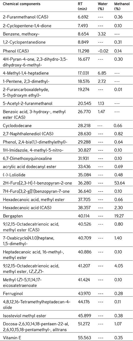
The chemical compositions of the water and methanol extracts of F. carica leaves were studied using GC-MS and the results are shown in Table 1. According to the GC-MS results, more different components were obtained in the methanol extract than in the aqueous extract of F. carica. Namely, six and 28 different compounds were determined in the water and methanol extracts, respectively. While the most abundant components are benzene, methoxy-(3.32%), 4-methyl-1,4-heptadiene (6.85%), 1-pentene, 2,3-dimethyl-(2.72%) for the water extract, they were 2H-furo[2,3-H]-1-benzopyran-2-one (53.64%), bergapten (19.27%), 9,12,15-octadecatrienoic acid, methyl ester, (Z,Z,Z)-(4.05%) for the methanol extract.
Table 1. Chemical components of the water and methanol extracts from F. carica.

When the extracts of the F. carica leaves were compared, we can see that the solubility of the methanol extracts was much greater than that of the water extracts, because the number of components is much higher in the methanol extracts. However, when we compare the enzyme inhibition activities, the water extracts showed higher inhibition activities than the methanol extracts (Table 2). This is most likely caused by the water extracts’ components. It is also interesting that almost none of these components are present in the methanol extract. Likewise, in the study conducted by Konyalıoğlu,31 the amount of alpha-tocopherol in fig leaves was determined by HPLC and correlated with antioxidant activity. In our study, GC-MS analysis of the F. carica leaves shows that the antioxidant vitamin alpha-tocopherol (vitamin E) was found in the methanol extract.
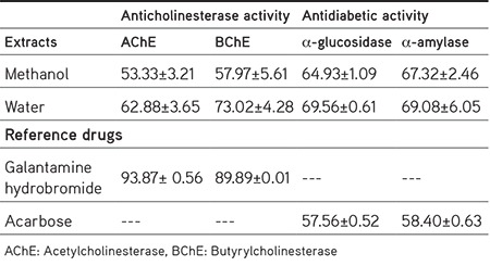
Table 2. Enzyme inhibitory activity (%) of the water and methanol extracts from F. carica leaves (at 2 mg/mL concentrations).

In vitro antioxidant activity
In vitro radical scavenging activity
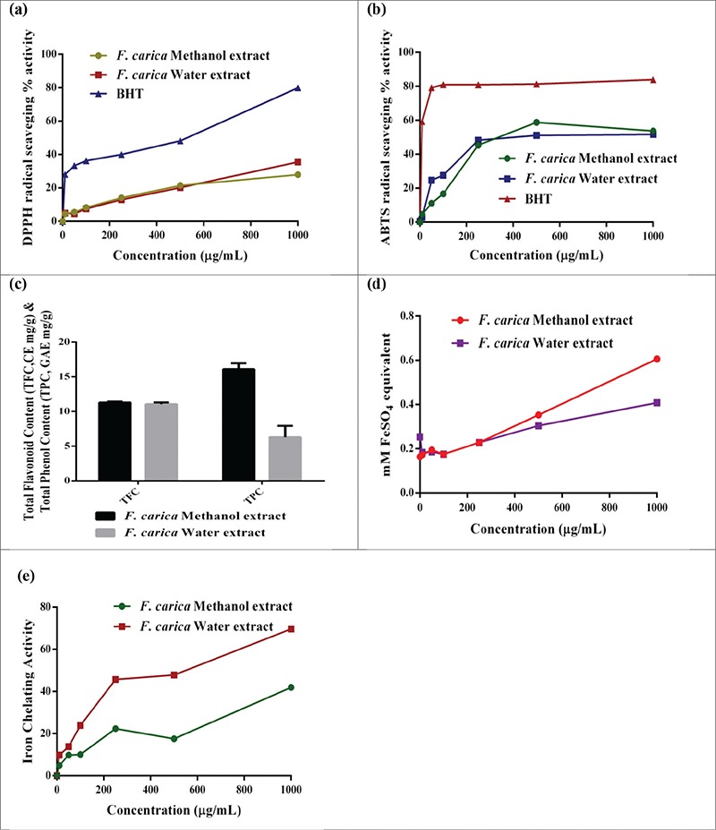
In some physiopathologic circumstances, there is excessive production of free radicals, leading to the occurrence of oxidative stress. This later is related to the appearance of many diseases including Alzheimer’s diseases, cardiovascular disease, and cancer.7,8 Natural antioxidants inhibit their activity by different mechanisms such as scavenging of reactive oxygen species, metal chelating, activation of antioxidant enzymes, and inhibition of oxidase. Therefore, it is necessary to use different methods to evaluate the antioxidant activity of extracts in plants. Previous studies have shown that the fig of F. carica has antioxidant activity.32 In our study, leaf extract of F. carica scavenged DPPH and ABTS radicals in a concentration-dependent manner. As shown in Figure 1a and 1b, the IC50 of ABTS radical scavenging activity of the methanol and water extract was 559.39 µg/mL and 428.51 µg/mL, while DPPH scavenging activity was 1.45 mg/mL and 1.83 mg/mL, respectively.
Figure 1.

In vitro antioxidant activity of the methanol and water extracts from F. carica leaves; a) DPPH radical scavenging activity; b) ABTS radical scavenging activity; c) total phenol and total flavonoid contents; d) ferric reducing power as FeSO4 equivalent; e) iron chelating activity.
The total phenolics (mg GAE/g of sample) and flavonoid (mg CE/g of sample) in the different extract of the F. carica leaves are exhibited in Figure 1c. F. carica leaf methanol extract (16.11 mg GAE/g) exhibited higher phenolic contents as compared to water extract (6.29 mg GAE/g), while the total flavonoid content was almost the same as that of methanol (11.29 mg CE/g) and water (11.06 mg CE/g) extract. The phenolic compounds in fig leaves were quantitatively determined using HPLC-DAD by Teixeira et al.33 We also achieved similar results using a different method in our study. In another study, by Ali et al.,34 it was shown that antioxidant and anti-inflammatory activities of fig leaves are associated with flavonoids and phenolic compounds found in the leaves.
It is well known that the ferrous and cupric ions stimulate lipid oxidation by breaking down hydrogen and lipid peroxides to reactive free radicals via the Fenton reaction. Therefore, metal chelating agents play an important role in terms of retarding the radical degradation by reducing the concentration of transition metal.35 According to our results, water extracts exhibited better iron chelating activity than methanol extract (Figure 1e).
In the FRAP assay, the reductants (antioxidants) present in the extract reduce a Fe3+–TPTZ complex to form blue Fe2+–TPTZ. The change in absorbance at 593 nm is proportional to the FRAP value of the antioxidants in the sample.36 The results of the FRAP assay are given in Figure 1d. In this assay, the higher activity was noted for methanol extract than water extract at higher concentration, but the ferric reducing power was the same at the lower concentration.
AChE and BChE inhibition activity
The methanol and water extracts prepared from F. carica leaves were evaluated for their inhibitory effects against AChE and BChE, which are Alzheimer’s disease-related enzymes. The water extract exhibited stronger activity and showed 63% and 73% inhibition of AChE and BChE, which was lower than the standard drug galantamine (with 93% and 90% inhibition) at the same concentration (Table 2). According to the report by Ahmad et al.37, the n-butanol fractions displayed the best anti-AChE activity, while ethyl acetate soluble fraction demonstrated the best anti-BChE activity among different solvent fractions of F. carica fruit. In the study by Orhan et al.,38 the n-hexane and acetone extracts of leaves exhibited notable inhibition activity against both AChE and BChE. However, in our study, the aqueous extract was found to be more active than the methanol in terms of these two enzyme inhibitions. This may be due to the more polar compounds present in the aqueous extract active against AChE and BChE enzyme inhibition.
In vitro α-glucosidase and α-amylase enzyme inhibition activity
It is known that α-amylase and α-glucosidase are enzymes that catalyze the hydrolysis of polysaccharides and disaccharides to monosaccharides. The inhibition of these two enzymes hinders the rapid uptake of blood glucose levels by delaying the digestion of carbohydrates.39 The results of the inhibitory activity of the F. carica leaf methanol and water extracts against α-glucosidase and α-amylase enzyme are presented in Table 2. When compared to each other, the water extract (69.56% and 69.08%) was found to be higher than the methanol extract (64.93% and 67.32%) in inhibiting α-glucosidase and α-amylase enzyme activity, and both extracts were found to be potential inhibitors against α-glucosidase and α-amylase compared with the standard antidiabetic drug acarbose (57.56% and 58.4%) at the same concentration (2 mg/mL). In a recent study, similar antidiabetic activities were reported for the ethyl acetate and ethanol extracts of F. carica fruit.39 In another study, the ethyl acetate extract of F. carica leaves showed antidiabetic activity by stimulating insulin production from regenerated pancreas beta cells.40 Similar results were reported for the water and methanol extracts of F. carica leaves in our study.
Antimicrobial activity
The antimicrobial activities of F. carica methanol and water extracts against S. aureus, E. faecalis, P. aeruginosa, E. coli, K. pneumoniae, and C. albicans were detected using the microdilution technique at the concentration range 0.156 to 2.5 mg/mL (Table 3). It has been reported that the antimicrobial activity of plant extracts was evaluated as significant with MIC value less than or equal to 0.1 mg/mL, moderate with 0.1< MIC ≤0.625 mg/mL, and weak with MIC value greater than 0.625 mg/mL.41 According to these criteria, the methanol extract of F. carica showed moderate antimicrobial activities against E. coli (0.625 mg/mL) and S. aureus (0.156 mg/mL) and weak antimicrobial activity against the other bacteria and C. albicans (≥2.5 mg/mL). Similarly, the water extract of F. carica displayed moderate antimicrobial activity against S. aureus (0.625 mg/mL) and weak antimicrobial activity against the other bacteria and C. albicans (≥2.5 mg/mL).
Table 3. Antimicrobial activity results of F. carica methanol and water extracts.

F. carica methanol extract has been studied against various bacteria and showed moderate to strong antibacterial activity. In an in vitro study, Jeong et al.42 reported that F. carica methanol extract had strong antibacterial activity on oral bacteria. In another study, Keskin et al.43 investigated the antimicrobial activity of different extracts of F. carica. Their study reported that the MIC values of the methanol and aqueous extracts of F. carica against bacteria and C. albicans were MIC 25-400< µg/mL and MIC 200-400< µg/mL, respectively.43 In the present study, E. coli and S. aureus were more susceptible to the methanol extract. Our results revealed that the methanol and water extracts of F. carica exhibited weak antimicrobial effects against other bacteria and C. albicans.
Cell viability
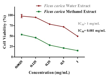
The XTT cell proliferation assay was used to evaluate the antiproliferative effects of the water and methanolic extracts of F. carica on MDA-MB-231 and L929 cell lines. As shown in Figure 2, the methanol extract at all concentrations significantly inhibited MDA-MB-231 cell proliferation (p<0.05) in a dose-dependent manner (IC50=0.081 mg/mL). On the other hand, concentration of 1 mg/mL of the water extract moderately decreased the cell viability (IC50>1 mg/mL) (p<0.05). However, neither extract exhibited any significant cytotoxicity on the L929 cell line in the concentrations range (1-0.0625 mg/mL).
Figure 2.

Effects of the water and methanol extracts from F. carica on the viability of MDA-MB-231 cell line, after treatment with various concentrations (range: 0.065-1 mg/mL) for 24 h. Both extracts showed no toxicity in normal cells
Our cytotoxicity results clearly indicated that the methanol extract is more toxic than the water extract of F. carica. This may be due to the fact that the methanol extract has richer active ingredients than the water extract, as shown in Table 1. Additionally, the anticancer effects may be associated with antioxidant features due to its polyphenolic components quantity (Figure 1). To the best of our knowledge, this is the first study of the anticancer effect of fig leaf extracts on MDA-MB-231. However, different parts of F. carica and different extracts of fig leaf have already been found to be cytotoxic on various cancer cells such as the stomach and cervix.6,44
CONCLUSION
Overall, in this study, the components and antioxidant, antimicrobial, anticancer, enzyme inhibition, and antidiabetic effects of F. carica leaf methanol and water extracts were investigated. Despite the several antioxidant activities of F. carica leaves, to the best of our knowledge there are no reports on the comparative study of extracts with different polarity as well as other antioxidant methods such as iron chelating and ferric reducing power. Our results indicated that especially the methanol extract has strong anticancer, antioxidant, and antidiabetic activities. There is a correlation between anticancer and antioxidant activity and total phenolic content. Moreover, the richer chemical content of the methanol extract may be associated with higher biological activity. Consequently, the methanolic extract of the leaf of F. carica may be considered a potential therapeutic agent in cancer and diabetes mellitus. However, further studies, particularly in vivo experiments, are needed to verify these effects.
Footnotes
Conflicts of interest: No conflict of interest was declared by the authors.
References
- 1.Manda SC, Mukherjee KP, Saha K, Das J, Pal M, Saba PB. Hypoglycemic activity of Ficus carica L. leaves in streptozotocin-induced diabetic rats. Nat Prod Sci. 1997;3:38–41. [Google Scholar]
- 2.Alamgeer Iman S, Asif H, Saleem M. Evaluation of antihypertensive potential of Ficus carica fruit. Pharm Biol. 2017;55:1047–1053. doi: 10.1080/13880209.2017.1278611. [DOI] [PMC free article] [PubMed] [Google Scholar]
- 3.Turkoglu M, Pekmezci E, Kilic S, Dundar C, Sevinc H. Effect of Ficus carica leaf extract on the gene expression of selected factors in HaCaT cells. J Cosmet Dermatol. 2017;16:54–58. doi: 10.1111/jocd.12344. [DOI] [PubMed] [Google Scholar]
- 4.Joseph B, Raj SJ. Pharmacognostic and phytochemical properties of Ficus carica Linn - An overview. Int.J. PharmTech Res. 2011;3:8–12. [Google Scholar]
- 5.Buenrostro-Figueroa JJ, Velázquez M, Flores-Ortega O, Ascacio-Valdés JA, Huerta-Ochoa S, Aguilar CN, Prado-Barragán LA. Solid state fermentation of fig (Ficus carica L.) by-products using fungi to obtain phenolic compounds with antioxidant activity and qualitative evaluation of phenolics obtained. Process Biochem. 2017;62:16–23. [Google Scholar]
- 6.Hashemi SA, Abediankenari S, Ghasemi M, Azadbakht M, Yousefzadeh Y, Dehpour AA. The effect of fig tree latex (Ficus carica) on stomach cancer line. Iran Red Crescent Med J. 2011;13:272–275. [PMC free article] [PubMed] [Google Scholar]
- 7.Park EJ, Choi KS, Kwon TK. β-Lapachone-induced reactive oxygen species (ROS) generation mediates autophagic cell death in glioma U87 MG cells. Chem Biol Interact. 2011;189:37–44. doi: 10.1016/j.cbi.2010.10.013. [DOI] [PubMed] [Google Scholar]
- 8.Amessis-Ouchemoukh N, Ouchemoukh S, Meziant N, Idiri Y, Hernanz D, Stinco CM, Rodríguez-Pulido FJ, Heredia FJ, Madani K, Luis J. Bioactive metabolites involved in the antioxidant, anticancer and anticalpain activities of Ficus carica L., Ceratonia siliqua L. and Quercusilex L. extracts. Industrial Crops and Products. 2017;95:6–17. [Google Scholar]
- 9.Kumkrai P, Weeranantanapan O, Chudapongse N. Antioxidant, α-glucosidase inhibitory activity and sub-chronic toxicity of Derris reticulata extract: its antidiabetic potential. BMC Complement Altern Med. 2015;15:35. doi: 10.1186/s12906-015-0552-4. [DOI] [PMC free article] [PubMed] [Google Scholar]
- 10.Rhee IK, van de Meent M, Ingkaninan K, Verpoorte R. Screening for acetylcholinesterase inhibitors from Amaryllidaceae using silica gel thin-layer chromatography in combination with bioactivity staining. J Chromatogr A. 2001;915:217–223. doi: 10.1016/s0021-9673(01)00624-0. [DOI] [PubMed] [Google Scholar]
- 11.Tayoub G, Al-Odat M, Amer A, Aljapawe A, Ekhtiar A. Antiproliferative effects of Pancratium maritimum extracts on normal and cancerous cells. Iran J Med Sci. 2018;43:52–64. [PMC free article] [PubMed] [Google Scholar]
- 12.Birjandiana E, Motameda N, Yassa N. Crude methanol extract of Echinophora platyloba induces apoptosis and cell cycle arrest at S-phase in human breast cancer cells. Iran J Pharm Res. 2018;17:307–316. [PMC free article] [PubMed] [Google Scholar]
- 13.Sengupta P, Raman S, Chowdhury R, Lohitesh K, Saini H, Mukherjee S, et al. Evaluation of apoptosis and autophagy inducing potential of Berberis aristata, Azadirachta indica, and their synergistic combinations in parental and resistant human osteosarcoma cells. Front Oncol. 2017;7:296. doi: 10.3389/fonc.2017.00296. [DOI] [PMC free article] [PubMed] [Google Scholar]
- 14.Jeong MR, Kim HY, Cha JD. Antimicrobial activity of methanol extract from Ficus carica leaves against oral bacteria. J Bacteriol Virol. 2009;39:97–102. [Google Scholar]
- 15.Wang G, Wang H, Song Y, Jia C, Wang Z, Xu H. Studies on anti-HSV effect of Ficus carica leaves. Zhong Yao Cai. 2004;27:754–756. [PubMed] [Google Scholar]
- 16.Jeong MR, Cha JD, Lee YE. Antibacterial activity of Korean Fig (Ficus carica L.) against food poisoning bacteria. Korean J Food Cookery Sci. 2005;21:84–93. [Google Scholar]
- 17.Rashid KI, Mahdi NM, Alwan MA, Khalid LB. Antimicrobial activity of fig (Ficus carica Linn.) leaf extract as compared with latex extract against selected bacteria and fungi. JUBPAS. 2014;22:1620–1626. [Google Scholar]
- 18.Mawa S, Husain K, Jantan I. Ficus carica L. (Moraceae): Phytochemistry, Traditional uses and Biological Activities. Evid Based Complement Alternat Med. 2013;2013:974256. doi: 10.1155/2013/974256. [DOI] [PMC free article] [PubMed] [Google Scholar]
- 19.Sacchetti G, Maietti S, Muzzoli M, Scaglianti M, Manfredini S, Radice M, Bruni R. Comparative evaluation of 11 essential oils of different origin as functional antioxidants, antiradicals and antimicrobials in foods. Food Chem. 2005;91:621–632. [Google Scholar]
- 20.Miser Salihoglu E, Akaydin G, Caliskan Can E, Yardim Akaydin S. Evaluation of antioxidant activity of various herbal folk medicine. J Nutr Food Sci. 2013;3:1–9. [Google Scholar]
- 21.Re R, Pellegrini N, Proteggente A, Pannala A, Yang M, Rice-Evans C. Antioxidant activity applying an improved ABTS radical cation decolorization assay. Free Radic Biol Med. 1999;26:1231–1237. doi: 10.1016/s0891-5849(98)00315-3. [DOI] [PubMed] [Google Scholar]
- 22.Clarke G, Ting KN, Wiart C, Fry J. High correlation of 2,2-diphenyl-1-picrylhydrazyl (DPPH) radical scavenging, ferric reducing activity potential and total phenolics content indicates redundancy in use of all three assays to screen for antioxidant activity of extracts of plants from the Malaysian rainforest. Antioxidants (Basel). 2013;2:1–10. doi: 10.3390/antiox2010001. [DOI] [PMC free article] [PubMed] [Google Scholar]
- 23.Molan AL, Mahd AS. Iraqi medicinal plants: Total flavonoid contents, free-radical scavenging and bacterial beta-glucuronidase inhibition activities. IOSR-JDMS. 2014;13:72–77. [Google Scholar]
- 24.Chai T, Mohan M, Ong H, Wong F. Antioxidant, iron-chelating and anti-glucosidase activities of Typha domingensis Pers (Typhaceae) Trop J Pharm Res. 2014;13:67–72. [Google Scholar]
- 25.Wojdylo A, Oszmianski J, Czemerys R. Antioxidant activity and phenolic compounds in 32 selected herbs. Food Chemistry. 2007;105:940–949. [Google Scholar]
- 26.Yang H, Dong Y, Du H, Shi H, Peng Y, Li X. Antioxidant compounds from propolis collected in Anhui, China. Molecules. 2011;16:3444–3455. doi: 10.3390/molecules16043444. [DOI] [PMC free article] [PubMed] [Google Scholar]
- 27.Ellman GL, Courtney KD, Andres Jr V, Featherstone RM. A new and rapid colorimetric determination of acetylcholinesterase activity. Biochemical Pharmacology. 1961;7:88–95. doi: 10.1016/0006-2952(61)90145-9. [DOI] [PubMed] [Google Scholar]
- 28.Kumar D, Kumar H, Vedasiromoni JR, Pal BC. Bio-assay guided isolation of α-glucosidase inhibitory constituents from Hibiscus mutabilis leaves. Phytochem Anal. 2012;23:421–425. doi: 10.1002/pca.1375. [DOI] [PubMed] [Google Scholar]
- 29.Kumar D, Gupta N, Ghosh R, Gaonkar RH, Pal BC. α-Glucosidase and α-amylase inhibitory constituent of Carex baccans: Bio-assay guided isolation and quantification by validated RP-HPLC-DAD. J Funct Foods. 2013;5:211–218. [Google Scholar]
- 30.Eloff JN. A sensitive and quick microplate method to determine the minimal inhibitory concentration of plant extracts for bacteria. Planta Med. 1998;64:711–713. doi: 10.1055/s-2006-957563. [DOI] [PubMed] [Google Scholar]
- 31.Konyalıoğlu S, Sağlam H, Kıvçak B. α-Tocopherol, flavonoid, and phenol contents and antioxidant activity of Ficus carica leaves. J Pharm Biol. 2005;43:683–686. [Google Scholar]
- 32.Harzallah A, Bhouri AM, Amri Z, Soltana H, Hammami M. Phytochemical content and antioxidant activity of different fruit parts juices of three figs (Ficus carica L.) varieties grown in Tunisia. Ind Crops Prod. 2016;83:255–267. [Google Scholar]
- 33.Teixeira DM, Canelas VC, de Canto AM, Teixeira JMG, Dias CB. HPLC-DAD quantification of phenolic compounds contributing to the antioxidant activity of Maclura pomifera, Ficus carica and Ficus elastica extracts. J Anal Lett. 2009;42:2986–3003. [Google Scholar]
- 34.Ali B, Mujeeb M, Aeri V, Mir SR, Faiyazuddin M, Shakeel F. Anti-inflammatory and antioxidant activity of Ficus carica Linn. leaves. Nat Prod Res. 2012;26:460–465. doi: 10.1080/14786419.2010.488236. [DOI] [PubMed] [Google Scholar]
- 35.Halliwell B, Gutteridge JM. Oxygen toxicology, oxygen radicals, transition metals and disease. Biochem J. 1984;219:1–14. doi: 10.1042/bj2190001. [DOI] [PMC free article] [PubMed] [Google Scholar]
- 36.Szeto YT, Tomlinson B, Benzie IF. Total antioxidant and ascorbic acid content of fresh fruits and vegetables: implications for dietary planning and food preservation. Br J Nutr. 2002;87:55–59. doi: 10.1079/BJN2001483. [DOI] [PubMed] [Google Scholar]
- 37.Ahmad S, Bhatti FR, Khaliq FH, Younas T, Madni MA, Latif A. In vitro enzymatic investigation of Ficus carica (Fruit) Pak. J. Pharm. Sci. 2016;29:1541–1544. [PubMed] [Google Scholar]
- 38.Orhan IE, Ustün O, Sener B. Estimation of cholinesterase inhibitory and antioxidant effects of the leaf extracts of Anatolian Ficus carica var. domestica and their total phenol and flavonoid contents. Nat Prod Commun. 2011;6:375–378. [PubMed] [Google Scholar]
- 39.Mopuri R, Ganjayi M, Meriga B, Koorbanally NA, Islam MS. The effects of Ficus carica on the activity of enzymes related to metabolic syndrome. J Food Drug Anal. 2018;26:201–210. doi: 10.1016/j.jfda.2017.03.001. [DOI] [PMC free article] [PubMed] [Google Scholar]
- 40.Stephen Irudayaraj S, Christudas S, Antony S, Duraipandiyan V, Naif Abdullah AD, Ignacimuthu S. Protective effects of Ficus carica leaves on glucose and lipids levels, carbohydrate metabolism enzymes and β-cells in type 2 diabetic rats. Pharm Biol. 2017;55:1074–1081. doi: 10.1080/13880209.2017.1279671. [DOI] [PMC free article] [PubMed] [Google Scholar]
- 41.Kuete V. Potential of Cameroonian plants and derived products against microbial infections: A review. Planta Med. 2010;76:1479–1491. doi: 10.1055/s-0030-1250027. [DOI] [PubMed] [Google Scholar]
- 42.Jeong MR, Kim HY, Cha JD. Antimicrobial activity of methanol extract from Ficus carica leaves against oral bacteria. J Bacteriol Virol. 2009;39:97–102. [Google Scholar]
- 43.Keskin D, Ceyhan Guvensen N, Zorlu Z, Ugur A. Phytochemical analysis and antimicrobial activity of different extracts of fig leaves (Ficus carica L.) from West Anatolia against some pathogenic microorganisms. J Pure Appl Microbiol. 2012;6:1105–1110. [Google Scholar]
- 44.Khodarahmi GA, Ghasemi N, Hassanzadeh F, Safaie M. Cytotoxic effects of different extracts and latex of Ficus carica L. on HeLa cell line. Iran J Pharm Res. 2011;10:273–277. [PMC free article] [PubMed] [Google Scholar]


