Abstract
Bilateral agenesis of the long head of the biceps brachial tendon (LHB) is a very rare variation of the anatomy. We report a case of an 18-year-old man with bilateral agenesis of the long head of the biceps brachii tendon. We present initial findings, radiographical examinations and the follow-up of an unusual entity. Diagnosis of agenesis of the LHB can be challenging especially in cases of traumatic shoulder pain. It is not a very known entity because of its rareness. However, it could be associated with other congenital anomalies. The absence of the LHB is easily ignored in the diagnostic process. Clinical examination should be a pitfall, radiological examination is helpful to confirm the suspicion of LHB absence. MRI is often the first choice, although ultrasonography is cheaper and much easier to access and it is an excellent tool to visualise this anatomic variation with empty or shallow intertubercular groove.
Keywords: musculoskeletal and joint disorders, emergency medicine, trauma, orthopaedics, radiology
Background
The biceps brachii muscle has anatomical diversity in human’s body. It has two tendinous origins, the long head takes origin at the supraglenoid tubercle of the scapula while the short head origin is at the coracoid process. Muscle bellies of the biceps join in the middle upper arm to form a single muscle mass unite to a single tendinous insertion on the radial tuberosity of the proximal radius. Congenital absence or capsular insertion of the long head of the biceps tendon is rare with an unknown incidence.1–4 The long head of the biceps brachial tendon (LHB) variations can be misleading, especially in cases of traumatic shoulder pain. Its rareness makes it difficult to distinguish it from ruptures of the LHB on physical examination. Clinical diagnosis is extremely challenging and somewhat impossible due to the rarity of the condition.5 The LHB plays a functional role in shoulder stability acting dynamically through the range of motion as well as via depression of the humeral head and serving as anchorage point of the superior glenoid labrum.6 To the best of our knowledge, few cases have been described so far.7 Clinical, radiological and peroperative findings are exposed.
Case presentation
We report a rare case of a healthy 18-year-old man without any other health disease or anatomical abnormality, presenting himself to the emergency department after a fall on the right shoulder during playing football. Physical examination shows a normal contour and pain in the shoulder with limitation of the range of motion (flexion 90°, abduction 90° and free rotations) associated to a painful Jobe’s test. He had full strength (5/5) with resisted external rotation, belly press, lift off and bear hug tests. He did not demonstrate signs of laxity or hypermobility in his upper extremities (negative sulcus sign). Relocation test was also negative. He has no ‘Popeye’ sign to reveal an LHB tendon torn. Clinical examination did not reveal any evidence of biceps pathology with a negative palm-up test and a negative O’Brien test. Acromioclavicular palpation or testing was pain free and there was no sign of subacromial inpingment. No X-rays were performed and the initial diagnosis was a simple contusion treated with paracetamol, non-steroidal anti-inflammatory drugs (NSAIDs) and a sling for comfort.
One week later, the patient returned to our emergency department because of the inability to work. Radiographs showed no abnormality (figure 1) and we decided to follow the initial treatment with paracetamol and NSAIDs. Two weeks after the trauma, on return follow-up, the patients shoulder was still painful with an increased limitation of the active range of motion (flexion 15°, abduction 15° and free rotations). Rest of the examination was the same as day 0. Regarding to the constant pain, an MRI was obtained, which showed what appeared to be a rupture of the long head of the biceps brachii tendon. A physiotherapy treatment associated with analgesia was prescribed.
Figure 1.
(A) Radiography of the right shoulder (Anteroposterior view in the standard radiological examination of the right shoulder). (B) Radiography of the right shoulder (Y view in the standard radiological examination of the right shoulder). (C) Radiography of the right shoulder (axillary view in the standard radiological examination of the right shoulder).
Outcome and follow-up
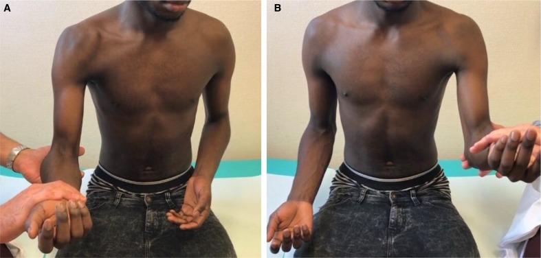
At 3 months after the trauma, follow-up examination showed no Popeye sign. O’Brien, palm-up and Speed’s tests were negative. A symmetric morphology of the two arms was observed and a free motion of the shoulder without any pain (figure 2). Repeated rotator cuff tests, acromioclavicular joint tests and subacromial impingement tests were all normal. Regarding to this uncommon case, repeat assessment of the MRI was done. It showed an agenesis of the LHB (figure 3). To complete the radiological examination, we proceed with a sonography of both intertubercular grooves (figure 4). Both grooves were empty with no evidence for the presence of LHB.
Figure 2.
(A) Clinical findings during physical examination while bending the right elbow against resistance and absence of any Popeye sign. (B) Clinical findings during physical examination while bending the left elbow against resistance and absence of any Popeye sign.
Figure 3.
(A and B) MRI of the right shoulder (axial view; blue arrow shows the empty intertubercular groove). (C) MRI of the right shoulder (coronal view; red arrow shows the absence of the long biceps tendon in the pulley). (D) MRI of the right shoulder (sagittal view; red arrow shows the absence of the long biceps tendon in the pulley).
Figure 4.
(A) Ultrasonographic images of the right shoulder showing the absence of the long biceps tendon in the intertubercular groove (blue arrow). (B) Ultrasonographic images of the left shoulder showing the absence of the long biceps tendon in the intertubercular groove (blue arrow).
Discussion
Bilateral agenesis of the LHB is a rare variation of the anatomy. Most of the cases reported are described after shoulder instability investigation.1 8 Congenital anomalies have been reported in five out of 11 patients so far in the literature (45%).8 Fetal insult at the sixth or seventh week of gestation, during biceps differentiation could explain the association with other congenital anomalies.3 9 This common associated finding in a rare condition should raise the surgeon’s index of suspicion for the possibility of absence of the LHB tendon when there is a history of congenital anomalies and shoulder pain and/or instability. Medical history should be reviewed accurately to detect other congenital anomalies when there is a diagnosis of absence of the LHB tendon. Biceps tests, such as palm-up, Speed’s and O’Brien, are not reliable for detecting intraoperative biceps pathology regarding to prevalent studies.10 11 The clinical examination could be a pitfall when suspicion arises for a rupture of LHB. Comparison of both sides is mandatory and can reveal reassuring symmetries.
Diagnosing pathology of the LHB provides challenges both on clinical and radiographical examination as was shown in our case. MRI is often the first choice, although ultrasonography is less expensive and much easier to access. It is an excellent tool to visualise anatomics variations with, for example, empty or shallow intertubercular groove as seen in cases of agenesis of the long head of the biceps brachii.
Shoulder arthroscopy has been described as the gold standard modality for diagnosis; its hallmark is the absence of the intra-articular portion of LHB in the presence of a shallow or absent bicipital groove.3 12
Furthermore, the detection of LHB agenesis calls into question the mechanical role of the LHB throughout the evolution. Since DePalma’s article about the Origin and Comparative Anatomy of the Pectoral Limb,13 we might suppose that maybe the increasing prominence of the coracoid process makes it more likely to encounter this kind of anatomical variation
Patient’s perspective.
I do not have pain, at rest and when using the arm.
I am happy with the result and have returned to all activities.
Learning points.
This variation is poorly understood because of its rarity.
It could be associated with other congenital anomalies.
Clinical examination should be thorough; radiological examination is helpful to confirm the suspicion.
Diagnosis of variation of the long head of the biceps brachial tendon may be more difficult or missed as the clinician may not be thinking of this condition when there are no congenital conditions existing that often associate with biceps agenesis.
Diagnosis can be challenging, especially in cases of traumatic shoulder pain.
Acknowledgments
We kindly thank our colleagues Patrick Goetti, Stefan Bauer and Ludovic Tapparel.
Footnotes
Contributors: AT: discuss planning and conception. KP: follow-up and writing. NG: research and corrections. AF: conception and design.
Funding: The authors have not declared a specific grant for this research from any funding agency in the public, commercial or not-for-profit sectors.
Competing interests: None declared.
Patient consent for publication: Obtained.
Provenance and peer review: Not commissioned; externally peer reviewed.
References
- 1.Glueck DA, Mair SD, Johnson DL. Shoulder instability with absence of the long head of the biceps tendon. Arthroscopy 2003;19:787–9. 10.1016/S0749-8063(03)00392-X [DOI] [PubMed] [Google Scholar]
- 2.Koplas MC, Winalski CS, Ulmer WH, et al. Bilateral congenital absence of the long head of the biceps tendon. Skeletal Radiol 2009;38:715–9. 10.1007/s00256-009-0688-8 [DOI] [PubMed] [Google Scholar]
- 3.Stadnick ME. Pathology of the long head of the biceps tendon, Radsource MRI web clinic, 2014. Available: https://radsource.us/pathology-of-the-long-head-of-the-biceps-tendon/
- 4.Wahl CJ, MacGillivray JD. Three congenital variations in the long head of the biceps tendon: a review of pathoanatomic considerations and case reports. J Shoulder Elbow Surg 2007;16:e25–30. 10.1016/j.jse.2006.10.020 [DOI] [PubMed] [Google Scholar]
- 5.Audenaert EA, Barbaix EJ, Van Hoonacker P, et al. Extraarticular variants of the long head of the biceps brachii: a reminder of embryology. J Shoulder Elbow Surg 2008;17:S114–7. 10.1016/j.jse.2007.06.014 [DOI] [PubMed] [Google Scholar]
- 6.Ghalayini SRA, Board TN, Srinivasan MS. Anatomic variations in the long head of biceps: contribution to shoulder dysfunction. Arthroscopy 2007;23:1012–8. 10.1016/j.arthro.2007.05.007 [DOI] [PubMed] [Google Scholar]
- 7.Kumar CD, Rakesh J, Tungish B, et al. Congenital absence of the long head of biceps tendon & its clinical implications: a systematic review of the literature. Muscles Ligaments Tendons J 2017;7:562–9. 10.32098/mltj.03.2017.21 [DOI] [PMC free article] [PubMed] [Google Scholar]
- 8.Foad A, Faruqui S. Case report: absence of the long head of the biceps brachii tendon. Iowa Orthop J 2016;36:88–93. [PMC free article] [PubMed] [Google Scholar]
- 9.Gardner E, Gray DJ. Prenatal development of the human shoulder and acromioclavicular joints. Am J Anat 1953;92:219–76. 10.1002/aja.1000920203 [DOI] [PubMed] [Google Scholar]
- 10.Bennett WF. Specificity of the speed's test: arthroscopic technique for evaluating the biceps tendon at the level of the bicipital groove. Arthroscopy 1998;14:789–96. 10.1016/S0749-8063(98)70012-X [DOI] [PubMed] [Google Scholar]
- 11.Lafosse L, Reiland Y, Baier GP, et al. Anterior and posterior instability of the long head of the biceps tendon in rotator cuff tears: a new classification based on arthroscopic observations. Arthroscopy 2007;23:73–80. 10.1016/j.arthro.2006.08.025 [DOI] [PubMed] [Google Scholar]
- 12.Rego Costa F, Esteves C, Melão L. Bilateral congenital agenesis of the long head of the biceps tendon: the beginning. Case Rep Radiol 2016;2016:4309213. 10.1155/2016/4309213 [DOI] [PMC free article] [PubMed] [Google Scholar]
- 13.DePalma AF. Origin and comparative anatomy of the pectoral limb : DePalma AF, Surgery of the shoulder. Philadelphia, PA: Lippincott Williams & Wilkins, 1950: 1–14. [DOI] [PMC free article] [PubMed] [Google Scholar]


