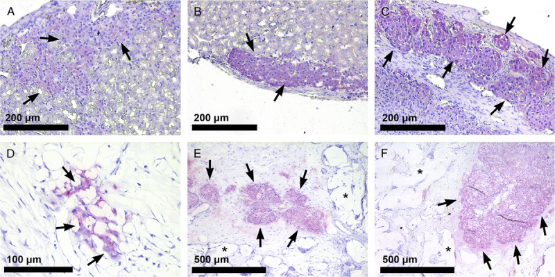
FIGURE 4.

Histological analysis of islets transplanted under the kidney capsule or in the PDLLCL scaffold. Insulin staining (SIGMAFAST Fast Red, arrows) of 400 (A), 800 (B), and 1200 (C) rat islets under the kidney capsule (20×) 10 weeks after transplantation. Insulin positive cells (40×) 10 weeks after transplantation of 400 islets into the subcutaneous PDLLCL scaffold (D) and insulin-positive islets (10×) after transplantation of 800 (E) and 1200 islets (F) in the polymer (*) scaffold.
