Dear Editor
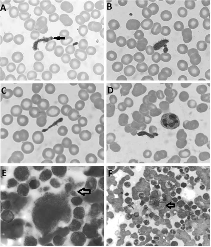
We report a 62-year-old Punjabi woman with past history of ischemic stroke and hypertension and was found to have thrombocytopenia (Platelet count- 18 × 109/L) on routine complete blood counts (CBCs). She did not have family history of any hematological illness. She presented with no bleeding manifestations and was receiving low dose Aspirin and anti-hypertensive medications (chlorthalidone for last 7 years) for her medical condition. Her previous platelet count which was available was 96 × 109/L, 3 years back when she suffered from stroke. The renal and liver function tests were within normal limits. Viral screen for Hepatitis B, Hepatitis C and Dengue virus was negative. The autoimmune work-up including anti-centromere, anti-Jo 1, anti-Scl-70, anti-Sm, anti-SSA/Ro, anti-SSB/Ra and anti-nRNP was negative. Peripheral smear and bone marrow examination was undertaken to further investigate thrombocytopenia. The peripheral smear showed many large and elongated proplatelets with staining characteristic similar to the mature platelets (Fig. 1a–d). The approximate platelet count including proplatelets on the peripheral smear was 40 × 109/L.
Fig. 1.
Proplatelets in the peripheral blood (a, b, c, d, Leishman stain, original magnification × 1000). a Nascent platelet bud at end of a proplatelet. e Megakaryocyte with psuedopodial extensions with branching (May Grunwald Giemsa, original magnification × 1000). f Sea-blue histiocyte (May Grunwald Giemsa, original magnification × 1000)
The bone marrow was mildly hypercellular and no dysplasia was noted in any of the major lineages. The megakaryocytes were mildly increased in number with mild increase of stage I megakaryocytes. Occasional megakaryocytes show branching psuedopodial cytoplasmic extensions (Fig. 1e). In addition occasional scattered sea-blue histiocytes were also noted (Fig. 1f).
The cytogenetic studies, conventional karyotyping and fluorescence in-situ hybridization (FISH) studies (Deletion 5q31, 7q31, 20q12 & trisomy 8) done to rule out any possibility of myelodysplastic syndrome doesn’t reveal any abnormality.
Patient was shifted from chlorthalidone to Amlodipine in view of possibility of drug induced thrombocytopenia. But platelet counts did not improve after changing the medicine even after 1 month. A final diagnosis of immune thrombocytopenia was made (ITP).
The proplatelets in peripheral blood have been reported in the past [1]. Sea-blue histiocytes have been reported earlier in association with ITP [2].
The mechanism of platelet generation is still not completely understood. The most accepted mechanism is modified flow model of platelet generation. In this model, platelets are generated through intermediate stage of proplatelet formation (psuedopodial extensions of megakaryocytes cytoplasm). Proplatelets are described as uniform, elongated tubular structures that have periodic platelet sized swellings along the length. The proplatelet formation starts with erosion of one pole of megakaryocyte to generate psuedopodial structures that elongate and branch multiple times to give rise to multiple free ends (Fig. 1e). In the end, the proplatelets tip acquires a single microtubule derived from the microtubule bundles of the proplatelet shaft and forms a circumferential coil that defines the territory of a nascent platelet bud (Fig. 1a). The nascent platelet bud acquires platelet specific substances and finally mature platelets are released from the protoplatelet end. The proplatelets can dissociate from the megakaryocyte body before a significant conversion into platelets and released proplatelets retain the capacity to generate further platelets. The platelet generation takes place from the both ends of proplatelet [3, 4].
This case beautifully highlights the various stages of platelet release. Also increased number of proplatelets in the peripheral blood can interfere with impedance based platelet counts by some hematology analyzers giving rise to spurious low platelet counts as in the present case. In addition, presence of increased proplatelets might indicate response to therapy in cases of immune thrombocytopenia indicating increased thrombopoiesis.
Conflict of interest
The authors declare that they have no conflict of interest.
Footnotes
Publisher's Note
Springer Nature remains neutral with regard to jurisdictional claims in published maps and institutional affiliations.
References
- 1.Bourrel A, Clauser S. Circulating proplatelet. Blood. 2013;122(22):3559. doi: 10.1182/blood-2013-06-511477. [DOI] [PubMed] [Google Scholar]
- 2.Rywlin AM, Hernandez JA, Chastain DE, Pardo V. Ceroid histiocytosis of spleen and bone marrow in idiopathic thrombocytopenic purpura (ITP): a contribution to the understanding of the sea-blue histiocyte. Blood. 1971;37(5):587–593. doi: 10.1182/blood.V37.5.587.587. [DOI] [PubMed] [Google Scholar]
- 3.Italiano JE, Jr, Lecine P, Shivdasani RA, Hartwig JH. Blood platelets are assembled principally at the ends of proplatelet processes produced by differentiated megakaryocytes. J Cell Biol. 1999;147(6):1299–1312. doi: 10.1083/jcb.147.6.1299. [DOI] [PMC free article] [PubMed] [Google Scholar]
- 4.Italiano JE, Jr, Patel-Hett S, Hartwig JH. Mechanics of proplatelet elaboration. J Thromb Haemost. 2007;5(Suppl 1):18–23. doi: 10.1111/j.1538-7836.2007.02487.x. [DOI] [PubMed] [Google Scholar]


